. 2021 Apr;28(3-4):162-176.
doi: 10.1038/s41434-020-00200-2. Epub 2020 Oct 22.

Sensitization of ON-bipolar cells with ambient light activatable multi-characteristic opsin rescues vision in mice

Affiliations

Sensitization of ON-bipolar cells with ambient light activatable multi-characteristic opsin rescues vision in mice

Subrata Batabyal et al. Gene Ther. 2021 Apr.

Abstract

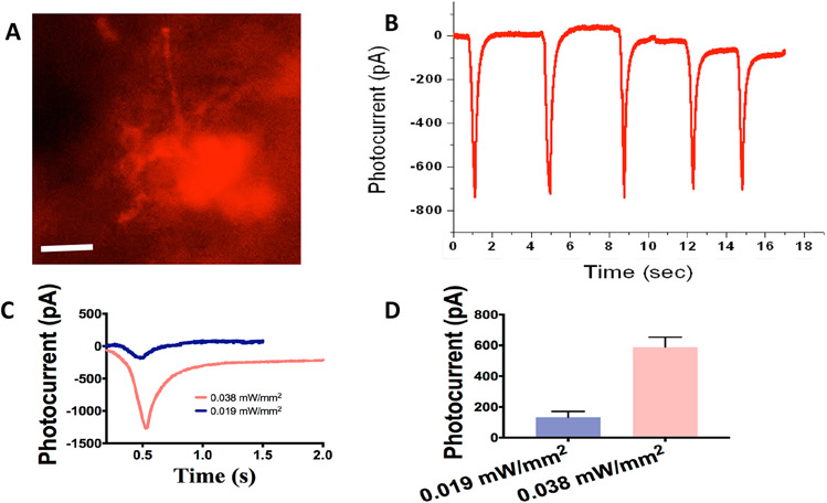
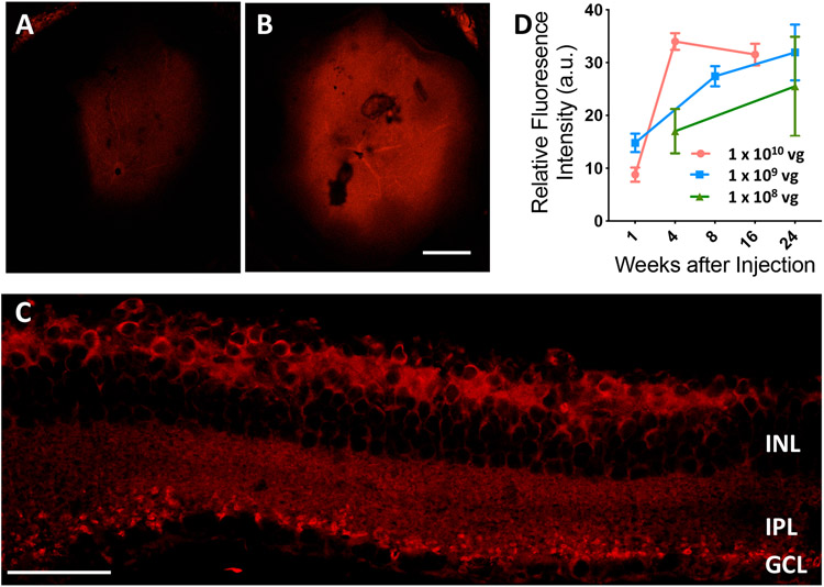
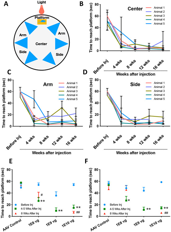
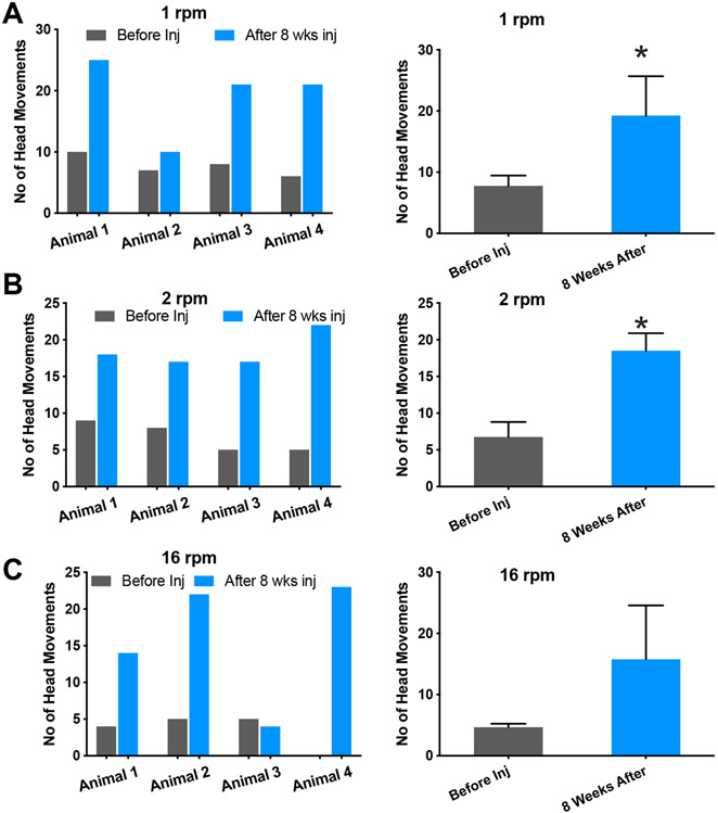
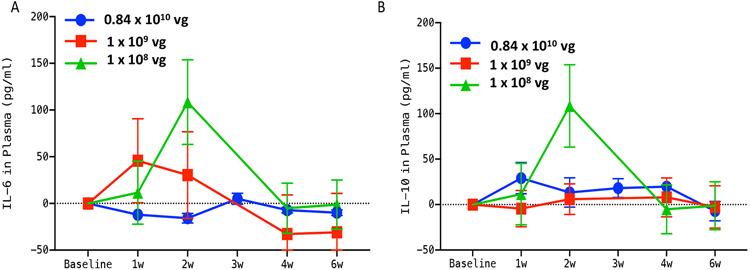
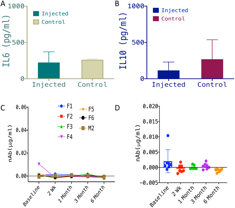
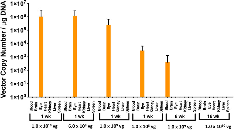
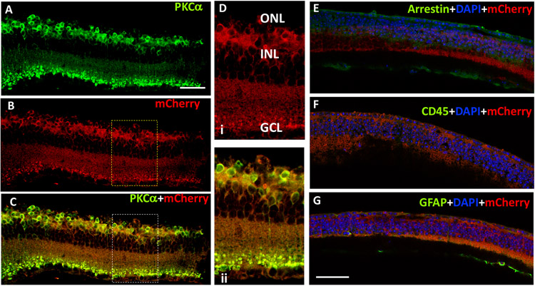
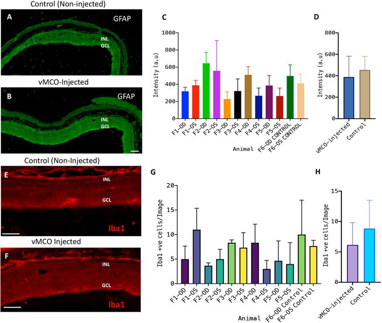
Gene therapy-based treatment such as optogenetics offers a potentially powerful way to bypass damaged photoreceptors in retinal degenerative diseases and use the remaining retinal cells for functionalization to achieve photosensitivity. However, current approaches of optogenetic treatment rely on opsins that require high intensity light for activation thus adding to the challenge for use as part of a wearable device. Here, we report AAV2 assisted delivery of highly photosensitive multi-characteristic opsin (MCO1) into ON-bipolar cells of mice with retinal degeneration to allow activation by ambient light. Rigorous characterization of delivery efficacy by different doses of AAV2 carrying MCO1 (vMCO1) into targeted cells showed durable expression over 6 months after delivery as measured by reporter expression. The enduring MCO1 expression was correlated with the significantly improved behavioral outcome, that was longitudinally measured by visual water-maze and optomotor assays. The pro/anti-inflammatory cytokine levels in plasma and vitreous humor of the vMCO1-injected group did not change significantly from baseline or control group. Furthermore, biodistribution studies at various time points after injection in animal groups injected with different doses of vMCO1 showed non-detectable vector copies in non-targeted tissues. Immunohistochemistry of vMCO1 transfected retinal tissues showed bipolar specific expression of MCO1 and the absence of immune/inflammatory response. Furthermore, ocular imaging using SD-OCT showed no change in the structural architecture of vMCO1-injected eyes. Induction of ambient light responsiveness to remaining healthy bipolar cells in subjects with retinal degeneration will allow the retinal circuitry to gain visual acuity without requiring an active stimulation device.


Figures

Fig. 1:
Fig. 1:. Expression of ambient light activatable Multi-Characteristic Opsin (MCO1) in retinal explant of rd10 mice led to significant photocurrent.
(A) Representative image showing MCO1 expression in the mice (12 weeks old) retinal explants (4 days after transfection by lipofection). Scale bar: 20 μm. (B) Representative inward photocurrent induced by light pulse (100 ms) train at intensity of 0.038 mW/mm2. (C) Representative profiles of inward photocurrent in retinal cell expressing MCO1 at two different light intensities. (D) Variation of photocurrent as a function of stimulation light intensity. Average ± S.D. N= 3 cells (n= 7-14 sweeps/ intensity).
Fig. 2:
Fig. 2:. Kinetics of vMCO1-dose dependent expression in mouse retina.
Representative low magnification (4X) fluorescence confocal image of retina cup after (A) 1 week, (B) 8 weeks after intravitreal injection of vMCO1 (1.0 x 10¹⁰ vg in eye) into rd10 mice (12 weeks old). Scale bar: 200 μm. (C) Confocal mCherry fluorescence image of cross-section of retina transfected with vMCO1 at dose of 1.0 x 10¹⁰ vg. Scale bar: 50 μm. (D) Kinetics of MCO1 expression quantified by means of measured reporter gene (mCherry) expression in rd10 mice retina at three different doses of vMCO1. Average ± SD. N=4.
Fig. 3:
Fig. 3:. Intravitreal injection of vMCO1 in rd10 mice led to ambient-light guided locomotion in a longitudinal manner.
(A) Schematic of water maze set up representing different locations and platform. (B) Time to reach platform (latency) by the mice (injected at age of 12 weeks) from center of the maze (light intensity: 7 μW/mm2) as a function of 16-week post-injection period. (C) Latency to reach platform by the mice from near arm of the maze (light intensity: 1 μW/mm2) as a function of 16-week post-injection period. (D) Time to reach platform from the side of the maze (light intensity: 2 μW/mm2) as a function of 16-week post-injection period. Intravitreal vMCO1 dose (1.0x10¹⁰, 1.0x10⁹ and 1.0x10⁸ vg) response of mice along with AAV2 vehicle control measured by water maze latency score for center (E) and side (F) at baseline, 4-5 weeks and 8 weeks after injection. N=5; Average ± S.D., *p<0.05 between Before and 4-5 weeks, **p<0.01 between Before and 4-5 weeks, #p<0.05 between Before and 8 weeks, ##p<0.01 between Before and 8 weeks, No statistical difference between 4 weeks and 8 weeks all dose.
Fig. 4:
Fig. 4:. Improvement of optomotor response in vMCO1 treated rd10 mice at ambient light level.
Quantitative comparison of number of head movement at different speed of rotation of the vertical stripes: (A) 1 rpm, and (B) 2 rpm, between before and 8 weeks after intravitreal injection of vMCO1 (1.0 x 10¹⁰ vg) in 12 weeks old mice. vMCO1 treated rd10 mice shows improved optomotor response as reflected in the increase head movement after vMCO1 injection. Average + SD. N=4. *p< 0.05. The average light intensity at the center of the chamber was 1 μW/mm2.
Fig. 5:
Fig. 5:. No detectable increase in inflammatory response in plasma of rd10 mice after vMCO1 injection.
(A) Quantitative comparison of change in IL-6 (pro-inflammatory marker) from baseline in plasma between group-1 (1.0 x 10⁸ vg in eye), group-2 (1.0 x 10⁹ vg) and group-3 (0.84 x 10¹⁰ vg) rd10 mice at 1,2, 3, 4, and 6 weeks after vMCO1 injection at the age of 12 weeks. (B) Quantitative comparison of change in IL-10 (anti-inflammatory marker) from baseline between the groups. Average ± SD. N=5 for 1.0 x 10⁸ vg and 1.0 x 10⁹ vg and N=3 for 0.84 x 10¹⁰ vg.
Fig. 6:
Fig. 6:. No detectable inflammatory response in vitreous humor or immune response in plasma of vMCO1 injected mice.
Quantitative comparison of IL-6 (pro-inflammatory marker) (A) and IL-10 (anti-inflammatory marker) (B) in vitreous humor of rd10 mice injected with vMCO1 (1.0 x 10⁹ vg in eye) and non-injected control, 6 months after vMCO1 injection (at the age of 12 weeks). Average ± SD. N=7. (C) Longitudinal monitoring of neutralizing Antibody (nAb) level in serum of rd10 mice injected with vMCO1 (1.0 x 10⁹ vg in eye) before and after injection (F: Female; M: Male) at 12 weeks of age. (D) Scatter plot showing the mean and variation of measured neutralizing antibody concentration in serum of vMCO-010 injected rd10 mice at each time point. N=7.
Fig. 7:
Fig. 7:. Bio-distribution show non-detectable levels of the vector in non-targeted organs of intravitreally-injected rd10 mice.
Distribution of the AAV2 vector was quantified by qPCR in 7 different tissues, including the injected eyes of the mice (age: 12 weeks), at different time points and vMCO1 doses (1.0x10¹⁰, 6.0x10⁹, 1.0x10⁹ and 1.0x10⁸ vg/eye). qPCR analysis showed the presence of AAV2 in treated eyes after first week of injection. Negligible quantities of AAV2 vector copies were identified in some tissues (not visible in the graph). Average ± SD. N=5.
Fig. 8:
Fig. 8:. Intravitreal injection of vMCO1 led to ON-bipolar specific expression in rd10 mice retina without causing inflammatory-response.
The cryo-sectioned retinal slices were immunostained using primary antibodies with different dilution (PKCα- 1:200; mCherry-1: 500; Arrestin-1: 1000; GFAP-1: 500; CD45-1: 500) and DyLight 488 (1: 500 dilution) as a secondary antibody. PKCα stain (green, A) showing bipolar cells expressing MCO1 (visualized by intrinsic mCherry expression 16 weeks after injection of vMCO-1 (1.0 x 10¹⁰ vg) in 12 weeks old rd10 mice, B). High levels of colocalization with virally expressed MCO1 in bipolar cells, evident in (C). (D) Representative high-resolution zoomed picture showing membrane expression of MCO1 in bipolar cells and overlay picture of mCherry and PKCα confirming co-localization. (E) Absence of S-arrestin (green) confirms complete loss of photoreceptors. (F) Absence of CD45 (green) marker suggests no detectable immune cells after vMCO1 injection. (G) GFAP (green) observed as reported in photoreceptor degenerated retina. Scale: 50 μm.
Fig. 9:
Fig. 9:. Intravitreal injection of vMCO1 did not cause cellular inflammatory response in retina of rd10 mice.
The cryo-sectioned retinal slices were immunostained using primary antibodies (Iba1 for microglia and GFAP for glial cells) and secondary antibodies (DyLight 488 for GFAP and Alexa Fluor 568 for Iba1). Representative confocal fluorescence microscope image of GFAP-stained retina of (A) non-injected control and (B) vMCO1 (3.5 x 10⁹ vg/eye) injected mice eyes 6 months after bilateral injection. Scale: 50 μm. (C) Quantification of fluorescence intensity of GFAP-labeled retina for individual mice (F1-F5) showing inter-animal variability along with the non-injected negative control. Average ± SD. N=3 retina slices/mouse. (D) No statistically significant difference in fluorescence intensity of GFAP-labeled retina between the vMCO1-injected group and negative control. Representative confocal fluorescence microscope image of Iba1-stained retina of (E) non-injected control and (F) vMCO1(3.5 x 10⁹ vg/eye) injected mice eyes 6 months after bilateral injection. Scale: 50 μm. (G) Quantification of Iba1+ve cells for individual mice (F1-F5) showing inter-animal variability along with the non-injected negative control. Average ± SD. N=3 retina slices/mouse. (H) No statistically significant difference in Iba1+ cells between the vMCO1-injected group and negative control.

References

    1. Takeda A, Baffi JZ, Kleinman ME, Cho WG, Nozaki M, Yamada K, et al. CCR3 is a target for age-related macular degeneration diagnosis and therapy. Nature. 2009;460(7252):225–U87. - PMC - PubMed
    1. Schuchard RA, Naseer S, de Castro K. Characteristics of AMD patients with low vision receiving visual rehabilitation. J Rehabil Res Dev. 1999;36(4):294–302. - PubMed
    1. Yang ZL, Camp NJ, Sun H, Tong ZZ, Gibbs D, Cameron DJ, et al. A variant of the HTRA1 gene increases susceptibility to age-related macular degeneration. Science. 2006;314(5801):992–3. - PubMed
    1. Haines JL, Hauser MA, Schmidt S, Scott WK, Olson LM, Gallins P, et al. Complement factor H variant increases the risk of age-related macular degeneration. Science. 2005;308(5720):419–21. - PubMed
    1. Humayun M, Dorn J, da Cruz L. Interim results from the international trial of Second Sight's visual prosthesis. Ophthalmology. 2012;119:779–88. - PMC - PubMed

Publication types