Review
. 2021 Feb;19(2):110-121.
doi: 10.1038/s41579-020-00448-w. Epub 2020 Oct 21.

The structural basis of herpesvirus entry

Affiliations
Review

The structural basis of herpesvirus entry

Sarah A Connolly et al. Nat Rev Microbiol. 2021 Feb.

Abstract

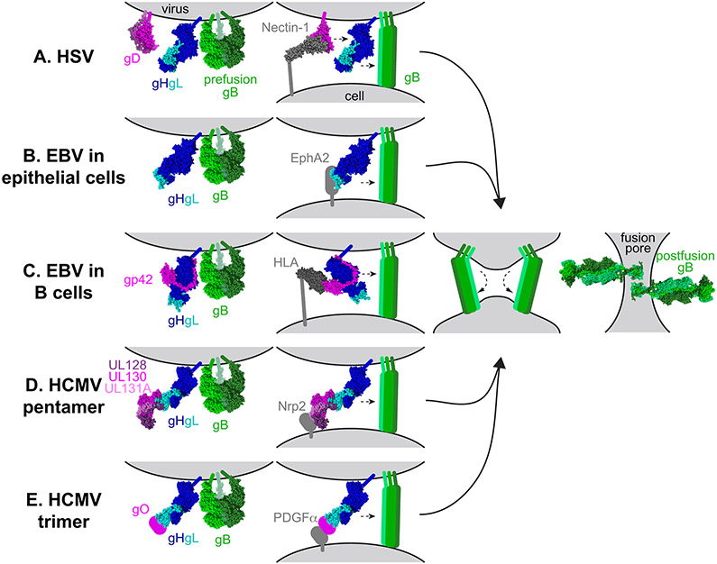
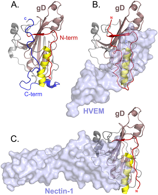
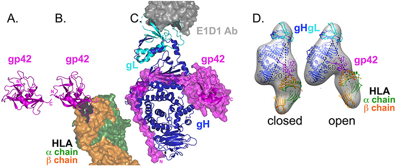
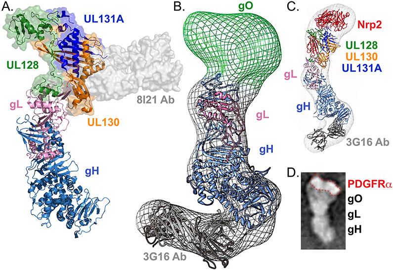
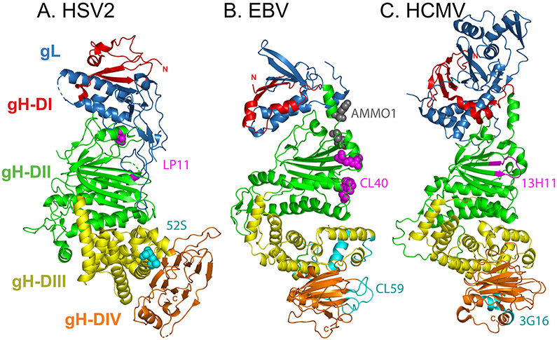
Herpesviruses are ubiquitous, double-stranded DNA, enveloped viruses that establish lifelong infections and cause a range of diseases. Entry into host cells requires binding of the virus to specific receptors, followed by the coordinated action of multiple viral entry glycoproteins to trigger membrane fusion. Although the core fusion machinery is conserved for all herpesviruses, each species uses distinct receptors and receptor-binding glycoproteins. Structural studies of the prototypical herpesviruses herpes simplex virus 1 (HSV-1), HSV-2, human cytomegalovirus (HCMV) and Epstein-Barr virus (EBV) entry glycoproteins have defined the interaction sites for glycoprotein complexes and receptors, and have revealed conformational changes that occur on receptor binding. Recent crystallography and electron microscopy studies have refined our model of herpesvirus entry into cells, clarifying both the conserved features and the unique features. In this Review, we discuss recent insights into herpesvirus entry by analysing the structures of entry glycoproteins, including the diverse receptor-binding glycoproteins (HSV-1 glycoprotein D (gD), EBV glycoprotein 42 (gp42) and HCMV gH-gL-gO trimer and gH-gL-UL128-UL130-UL131A pentamer), as well gH-gL and the fusion protein gB, which are conserved in all herpesviruses.


Figures

Fig. 1.
Fig. 1.. Model of the herpesvirus entry mechanism.
a∣ Herpes simplex viruses 1 and 2 (HSV-1 and HSV-2) fuse with a host cell at the plasma or endosomal membrane. The glycoprotein D (gD) dimer (pink), gH–gL heterodimer (dark and light blue), and gB trimer (green) are necessary and sufficient for entry (column 1). gD binds to one of several entry receptors, including nectin-1 (grey, column 2). Receptor binding displaces the carboxyl-terminus of the gD ectodomain and transmits a signal to gH–gL (small arrow). gH–gL activates the fusion protein gB (small arrow) to insert hydrophobic fusion loops into the cell membrane. b∣ Epstein–Barr virus (EBV) fuses with the plasma membrane of an epithelial cell. gH–gL (blue) and gB (green) are sufficient for fusion (column 1). The binding of gH–gL to EphA2 (grey) triggers gB to insert into the host cell (column 2). c∣ EBV fusion with B cells occurs in the endosome. A complex of gp42 (pink) and gH–gL (blue) binds to human leukocyte antigen (HLA) class II (grey). The binding triggers gB and may impact membrane orientation (column 2). d∣ Human cytomegalovirus (HCMV) entry into epithelial and endothelial cells occurs after endocytosis and requires a pentamer complex of gH–gL (blue) bound to UL128–UL130–UL131A (shades of pink) (column 1). Pentamer binding to Neuropilin-2 (Nrp2) triggers gB (column 2). e∣ HCMV entry into all cells requires a trimer complex comprised of gO and gH–gL (column 1). In fibroblasts, the trimer binds to platelet-derived growth factor receptor α (PDGFRα) and triggers gB at the plasma membrane (column 2). The reason that the trimer is required for entry into epithelial and endothelial cells is unclear currently. After inserting into the target cell membrane, gB folds back on itself (column 3). Fusion most likely requires more than one gB trimer to be triggered. The other entry glycoproteins are removed for the figure for clarity. As gB refolds into its postfusion conformation, the viral and cell membranes are fused, creating a fusion pore, through which the viral capsid can enter the cell (column 4).
Fig. 2.
Fig. 2.. Herpes simplex virus 1 glycoprotein D crystal structures.
a∣ A monomer from a dimeric form of glycoprotein D (gD) that was stabilized by introducing a disulfide bond at the carboxyl-terminus of the ectodomain (Protein Data Bank (PDB) ID 2C36). gD is oriented to show the receptor binding face. The core immunoglobulin fold of gD (pale pink) is flanked by amino-terminal (red) and C-terminal (blue) extensions. A α-helix (yellow) that supports the receptor binding site is shown and the N-termini and C-termini are marked. b∣ Herpesvirus entry mediator (HVEM) receptor (transparent surface rendering) bound to gD (PDB ID 1JMA). The N-terminus of gD (red) forms a loop that contains all of the contact residues for HVEM. c∣ Nectin-1 receptor (transparent surface rendering) bound to gD (PDB ID 3SKU). The HVEM and nectin-1 binding sites overlap. Binding of either HVEM or nectin-1 would displace the gD C-terminus.
Fig. 3.
Fig. 3.. Epstein–Barr virus glycoprotein 42 structures.
a∣ Crystal structure of glycoprotein 42 (gp42) alone (pink ribbons). The carboxyl-terminal C-type lectin domain (CTLD) is shown (Protein Data Bank (PDB) ID 3FD4). b∣ Crystal structure of gp42 (pink) bound to the human leukocyte antigen (HLA) class II receptor (green and orange surface rendering, partly shown) (PDB ID 1KG0). HLA binds to the gp42 CTLD and the gp42 amino-terminus is not resolved. HLA binding does not change the gp42 conformation drastically. c∣ Crystal structure of gp42–glycoprotein H (gH)–gL–E1D1 complex (PDB ID 5T1D). Although the CTLD of gp42 (pink ribbon with surface rendering) contacts gH–gL (blue and cyan ribbons, respectively), the majority of contacts lie in the gp42 N-terminal extension that extends down the length of gH–gL. Peptides from this gp42 N-terminus can inhibit entry into cells. E1D1 (grey surface rendering, partly shown) is a monoclonal antibody that binds to gL and partially neutralizes entry into epithelial cells but not B cells. d∣ Electron microscopy reconstructions of gp42–gH–gL–HLA complexes (grey surface). Fit inside the densities are the crystal structures of gH-gL (blue and cyan), gp42 (pink), and HLA (green and orange). Two conformations were observed: closed and open. In the closed conformation, HLA and gH–gL are arranged in a more parallel orientation than in the open conformation. Panel d is modified from ref. .
Fig. 4.
Fig. 4.. Structures of human cytomegalovirus glycoprotein H–glycoprotein L complexes.
a∣ Crystal structure of pentamer bound to mAb 8I21 (grey surface rendering). UL128 (green), UL130 (orange), and UL131A (dark blue) assemble on the distal tip of glycoprotein H (gH)–gL (light blue and pink, respectively), contacting an amino-terminal extension of gL that is unique to betaherpesviruses (Protein Data Bank (PDB) ID 5V0B). b∣ Electron microscopy (EM) reconstruction of gH–gL–gO trimer bound to monoclonal antibody (mAb) 3G16 (Electron Microscopy Data Bank entry EMD-6431). The gH–gL (blue and pink) and mAb (grey) structures have been fit inside the density. gO (green) maps to the distal end of gH–gL, in a position analogous to UL128–UL130–UL131A. c∣ EM reconstruction of pentamer (colored as in A) bound to mAb 3G16 (grey) and Neuropilin-2 (Nrp2) receptor (red) (EMD-8884). Crystal structures have been fit inside the EM density and Nrp2 maps to the distal tip of the complex, contacting the UL128–UL130–UL131A–gL portion of the complex. d∣ EM image of trimer bound to the receptor platelet-derived growth factor receptor α (PDGFRα). A comparison of gH–gL–gO with and without receptor demonstrates that the receptor binds at the gO side of the complex, outlined in red. Panel b is modified from ref. Panel c is modified from ref. . Panel d is modified from ref. .
Fig. 5.
Fig. 5.. Crystal structures of the glycoprotein H–glycoprotein L complex.
a∣ Herpes simplex virus 2 (HSV-2) glycoprotein H (gH)–gL (Protein Data Bank (PDB) ID 3M1C). Four progressive gH domains (DI–DIV) and gL are shown. gH DI (red) is intimately associated with gL (blue). gL requires gH for proper folding and anchoring to the membrane. DII (green) includes parallel β-sheets and helices. DIII (yellow) is mostly helical and DIV (orange) includes a β-sandwich. Although domain designations between the gH–gL structures can differ, specifically for distinction between DII and DIII, this figure uses the domain designations identified for Epstein–Barr virus (EBV) gH–gL. The carboxyl-terminus would extent from DIV into the transmembrane (TM) domain. The overall complex adopts a boot-like shape. Substitutions at HSV-1 gH residues 168 or 329 (magenta spheres) prevent binding of the neutralizing monoclonal antibody (nAb) LP11. On the opposite face, substitution mutations at gH residues 536 or 537 (cyan spheres) prevent binding of the nAb 52S (ref. ). b∣ EBV gH–gL (PDB 3PHF). The domains are colored as in part a and they adopt a more linear orientation. Co-crystallization revealed that nAb CL40 contacts gH residues 184, 239, 243, 284, and 286 (magenta spheres) and overlaps the binding site for the gp42 C-terminal domain. Electron microscopy reconstruction of bound nAb CL59 shows that this nAb binds to a distinct site within DIII and DIV (cyan ribbons; gH residues 406–415, 456–468, 494–503, 568–577, 623–626, and 645–656). The CL40 epitope partially overlaps with the nAb AMMO1 binding site (grey spheres; gH residues 73 and 76). c∣ Human cytomegalovirus (HCMV) gH–gL (PDB ID 5VOC) . Domains are colored as in part a. HCMV gL includes an amino-terminal extension that is absent from the other structures. Hydrogen deuterium exchange coupled to mass spectroscopy maps nAb 13H11 binding to gH residues 238–247 (magenta ribbons) and nAb 3G16 binding to gH residues 677–684 and 705–708 (cyan ribbons).
Fig. 6.
Fig. 6.. Glycoprotein B structures.
a∣ Crystal structure of full-length postfusion herpes simplex virus 1 (HSV-1) glycoprotein B (gB) (Protein Data Bank (PDB) ID 5V2S). The trimeric ectodomain is comprised of five domains (DI–DV). DI (blue) contains hydrophobic fusion loops (magenta sticks) that insert into the host cell membrane. DIII (yellow) includes an extended central trimeric coiled-coil, against which DV (red) packs in an anti-parallel orientation, as DV proceeds through DI, towards the transmembrane (TM) domain. The membrane proximal region (dark green), TM (dark purple), and cytoplasmic (pink) domains are shown. b∣ Crystal structure of postfusion Epstein–Barr (EBV) gB ectodomain (PDB 3FVC) . c∣ Crystal structure of postfusion human cytomegalovirus (HCMV) gB ectodomain (PDB ID 5CXF). For parts b and c, the domains are colored as in part a. For crystallization, residues in the fusion loops of EBV and HCMV gB were replaced with HSV-1 residues. d∣ Cryoelectron tomography (cryoET) reconstruction of the compact conformation of a mutant form of HSV-1 gB, 12 nm in height. Density fitting of the gB domains from the postfusion gB structure is shown. Domains are colored as in part a. The fusion loops of DI (light blue) are oriented towards the membrane. For comparison, the ectodomain of postfusion HSV-1 gB is shown at a similar scale on the left, at 16 nm in height. e∣ Alternative cryoET reconstruction of a compact form of HSV-1 gB, 9 nm in height. A pseudoatomic model of prefusion gB, based on the prefusion structure of vesicular stomatitis virus protein G (VSV G) (PDB ID 5I2S), was fit into the 3D electron microscopy (EM) reconstruction. Domains are colored as in part a. DV is excluded from the model. f∣ CryoET reconstruction of human cytomegalovirus (HCMV) gB (Electron Microscopy Data Bank entry EMD-9328), 13 nm in height. Domains from a single protomer are colored as in part a. Domain fitting using the postfusion HCMV structure placed DI (blue) near the membrane, with the fusion loops (magenta) oriented towards the membrane. As the EM reconstruction cannot accommodate the DIII postfusion conformation, the DIII fit was modeled on VSV G. DV is excluded from the model. Part d is modified from ref. . Part e is modified from ref. . Part f is modified from ref. .

Similar articles

Cited by

References

    1. Vallbracht M, Backovic M, Klupp BG, Rey FA & Mettenleiter TC Common characteristics and unique features: A comparison of the fusion machinery of the alphaherpesviruses Pseudorabies virus and Herpes simplex virus. Advances in virus research 104, 225–281, doi:10.1016/bs.aivir.2019.05.007 (2019). - DOI - PubMed
    1. Mohl BS, Chen J & Longnecker R Gammaherpesvirus entry and fusion: A tale how two human pathogenic viruses enter their host cells. Advances in virus research 104, 313–343, doi:10.1016/bs.aivir.2019.05.006 (2019). - DOI - PubMed
    1. Nishimura M & Mori Y Entry of betaherpesviruses. Advances in virus research 104, 283–312, doi:10.1016/bs.aivir.2019.05.005 (2019). - DOI - PubMed
    1. Atanasiu D, Saw WT, Cohen GH & Eisenberg RJ Cascade of events governing cell-cell fusion induced by herpes simplex virus glycoproteins gD, gH/gL, and gB. J Virol 84, 12292–12299, doi:10.1128/JVI.01700-10 (2010). - DOI - PMC - PubMed
    1. Nicola AV Herpesvirus Entry into Host Cells Mediated by Endosomal Low pH. Traffic 17, 965–975, doi:10.1111/tra.12408 (2016). - DOI - PMC - PubMed

Publication types

MeSH terms