. 2020 Nov;587(7832):121-125.
doi: 10.1038/s41586-020-2850-3. Epub 2020 Oct 21.

Cancer immunotherapy via targeted TGF-β signalling blockade in TH cells

Affiliations

Cancer immunotherapy via targeted TGF-β signalling blockade in TH cells

Shun Li et al. Nature. 2020 Nov.

Abstract

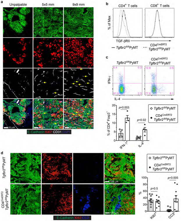
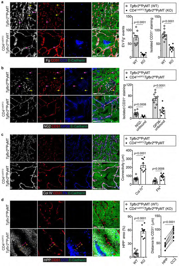
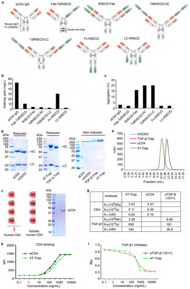
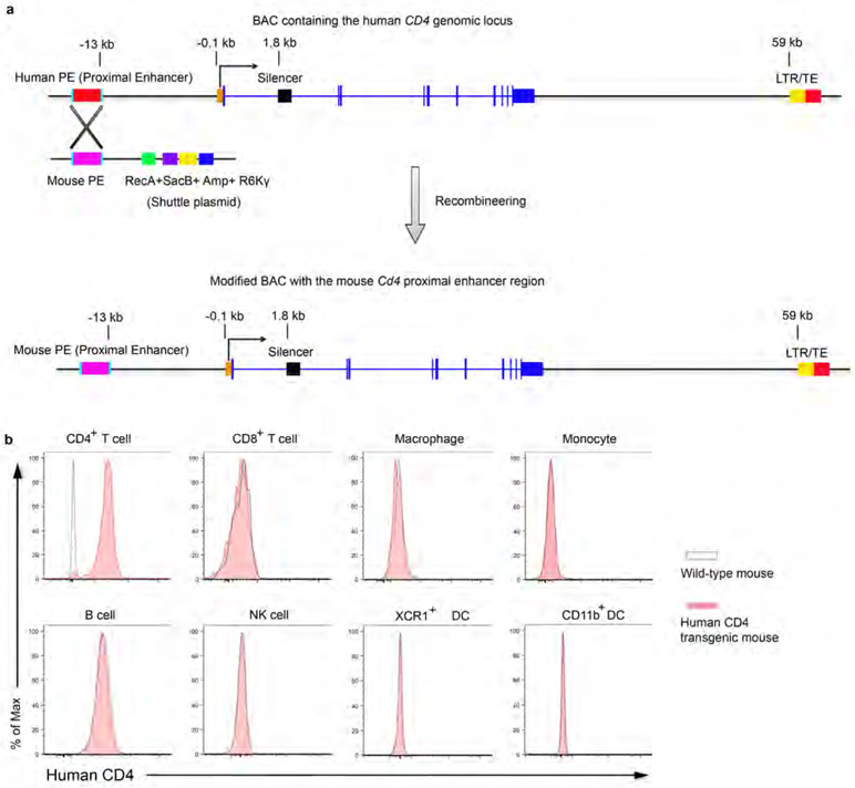
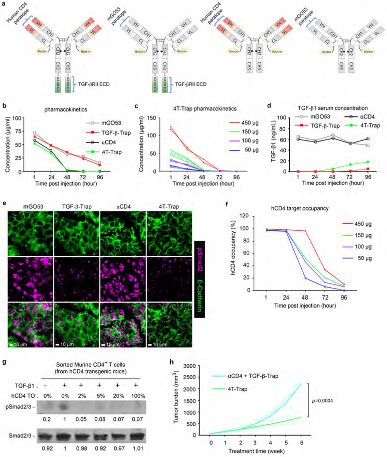
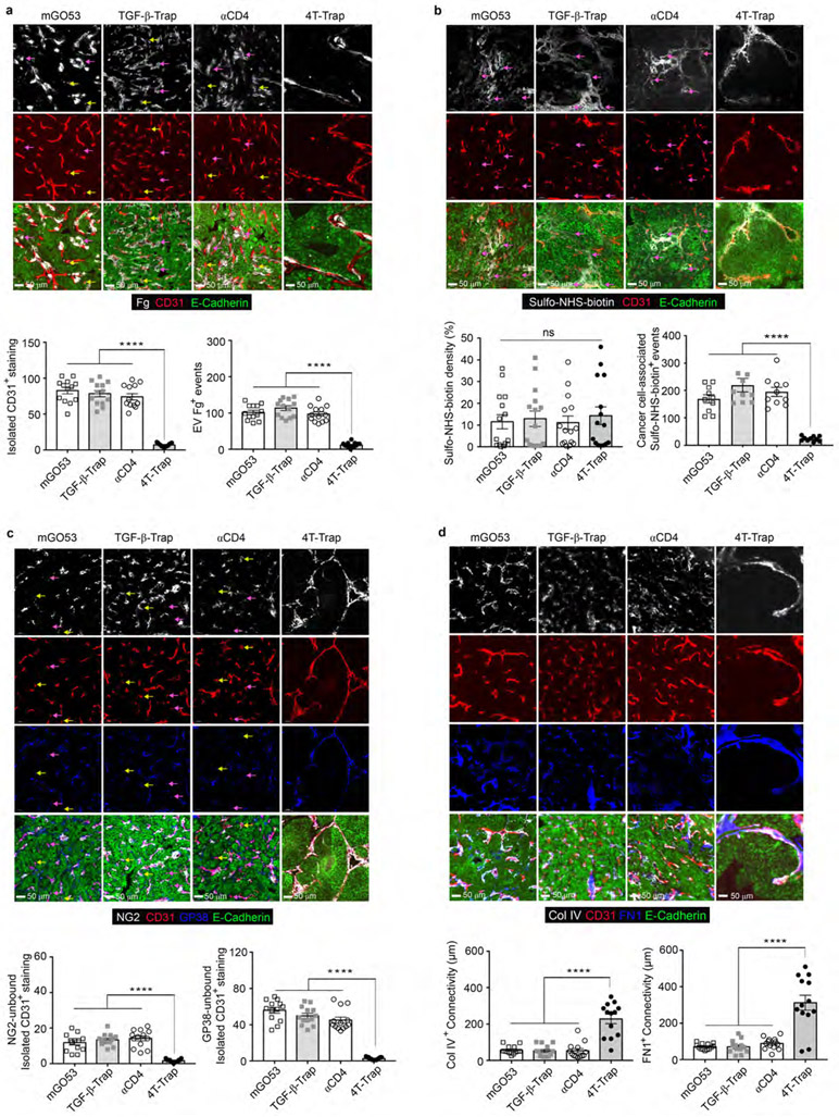
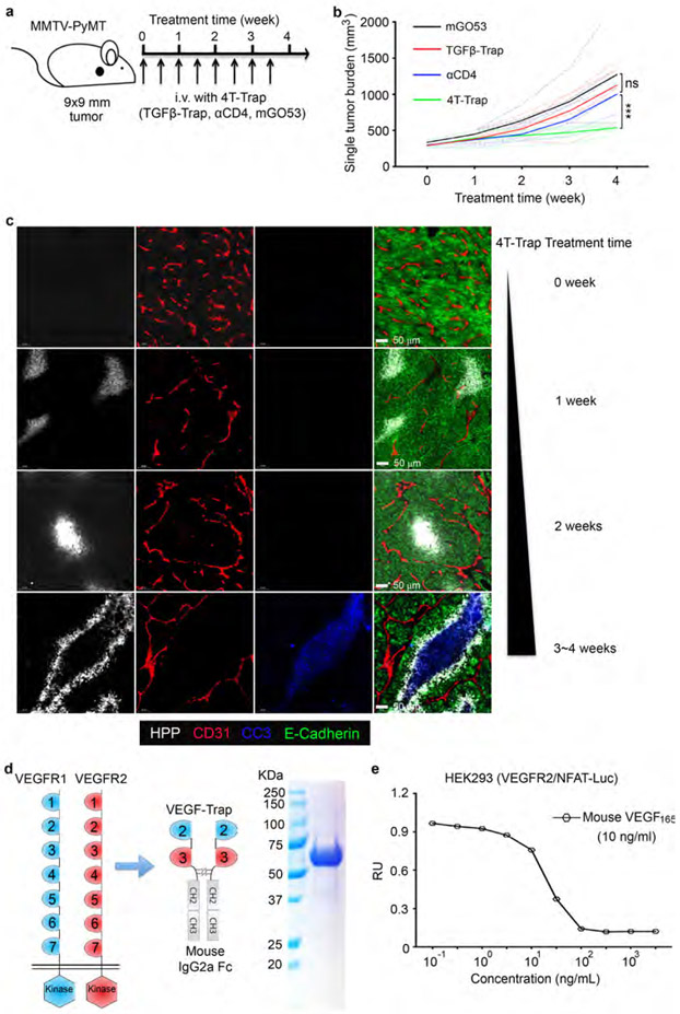
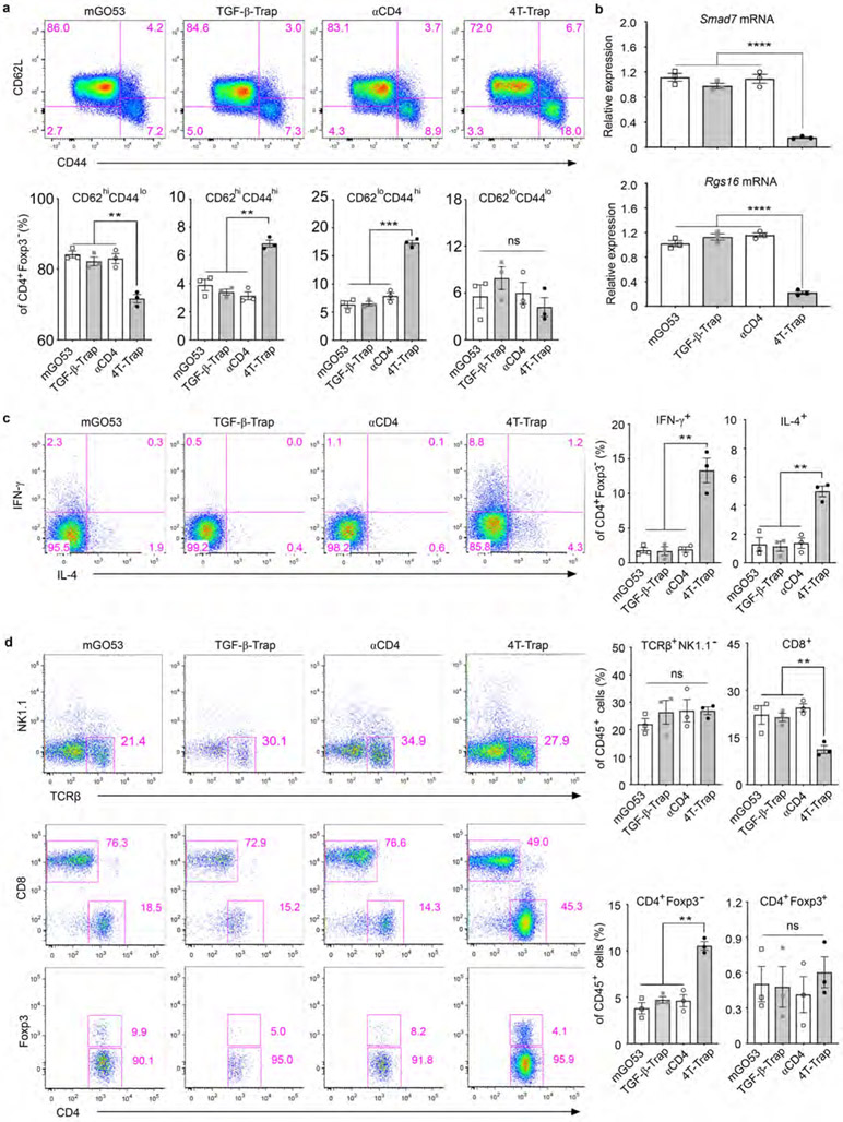
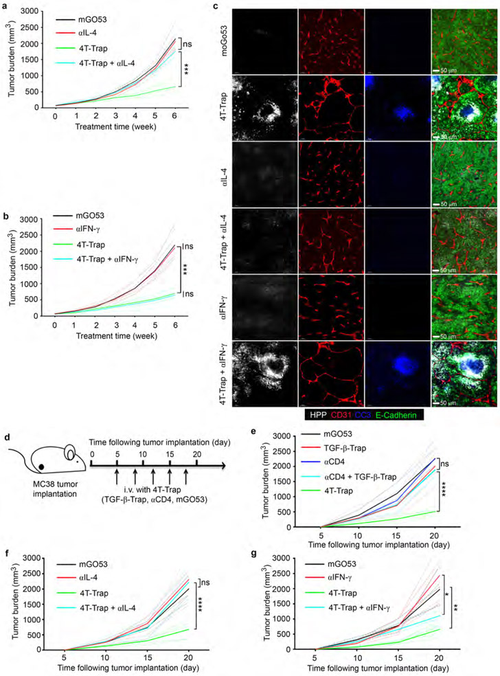
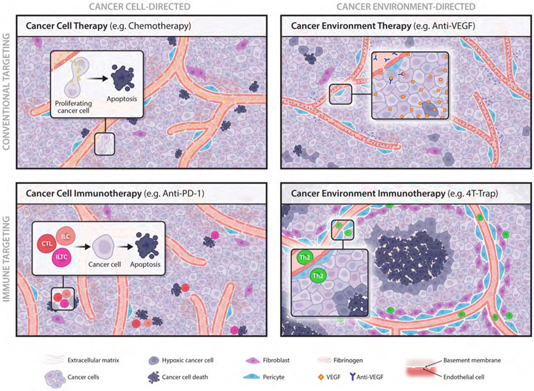
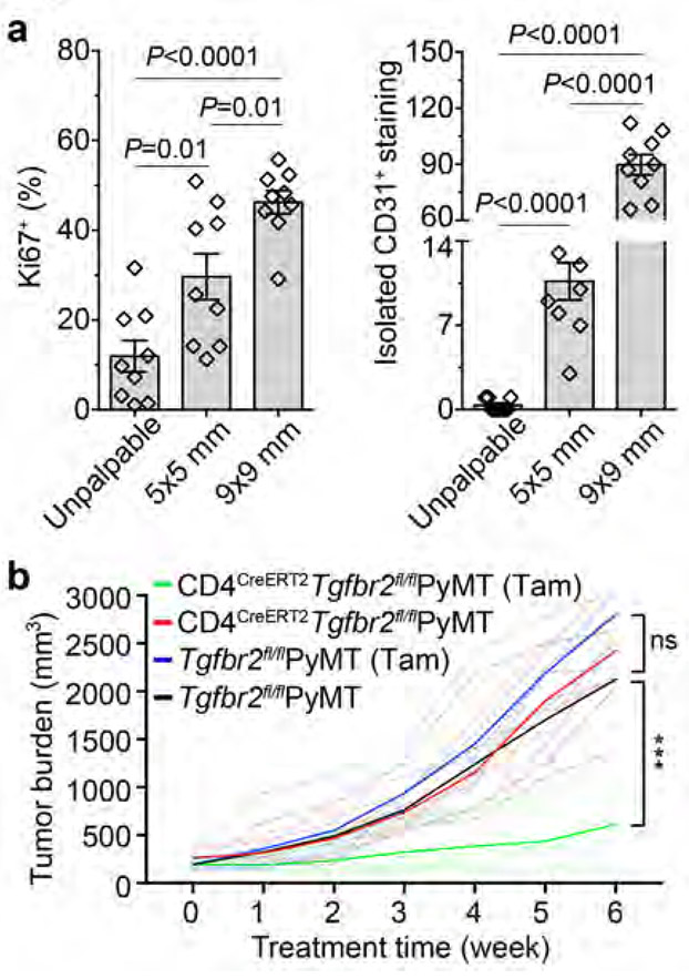
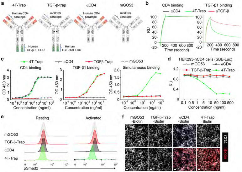
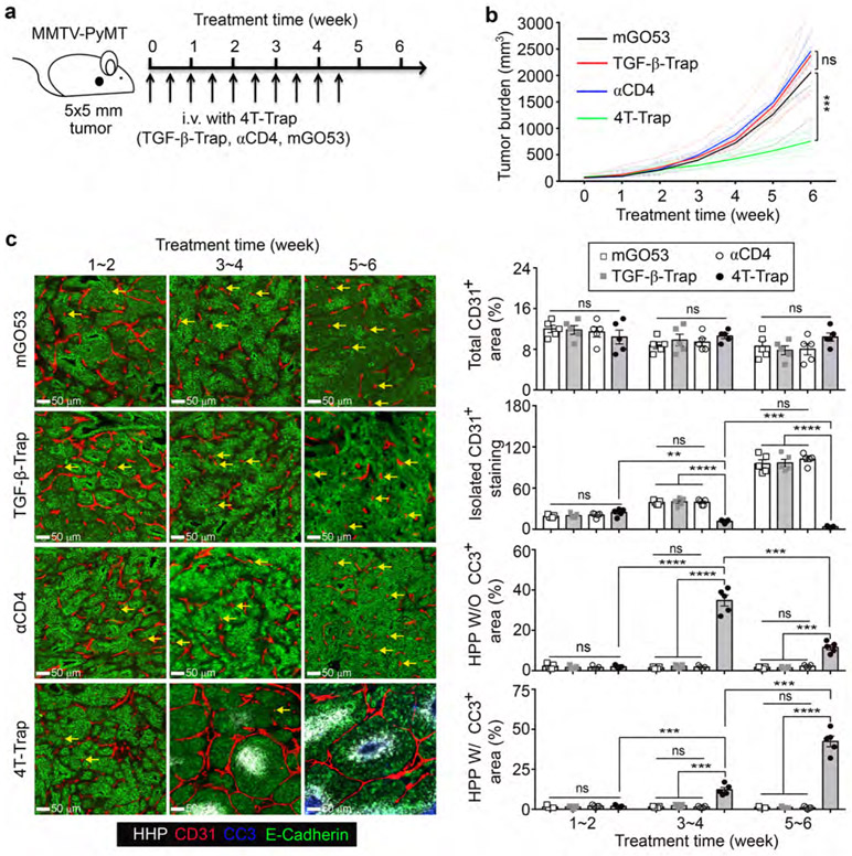
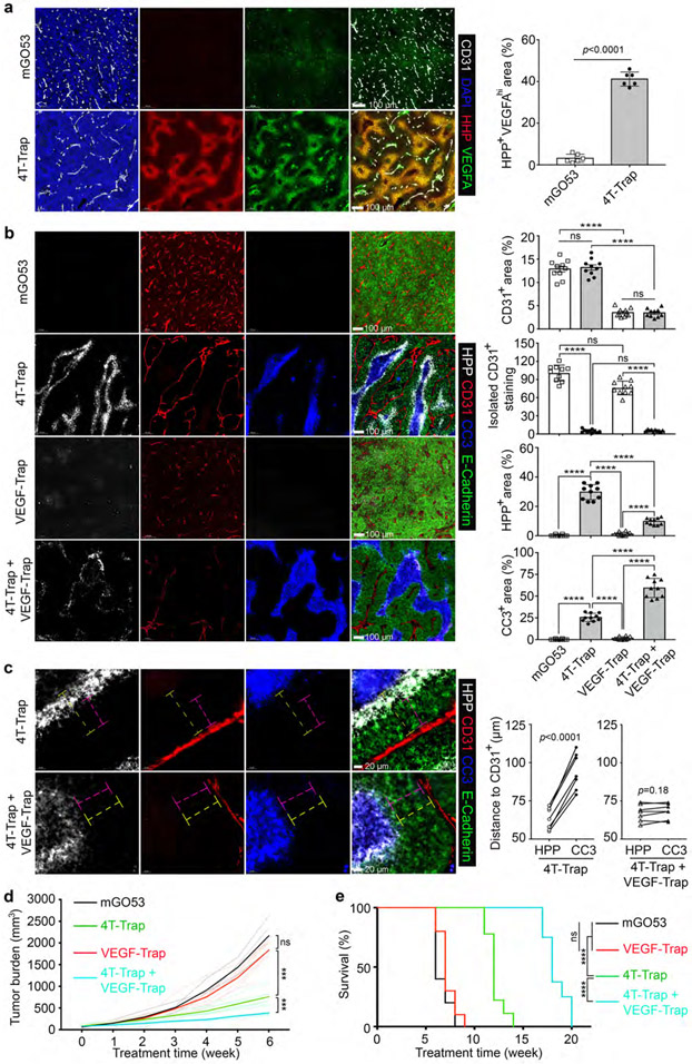
Cancer arises from malignant cells that exist in dynamic multilevel interactions with the host tissue. Cancer therapies aiming to directly kill cancer cells, including oncogene-targeted therapy and immune-checkpoint therapy that revives tumour-reactive cytotoxic T lymphocytes, are effective in some patients1,2, but acquired resistance frequently develops3,4. An alternative therapeutic strategy aims to rectify the host tissue pathology, including abnormalities in the vasculature that foster cancer progression5,6; however, neutralization of proangiogenic factors such as vascular endothelial growth factor A (VEGFA) has had limited clinical benefits7,8. Here, following the finding that transforming growth factor-β (TGF-β) suppresses T helper 2 (TH2)-cell-mediated cancer immunity9, we show that blocking TGF-β signalling in CD4+ T cells remodels the tumour microenvironment and restrains cancer progression. In a mouse model of breast cancer resistant to immune-checkpoint or anti-VEGF therapies10,11, inducible genetic deletion of the TGF-β receptor II (TGFBR2) in CD4+ T cells suppressed tumour growth. For pharmacological blockade, we engineered a bispecific receptor decoy by attaching the TGF-β-neutralizing TGFBR2 extracellular domain to ibalizumab, a non-immunosuppressive CD4 antibody12,13, and named it CD4 TGF-β Trap (4T-Trap). Compared with a non-targeted TGF-β-Trap, 4T-Trap selectively inhibited TH cell TGF-β signalling in tumour-draining lymph nodes, causing reorganization of tumour vasculature and cancer cell death, a process dependent on the TH2 cytokine interleukin-4 (IL-4). Notably, the 4T-Trap-induced tumour tissue hypoxia led to increased VEGFA expression. VEGF inhibition enhanced the starvation-triggered cancer cell death and amplified the antitumour effect of 4T-Trap. Thus, targeted TGF-β signalling blockade in helper T cells elicits an effective tissue-level cancer defence response that can provide a basis for therapies directed towards the cancer environment.


Figures

Extended Data Fig. 1 ∣
Extended Data Fig. 1 ∣. Inducible ablation of TGF-βRII in CD4+ T cells causes enhanced T helper cell responses and increased cancer cell death.
a, Representative immunofluorescence images of CD31 (white), Ki67 (red) and E-Cadherin (green) in mammary tumor tissues from PyMT mice harboring unpalpable, 5x5 mm, or 9x9 mm tumors. Isolated CD31+ staining in the tumor parenchyma (yellow arrows) is indicated. b, TGF-βRII expression on CD4+ T cells and CD8+ T cells from the tumor-draining lymph nodes of Tgfbr2fl/flPyMT and CD4CreERT2Tgfbr2fl/flPyMT mice treated with tamoxifen. c, Representative flow cytometry plots and statistical analyses of IL-4 and IFN-γ expression in CD4+Foxp3 T cells from the tumor-draining lymph nodes of Tgfbr2fl/flPyMT and CD4CreERT2Tgfbr2fl/flPyMT mice treated with tamoxifen. d, Representative immunofluorescence images of E-Cadherin (green), Ki67 (red) and cleaved Caspase 3 (CC3, blue) in mammary tumor tissues from Tgfbr2fl/flPyMT and CD4CreERT2Tgfbr2fl/flPyMT mice treated with tamoxifen. The percentage of Ki67+E-Cadherin+ cells over total E-Cadherin+ epithelial cells was calculated from 0.02 mm2 regions (n=9). The percentage of CC3+ areas over total E-Cadherin+ areas was calculated from 0.02 mm2 regions (n=10). All statistical data are shown as mean ± SEM. Two-tailed unpaired t-test (c, d). Data are pooled biological replicates (c) or representative of three independent experiments (a, b, d).
Extended Data Fig. 2 ∣
Extended Data Fig. 2 ∣. Inducible ablation of TGF-βRII in CD4+ T cells promotes tumor vessel reorganization, hypoxia and cancer cell death.
a, Representative immunofluorescence images of fibrinogen (Fg, white), CD31 (red), cleaved Caspase 3 (CC3, cyan) and E-Cadherin (green) in mammary tumor tissues from Tgfbr2fl/flPyMT and CD4CreERT2Tgfbr2fl/flPyMT mice treated with tamoxifen. Extravascular (EV) Fg deposition events (magenta arrows) were calculated from 1 mm2 regions (n=9 for each group). Isolated CD31+ staining (yellow arrows) was counted from 1 mm2 regions (n=9 for each group). b, Representative immunofluorescence images of NG2+ pericytes (white), CD31+ endothelial cells (red), GP38+ fibroblasts (blue) and E-Cadherin (green) in mammary tumor tissues from Tgfbr2fl/flPyMT and CD4CreERT2Tgfbr2fl/flPyMT mice treated with tamoxifen. NG2-unbound (magenta arrows) or GP38-unbound (yellow arrows) isolated CD31+ staining was counted from 1 mm2 regions (n=9 for each group). c, Representative immunofluorescence images of collagen IV (Col IV, white), CD31 (red), fibronectin (FN, cyan) and E-Cadherin (green) in mammary tumor tissues from Tgfbr2fl/flPyMT and CD4CreERT2Tgfbr2fl/flPyMT mice treated with tamoxifen. The average continuous lengths of Col IV and FN were measured in 1 mm2 regions (n=9 for each group). d, Representative immunofluorescence images of a hypoxia probe (HPP, white), CD31 (red), CC3 (cyan) and E-Cadherin (green) in mammary tumor tissues from Tgfbr2fl/flPyMT and CD4CreERT2Tgfbr2fl/flPyMT mice treated with tamoxifen. The percentage of HPP+E-Cadherin+ areas over E-Cadherin+ epithelial areas was calculated from 1 mm2 regions (n=9 for each group). The shortest distance of HPP+ regions (magenta dashed lines) or CC3+ regions (yellow dashed lines) to CD31+ endothelial cells was measured in tumor tissues from CD4CreERT2Tgfbr2fl/flPyMT mice treated with tamoxifen (n=9). All statistical data are shown as mean ± SEM. Two-tailed unpaired t-test (a-d) or paired t-test (d). Data are representative of three independent experiments (a-d).
Extended Data Fig. 3 ∣
Extended Data Fig. 3 ∣. Biochemical properties of 4T-Trap and control antibodies.
a, Schematic representation of ibalizumab Fab and TGF-βRII ECD fusion proteins in a murine IgG1 framework. The star indicates a D265A substitution in the CH2 domain, and the semicircle and moon shapes indicate knob-into-hole (KIH) modifications in the CH3 domain to enable heavy chain heterodimerization. The gray or colored parts indicate mouse or human sequences, respectively. b-c, Yield and aggregation percentage of ibalizumab Fab and TGF-βRII ECD fusion proteins produced in a FreeStyle HEK293-F cell transient expression system. FreeStyle HEK293-F cells transfected with plasmids encoding the indicated fusion antibodies were cultured for 4 days, and the supernatant was collected. Protein G affinity purification and size exclusion chromatography were used to purify these antibodies. d, Molecular weights of αCD4, mGO53, 4T-Trap and TGF-β-Trap antibodies detected by Coomassie Blue staining of samples run in a SDS-PAGE gel under non-reduced or reduced conditions. Molecular size markers (kDa) are shown on the left. HC, heavy chain; LC, light chain. e, Size exclusion chromatography analyses of mGO53, TGF-β-Trap, αCD4 and 4T-Trap antibodies. f, Schematic representation of human CD4 structure and purity examination of recombinant soluble CD4 (sCD4) by SDS-PAGE followed Coomassie Blue staining. g, The binding affinities of 4T-Trap and αCD4 to human CD4 as well as 4T-Trap and αTGF-β (1D11 clone) to human TGF-β1 were determined by surface plasmon resonance. h, Binding of 4T-Trap to human CD4 ectopically expressed on HEK293 cells. Cells were incubated with serial dilutions of 4T-Trap and αCD4 antibodies followed by a fluorophore-conjugated anti-mouse IgG secondary antibody. Samples were analyzed by flow cytometry. The measured mean fluorescence intensity (MFI) was quantified. i, TGF-β signaling inhibitory functions of 4T-Trap and αTGF-β. HEK293 cells transfected with a TGF-β/SMAD firefly luciferase reporter plasmid and a pRL-TK Renilla luciferase reporter plasmid were incubated with the indicated antibodies for 30 min and treated with 10 ng/mL recombinant human TGF-β1 for 12 hr before subject to the luciferase assay. RU, relative unit of normalized Firefly luciferase activity to Renilla luciferase activity. Data are representative of three independent experiments (b-f, h, i).
Extended Data Fig. 4 ∣
Extended Data Fig. 4 ∣. Generation and validation of human CD4 transgenic mice.
a, Recombineering a bacterial artificial chromosome (BAC) DNA containing the human CD4 locus with the proximal enhancer (PE) element replaced by its murine equivalent. The shuttle plasmid contains the mouse Cd4 PE flanked by two homologous arms of the human CD4 gene (250 bps), the E coli. RecA gene to mediate homologous recombination, the SacB gene to mediate negative selection on sucrose, an Ampicillin resistance locus to mediate positive selection and a conditional R6Kγ replication origin. b, Flow cytometry analyses of human CD4 expression on leukocyte populations from wild-type or human CD4 transgenic mice. CD4+ T cells (CD45+TCRβ+CD4+), CD8+ T cells (CD45+TCRβ+CD8+), NK cells (CD45+TCRγTCRβNKp46+NK1.1+) were isolated from lymph nodes. B cells (CD45+MHCII+Ly6CB220+), XCR1+ dendritic cells (DCs) (CD45+LinF4/80Ly6CCD11c+MHCII+XCR1+), CD11b+ DCs (CD45+LinF4/80Ly6CCD11c+MHCII+CD11b+), Monocytes (CD45+LinF4/80+Ly6C+CD11b+) and Macrophages (CD45+LinF4/80+CD11bLy6C) were isolated from spleens. Data are representative of three independent experiments (b).
Extended Data Fig. 5 ∣
Extended Data Fig. 5 ∣. Pharmacokinetics, pharmacodynamics and efficacy study of 4T-Trap and control antibodies.
a, Schematic representation of biotinylated 4T-Trap and control antibodies. b, Mice were administered with a single dose of 150 μg 4T-Trap, αCD4, TGF-β-Trap or mGO53 by intravenous injection. Antibody serum levels at different time points were measured by ELISA. c, Mice were administered with a single dose of 50 μg, 100 μg, 150 μg or 450 μg 4T-Trap by intravenous injection. Antibody serum levels were measured by ELISA. d, Mice were administered with a single dose of 150 μg 4T-Trap, αCD4, TGF-β-Trap or mGO53 by intravenous injection. TGF-β1 serum levels were measured by ELISA. e, Representative immunofluorescence images of E-Cadherin (green) and phosphorylated Smad2 (pSmad2, magenta) in mammary tumor tissues from mice treated with the indicated antibodies at 12 hr post injection. f, Mice were administered with a single dose of 50 μg, 100 μg, 150 μg or 450 μg 4T-Trap by intravenous injection. Percentage of human CD4 molecule occupancy was measured by flow cytometry. g, Immunoblotting analyses of TGF-β-induced SMAD2/3 phosphorylation in mouse CD4+ T cells isolated from human CD4 transgenic mice with different levels of 4T-Trap human CD4 (hCD4) target occupancy (TO). Numbers under lanes indicate SMAD2/3 or pSMAD2/3 band intensity. h, Tumor measurements from hCD4PyMT mice treated with 4T-Trap (n=4) or combination of αCD4 with TGF-β-Trap (n=5). Two-tailed unpaired t-test (h). Data are pooled biological replicates (h) or representative of three independent experiments (b-g).
Extended Data Fig. 6 ∣
Extended Data Fig. 6 ∣. 4T-Trap promotes the generation of a reorganized, nonporous and mature tumor vasculature.
a, Representative immunofluorescence images of fibrinogen (Fg, white), CD31 (red) and E-Cadherin (green) in mammary tumor tissues from hCD4PyMT mice treated with 4T-Trap, αCD4, TGF-β-Trap or mGO53 antibodies. Isolated CD31+ staining (yellow arrows) was counted from 1 mm2 regions (n=13 for each group). Extravascular (EV) Fg deposition events (magenta arrows) were calculated from 1 mm2 regions (n=13 for each group). b, Representative immunofluorescence images of sulfo-NHS-biotin (white), CD31 (red) and E-Cadherin (green) in mammary tumor tissues from hCD4PyMT mice treated with 4T-Trap, αCD4, TGF-β-Trap or mGO53 antibodies. The percentage of sulfo-NHS-biotin areas over E-Cadherin+ epithelial regions was calculated from 1 mm2 regions (n=15 for each group). Cancer cell-associated sulfo-NHS-biotin deposition events (magenta arrows) in highly perfused regions were calculated from 1 mm2 regions (n=10 for each group). c, Representative immunofluorescence images of NG2+ pericytes (white), CD31+ endothelial cells (red), GP38+ fibroblasts (cyan) and E-Cadherin (green) in mammary tumor tissues from hCD4PyMT mice treated with 4T-Trap, αCD4, TGF-β-Trap or mGO53 antibodies. NG2-unbound (magenta arrows) or GP38-unbound (yellow arrows) isolated CD31+ staining was counted from 1 mm2 regions (n=13 for each group). d, Representative immunofluorescence images of collagen IV (Col IV, white), CD31 (red), fibronectin (FN, cyan) and E-Cadherin (green) in mammary tumor tissues from hCD4PyMT mice treated with 4T-Trap, αCD4, TGF-β-Trap or mGO53 antibodies. The average continuous lengths of Col IV and FN were measured in 1 mm2 regions (n=13 for each group). All statistical data are shown as mean ± SEM. ****: P<0.0001 and ns: not significant (one-way ANOVA with post hoc Bonferroni t-test (a-d)). Data are representative of three independent experiments (a-d).
Extended Data Fig. 7 ∣
Extended Data Fig. 7 ∣. 4T-Trap repression of tumor growth and VEGF-Trap construction.
a, A schematic representation of treatment with 4T-Trap and control antibodies. hCD4PyMT mice bearing 9x9 mm tumors were administered with 100 μg antibodies by intravenous injection twice a week for 4 weeks. b, Singular tumor measurements from hCD4PyMT mice treated with 4T-Trap, αCD4, TGF-β-Trap or mGO53 (n=7, 6, 7 and 5). c, Representative immunofluorescence images of a hypoxia probe (HPP, white), CD31 (red), cleaved Caspase 3 (CC3, blue) and E-Cadherin (green) in mammary tumor tissues from mice treated with 4T-Trap at the indicated time points. d, Schematic representation of human VEGFR1, VEGFR2 and VEGF-Trap as well as purity examination of recombinant VEGF-Trap by SDS-PAGE followed by Coomassie Blue staining. e, VEGF signaling inhibitory function of VEGF-Trap. HEK293 cells transfected with a VEGF/NFAT firefly luciferase reporter plasmid, together with a VEGFR2 expression plasmid and a pRL-TK Renilla luciferase reporter plasmid, were incubated with different concentrations of VEGF-Trap for 30 min followed by 10 ng/mL recombinant human VEGF165 for 12 hr before subject to the luciferase assay. RU, relative unit of normalized Firefly luciferase activity to Renilla luciferase activity. ***: P<0.001 and ns, not significant (one-way ANOVA with post hoc Bonferroni t-test (b)). Data are pooled biological replicates (b) or representative of three independent experiments (c-e).
Extended Data Fig. 8 ∣
Extended Data Fig. 8 ∣. 4T-Trap induces T helper cell activation, differentiation and tumor infiltration.
a, Representative flow cytometry plots and statistical analyses of CD62L and CD44 expression in conventional CD4+Foxp3 T cells from the tumor-draining lymph nodes of hCD4PyMT mice treated with 4T-Trap, αCD4, TGF-β-Trap or mGO53 antibodies (n=3 for each group). b, Quantitative RT-PCR analyses of Smad7 and Rgs16 mRNA expression in effector/memory CD4+ T cells from the tumor-draining lymph nodes of hCD4PyMT mice treated with the indicated antibodies. c, Representative flow cytometry plots and statistical analyses of IFN-γ and IL-4 expression in conventional CD4+Foxp3 T cells from the tumor-draining lymph nodes of hCD4PyMT mice treated with 4T-Trap, αCD4, TGF-β-Trap or mGO53 antibodies (n=3 for each group). d, Representative flow cytometry plots and statistical analyses of TCRβ, NK1.1, CD4, CD8 and Foxp3 expression in tumor-infiltrating leukocytes from hCD4PyMT mice treated with 4T-Trap, αCD4, TGF-β-Trap or mGO53 antibodies (n=3 for each group). All statistical data are shown as mean ± SEM. **: P<0.01; ***: P<0.001; ****: P<0.0001 and ns: not significant (one-way ANOVA with post hoc Bonferroni t-test (a-d)). Data are shown as mean ± SEM of three independent biological replicates (a-d).
Extended Data Fig. 9 ∣
Extended Data Fig. 9 ∣. 4T-Trap-triggered anti-tumor immunity is dependent on IL-4.
a-b, Tumor measurements from hCD4PyMT mice treated with mGO53 or 4T-Trap in the absence or presence of an IL-4 neutralizing antibody (αIL-4) or an IFN-γ neutralizing antibody (αIFN-γ) (n=5 for each group). c, Representative immunofluorescence images of a hypoxia probe (HPP, white), CD31 (red), cleaved Caspase 3 (CC3, blue) and E-Cadherin (green) in mammary tumor tissues from hCD4PyMT mice treated with mGO53 or 4T-Trap in the absence or presence of αIL-4 or αIFN-γ. d, Schematic representation of treatment with 4T-Trap and control antibodies. hCD4 mice injected subcutaneously with MC38 cancer cells, were treated with 4T-Trap or control antibodies including TGF-β-Trap, αCD4 and mGO53 (100 μg/dose), for a total of 5 doses. e, Tumor measurements from hCD4 mice bearing MC38 cancer cells treated with 4T-Trap, TGF-β-Trap, αCD4, mGO53 and combination of αCD4 with TGF-β-Trap (n=5 for each group). f, Tumor measurements from hCD4 mice bearing MC38 cancer cells treated with mGO53, αIL-4, 4T-Trap and combination of αIL-4 with 4T-Trap (n=5 for each group). g, Tumor measurements from hCD4 mice bearing MC38 cancer cells treated with mGO53, αIFN-γ, 4T-Trap and combination of αIFN-γ with 4T-Trap (n=5 for each group). *: P<0.05; **: P<0.01; ***: P<0.001, ****: P<0.0001 and ns: not significant (two-way ANOVA with post hoc Bonferroni t-test (a, b, e-g)). Data are pooled biological replicates (a, b, e-g) or representative of three independent experiments (c).
Extended Data Fig. 10 ∣
Extended Data Fig. 10 ∣. Cancer therapy landscape.
Cancer therapies are grouped into four categories in terms of targets and targeting strategies. Cancer cell-directed therapies aim to directly destruct cancer cells, which include conventional ‘cancer cell therapy’ with targeting approaches such as chemotherapy to eliminate mitotic cancer cells, and ‘cancer cell immunotherapy’ to engage immune effectors such as cytotoxic T lymphocytes (CTLs), killer innate lymphocytes (ILCs) and killer innate-like T cells (ILTCs) to eradicate cancer cells. The cancer immunosurveillance function of CTLs can be revived by immune checkpoint inhibitors such as PD-1 antibodies (anti-PD-1). Cancer environment-directed therapies aspire to rectify the host tissue pathology that fosters tumor growth. A cancer environment hallmark is angiogenesis characterized by a leaky and immature blood vasculature. Conventional ‘cancer environment therapy’ includes anti-angiogenics such as VEGF antibodies (anti-VEGF) that diminish vasculature abundance. Blockade of TGF-β signaling in helper T (Th) cells with 4T-Trap results in enhanced Th2 cell differentiation that promotes vasculature remodeling and tumor tissue healing with cancer cell hypoxia and cancer cell death instigated in avascular regions. 4T-Trap defines a novel modality of ‘cancer environment immunotherapy’.
Fig. 1 ∣
Fig. 1 ∣. Inducible ablation of TGF-βRII in CD4+ T cells inhibits tumor growth.
a, Statistical analyses of cancer cell proliferation and angiogenesis in tumor tissues from PyMT mice harboring unpalpable, 5x5 mm or 9x9 mm tumors. The percentage of Ki67+E-Cadherin+ cells over total E-Cadherin+ epithelial cells was calculated from 0.02 mm2 regions (n=9 for each group). Isolated CD31+ staining in the tumor parenchyma was counted from 1 mm2 regions (n=9 for each group). b, Tgfbr2fl/flPyMT and CD4CreERT2Tgfbr2fl/flPyMT mice bearing 5x5 mm tumors were left untreated or treated with Tamoxifen (Tam) (n=5, 5, 5 and 6) twice a week for 6 weeks. Tumor growth was monitored. Statistical data are shown as mean ± SEM. ***: P<0.001 and ns: not significant (one-way (a) or two-way ANOVA (b) with post hoc Bonferroni t-test). Data are pooled biological replicates (b) or representative of three independent experiments (a).
Fig. 2 ∣
Fig. 2 ∣. 4T-Trap effectively represses TGF-β signaling in lymph node CD4+ T cells.
a, Schematic representation of antibody structures for 4T-Trap, TGF-β-rap, αCD4 and mGO53. b, SPR sensorgrams of 4T-Trap and αCD4 binding to immobilized CD4 (left panel) as well as 4T-Trap and αTGF-β binding to immobilized TGF-β1 (right panel). RU, response unit. c, Enzyme-linked immunosorbent assay to assess 4T-Trap, TGF-β-Trap, αCD4 and mGO53 binding to CD4, TGF-β1 or both molecules. Optical densities (OD) were detected at 450 nm. d, TGF-β signaling inhibitory functions of the indicated antibodies in HEK293-hCD4 cells transfected with a TGF-β/SMAD Firefly luciferase reporter plasmid and a pRL-TK Renilla luciferase reporter plasmid. RU, relative unit of normalized Firefly luciferase activity to Renilla luciferase activity. e, Flow cytometry analyses of pSmad2 expression on resting or activated CD4+ T cells from the tumor-draining lymph nodes of mice treated with the indicated antibodies. CD4+ T cells were left untreated (resting) or treated with PMA/ionomycin for 4 hr (activated) before pSmad2 staining. f, Representative immunofluorescence images of CD4 (white) and Biotin (red) staining in the tumor-draining lymph nodes of mice treated with the indicated antibodies. Data are representative of two (b) or three independent experiments (c-f).
Fig. 3 ∣
Fig. 3 ∣. 4T-Trap reprograms the tumor vasculature causing cancer cell hypoxia and cancer cell death.
a, Schematic representation of a treatment scheme with 4T-Trap and control antibodies. hCD4PyMT mice bearing 5x5 mm tumors were administered with 100 μg antibodies by intravenous injection twice a week for 5 weeks. b, Tumor measurements from hCD4PyMT mice treated with 4T-Trap, αCD4, TGF-β-Trap or mGO53 (n=8, 5, 10 and 8). c, Representative immunofluorescence images of a hypoxia probe (HPP, white), CD31 (red), cleaved Caspase 3 (CC3, blue) and E-Cadherin (green) in tumor tissues from mice treated with the indicated antibodies and time points. The percentage of CD31+ areas, HPP+ without (W/O) CC3+ areas or HPP+ with (W/) CC3+ areas over E-Cadherin+ epithelial regions was calculated from 1 mm2 regions (n=5 for each group). Isolated CD31+ staining (yellow arrows) was counted from 1 mm2 regions (n=5 for each group). Statistical data are shown as mean ± SEM. **: P<0.01; ***: P<0.001; ****: P<0.0001 and ns: not significant (one-way ANOVA with post hoc Bonferroni t-test (b, c)). Data are pooled biological replicates (b) or representative of three independent experiments (c).
Fig. 4 ∣
Fig. 4 ∣. 4T-Trap synergizes with VEGF-Trap to induce cancer cell death and suppress tumor growth.
a, Representative immunofluorescence images of CD31 (white), a hypoxia probe (HPP, red) and VEGFA (green) in tumor tissues from hCD4PyMT mice treated with mGO53 or 4T-Trap. The percentage of HPP+VEGFAhi areas was calculated from 1 mm2 regions (n=6 for each group). b, Representative immunofluorescence images of HPP (white), CD31 (red), cleaved Caspase 3 (CC3, blue) and E-Cadherin (green) in tumor tissues from mice treated with the indicated antibodies. The percentage of CD31+ areas, HPP+ areas or CC3+ areas over E-Cadherin+ epithelial regions was calculated from 1 mm2 regions (n=10 for each group). Isolated CD31+ staining was counted from 1 mm2 regions (n=10 for each group). c, Representative high magnification immunofluorescence images of HPP (white), CD31 (red), CC3 (blue) and E-Cadherin (green) in tumor tissues from mice treated with the indicated antibodies. The shortest distance of HPP+ regions (magenta dashed lines) or CC3+ regions (yellow dashed lines) to CD31+ endothelial cells was measured and plotted (n=7 for each group). d, Tumor measurements from hCD4PyMT mice treated with the indicated antibodies (n=7 for each group). e, Kaplan-Meier survival curve of hCD4PyMT mice treated with mGO53, 4T-Trap, VEGF-Trap or 4T-Trap and VEGF-Trap (n=10, 9, 10 and 8). Statistical data are shown as mean ± SEM. ***: P<0.001; ****: P<0.0001 and ns: not significant (two-tailed unpaired t-test (a) or paired t-test (c), two-way ANOVA with post hoc Bonferroni t-test (b, d) or long-rank test (e)). Data are pooled biological replicates (d, e) or representative of three independent experiments (a, b, c).

Comment in

References

    1. Sawyers CL Shifting paradigms: the seeds of oncogene addiction. Nature medicine 15, 1158–1161, doi:10.1038/nm1009-1158 (2009). - DOI - PubMed
    1. Allison JP Checkpoints. Cell 162, 1202–1205, doi:10.1016/j.cell.2015.08.047 (2015). - DOI - PubMed
    1. Sabnis AJ & Bivona TG Principles of Resistance to Targeted Cancer Therapy: Lessons from Basic and Translational Cancer Biology. Trends in molecular medicine 25, 185–197, doi:10.1016/j.molmed.2018.12.009 (2019). - DOI - PMC - PubMed
    1. Sharma P, Hu-Lieskovan S, Wargo JA & Ribas A Primary, Adaptive, and Acquired Resistance to Cancer Immunotherapy. Cell 168, 707–723, doi:10.1016/j.cell.2017.01.017 (2017). - DOI - PMC - PubMed
    1. Folkman J Tumor angiogenesis: therapeutic implications. The New England journal of medicine 285, 1182–1186, doi:10.1056/NEJM197111182852108 (1971). - DOI - PubMed

Methods References

    1. Ouyang W, Beckett O, Ma Q & Li MO Transforming growth factor-beta signaling curbs thymic negative selection promoting regulatory T cell development. Immunity 32, 642–653, doi:10.1016/j.immuni.2010.04.012 (2010). - DOI - PMC - PubMed
    1. Sarkar A, Donkor MK & Li MO T cell- but not tumor cell-produced TGF-beta1 promotes the development of spontaneous mammary cancer. Oncotarget 2, 1339–1351, doi:10.18632/oncotarget.403 (2011). - DOI - PMC - PubMed
    1. Killeen N, Sawada S & Littman DR Regulated expression of human CD4 rescues helper T cell development in mice lacking expression of endogenous CD4. The EMBO journal 12, 1547–1553 (1993). - PMC - PubMed
    1. Franklin RA et al.The cellular and molecular origin of tumor-associated macrophages. Science 344, 921–925, doi:10.1126/science.1252510 (2014). - DOI - PMC - PubMed
    1. Oh SA et al.Foxp3-independent mechanism by which TGF-beta controls peripheral T cell tolerance. Proceedings of the National Academy of Sciences of the United States of America 114, E7536–E7544, doi:10.1073/pnas.1706356114 (2017). - DOI - PMC - PubMed

Publication types

MeSH terms

Substances