

Electrochemical Reduction and Oxidation of Eight Unnatural 2'-Deoxynucleosides at a Pyrolytic Graphite Electrode
- PMID: 33087943
- PMCID: PMC7571600
- DOI: 10.1016/j.electacta.2020.137210
Electrochemical Reduction and Oxidation of Eight Unnatural 2'-Deoxynucleosides at a Pyrolytic Graphite Electrode
Abstract
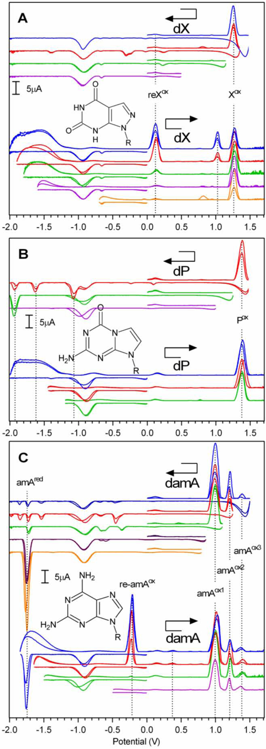
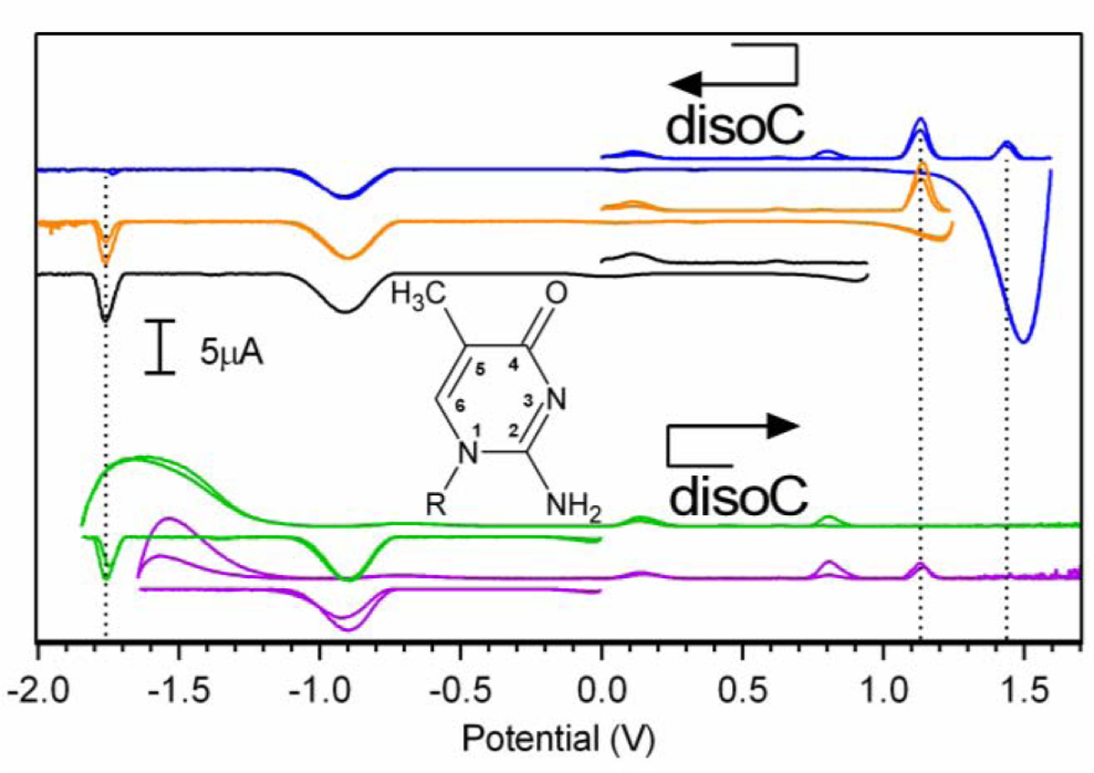
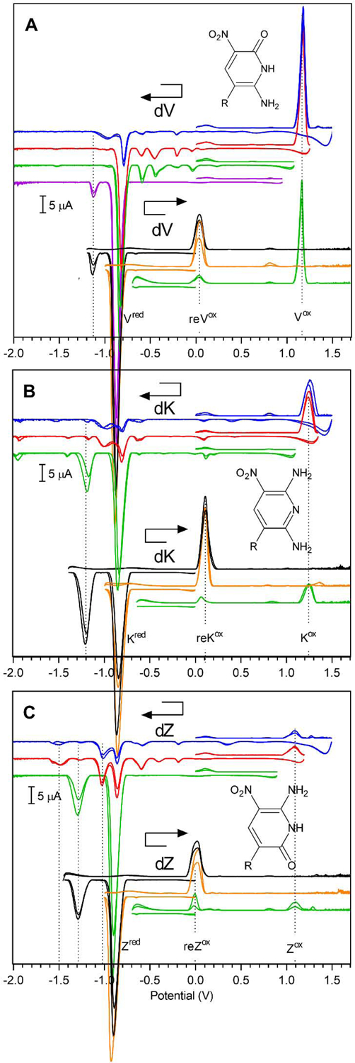
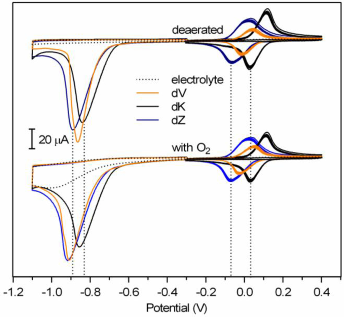
Recently we showed the reduction and oxidation of six natural 2'-deoxynucleosides in the presence of the ambient oxygen using the very broad potential window of a pyrolytic graphite electrode (PGE). Using the same procedure, 2'-deoxynucleoside analogs (dNs) that are parts of an artificially expanded genetic information system (AEGIS) were analyzed. Seven of the eight tested AEGIS dNs provided specific signals (voltammetric redox peaks). These signals, described here for the first time, will be used in future work to analyze DNA built from expanded genetic alphabets, helping to further develop AEGIS technology and its applications. Comparison of the electrochemical behavior of unnatural dNs with the previously documented behaviors of natural dNs also provides insights into the mechanisms of their respective redox processes.
Keywords: electrochemical oxidation; electrochemical reduction; electrochemistry; pyrolytic graphite; unnatural DNA.
Conflict of interest statement
Declaration of interests The authors declare that they have no known competing financial interests or personal relationships that could have appeared to influence the work reported in this paper.
Figures
Similar articles
-
Interaction of nucleic acids with electrically charged surfaces. VI. A comparative study on the electrochemical behaviour of native and denatured DNAs at graphite electrodes.Biophys Chem. 1979 May;9(4):289-97. Biophys Chem. 1979. PMID: 465642
-
Interaction of nucleic acids with electrically charged surfaces. VI. A comparative study on the electrochemical behaviour of native and denatured DNAs at graphite electrodes.Biophys Chem. 1975 May;9(4):289-97. Biophys Chem. 1975. PMID: 16997196
-
Highly sensitive voltammetric determination of lamotrigine at highly oriented pyrolytic graphite electrode.Bioelectrochemistry. 2012 Apr;84:38-43. doi: 10.1016/j.bioelechem.2011.10.008. Epub 2011 Nov 4. Bioelectrochemistry. 2012. PMID: 22137203
-
Voltammetric studies of sumatriptan on the surface of pyrolytic graphite electrode modified with multi-walled carbon nanotubes decorated with silver nanoparticles.Talanta. 2009 Nov 15;80(1):31-8. doi: 10.1016/j.talanta.2009.06.019. Epub 2009 Jun 16. Talanta. 2009. PMID: 19782189
-
Electrochemical behaviour of human adrenodoxin on a pyrolytic graphite electrode.Bioelectrochemistry. 2003 Apr;59(1-2):41-7. doi: 10.1016/s1567-5394(02)00188-3. Bioelectrochemistry. 2003. PMID: 12699818
Cited by
-
Reduced graphene oxide-based absorbance biosensors for detecting Escherichia coli DNA.Sci Rep. 2025 Aug 12;15(1):29548. doi: 10.1038/s41598-025-14189-4. Sci Rep. 2025. PMID: 40796589 Free PMC article.
References
-
- Hoshika S, Leal NA, Kim MJ, Kim MS, Karalkar NB, Kim HJ, Bates AM, Watkins NE, SantaLucia HA, Meyer AJ, DasGupta S, Piccirilli JA, Ellington AD, SantaLucia J, Georgiadis MM, Benner SA, Hachimoji DNA and RNA: A genetic system with eight building blocks, Science (80-. ). 363 (2019) 884–887. 10.1126/science.aat0971. - DOI - PMC - PubMed
Grants and funding
LinkOut - more resources
Full Text Sources