

Feasibility of Treatment Planning System in Localizing the COVID-19 Pneumonia Lesions and Evaluation of Volume Indices of Lung Involvement
- PMID: 33088245
- PMCID: PMC7545774
- DOI: 10.1177/1559325820962600
Feasibility of Treatment Planning System in Localizing the COVID-19 Pneumonia Lesions and Evaluation of Volume Indices of Lung Involvement
Abstract
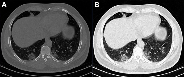
Background and purpose: To assess the feasibility of a treatment planning system in localizing, contouring, and targeting lung lesions along with an evaluation of volume indices of lung involvement in patients with COVID-19 pneumonia.
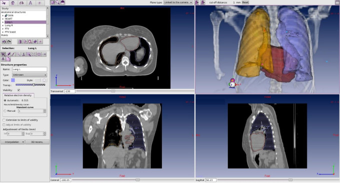
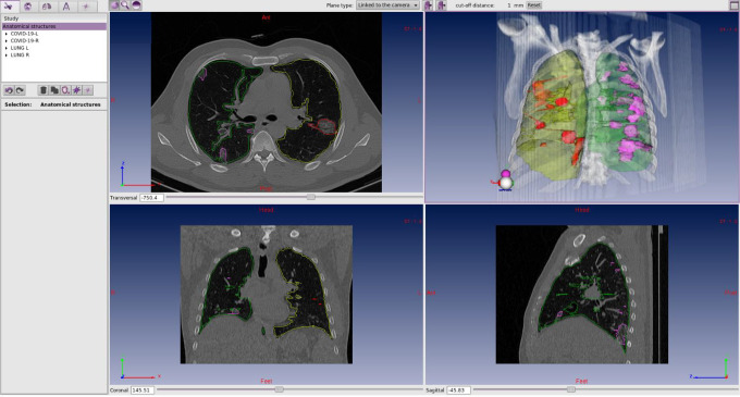
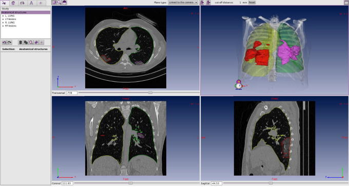
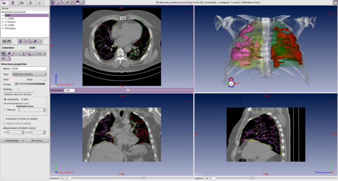
Methods: We evaluated 10 patients with PCR-confirmed COVID-19 pneumonia. The CT images were imported into the ISOgray® treatment planning system to anatomically define and contour the volumes of the pulmonary lesions, the lungs, and other nearby organs.
Results: The ratio of lung lesion volume to lung volume in this study was 0.11 ± 0.13 (11.13%). The highest mean biosynthesis ratio of lung lesions was 0.36. The ratio of lesion volume in the left lung of patients with the highest volume of involvement, was 0.44, and the ratio of lesion volume in the right lung of these patients was 0.27 (approximately 1.5 times more in the left lung than the right lung). On average, CTDIvol and DLP for all patients studied in our study were 11.22 ± 2.47 mGy and 354.20 ± 65.11 mGy.cm.
Conclusion: We reported the feasibility of using a treatment planning system in localizing COVID-19 pulmonary lesions and its validity in the volumetric assessment of infected lung regions.
Keywords: 3 dimensional (3D) conformal radiation therapy; COVID-19; SARS-CoV-2; acute respiratory distress syndrome (ARDS); low dose radiotherapy; treatment planning system.
© The Author(s) 2020.
Conflict of interest statement
Declaration of Conflicting Interests: The author(s) declared no potential conflicts of interest with respect to the research, authorship, and/or publication of this article.
Figures
References
-
- World Health Organization. Report of the WHO-China Joint Mission on Coronavirus Disease 2019 (COVID-19). February; 2020.
-
- Calabrese EJ. X-Ray treatment of carbuncles and furuncles (boils): a historical assessment. Hum Exp Toxicol. 2013;32(8):817–827. doi:10.1177/0960327112467046 [PubMed:23821639]. - PubMed
LinkOut - more resources
Full Text Sources
Miscellaneous
