Review
. 2020 Sep 3:12:254.
doi: 10.3389/fnagi.2020.00254. eCollection 2020.

Redox Post-translational Modifications of Protein Thiols in Brain Aging and Neurodegenerative Conditions-Focus on S-Nitrosation

Affiliations
Review

Redox Post-translational Modifications of Protein Thiols in Brain Aging and Neurodegenerative Conditions-Focus on S-Nitrosation

Mattéa J Finelli. Front Aging Neurosci. .

Abstract

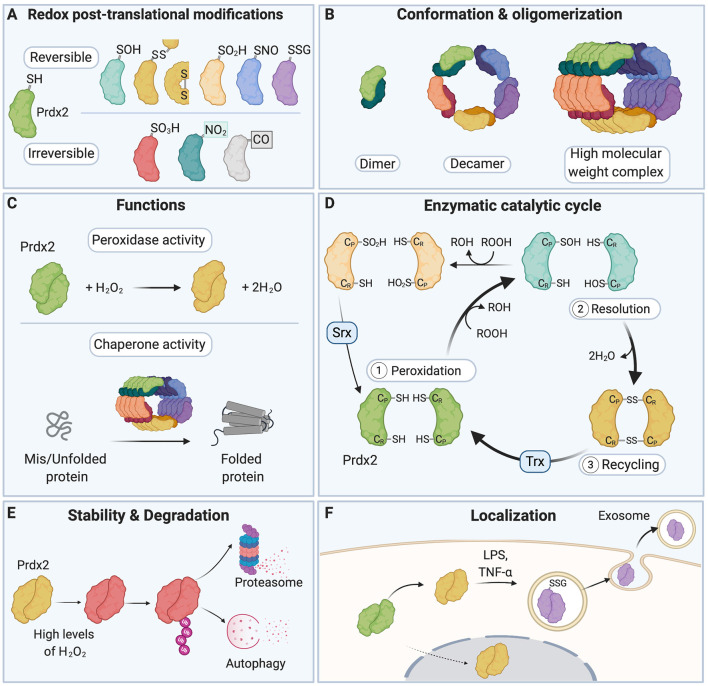
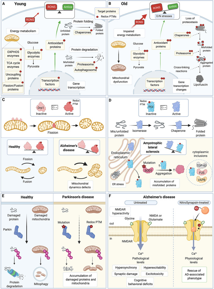
Reactive oxygen species and reactive nitrogen species (RONS) are by-products of aerobic metabolism. RONS trigger a signaling cascade that can be transduced through oxidation-reduction (redox)-based post-translational modifications (redox PTMs) of protein thiols. This redox signaling is essential for normal cellular physiology and coordinately regulates the function of redox-sensitive proteins. It plays a particularly important role in the brain, which is a major producer of RONS. Aberrant redox PTMs of protein thiols can impair protein function and are associated with several diseases. This mini review article aims to evaluate the role of redox PTMs of protein thiols, in particular S-nitrosation, in brain aging, and in neurodegenerative diseases. It also discusses the potential of using redox-based therapeutic approaches for neurodegenerative conditions.

Keywords: S-nitrosation; aging; brain; cysteine residues; neurodegenerative diseases; post-translational modifications; redox.


Figures

Figure 1
Figure 1
A redox switch modulates the multiple functions of Peroxiredoxin 2 (Prdx2). (A) The various residues of Prdx2 can be modified by S-sulfenylation (–SOH), intermolecular and intramolecular disulfide bonds (–SS–), S-sulfinylation (–SO2H), S-nitrosation (–SNO), S-glutathionylation (–SSG), S-sulfonylation (–SO3H), nitration (–NO2), and carbonylation (–CO: carbonyl groups). Only a subset of all the post-translational modifications (PTMs) that have been described for Prdx2 is shown here for clarity. (B) The redox state of the Cys residues of Prdx2 regulates its conformation as a dimer, decamer, or high molecular weight complex: the reduced and overoxidized dimers strongly tend to form decamers or high molecular weight complex, while oxidized forms are preferentially present as dimers. (C) Depending on its redox PTMs and conformation, Prdx2 can act as an antioxidant enzyme or as a chaperone. Multiple redox PTMs can coordinately regulate Prdx2 functions. (D) As part of Prdx2 catalytic cycle, its conserved peroxidatic cysteine (CP) first reduces H2O2 or ROOH (peroxidation step). Then a second free thiol (CR or resolving cysteine) forms a disulfide bond with its CP residue (resolution step). The catalytic cycle is completed when the disulfide bond is recycled, typically by a thioredoxin-like molecule (Trx), regenerating the free thiol forms of the CP and the CR residues (recycling step). CP-SOH Prdx2 can react with a second ROOH molecule before it can react with CR-SH, becoming overoxidized (CP-SO2H); this in turn inactivates Prdx2 peroxidase activity. CP-SO2H can be reduced by sulfiredoxin (Srx). (E) Oxidation of Prdx2 active site (Cys51) triggers conformational changes that bring to the protein surface of Prdx2 a flexible C-terminus that is subsequently polyubiquitinated; this leads to degradation of oxidized Prdx2 by the proteasome and autophagy. (F) Redox PTMs of Prdx2 can also regulate its secretion in the extracellular space. Upon exposure to inflammatory stimuli (e.g., LPS or TNF-α), Prdx2 Cys51 and Cys172 residues are oxidized forming disulfide-linked homodimers and mixed disulfides with glutathione, which triggers its secretion in exosomes into the extracellular milieu. Prdx2, peroxiredoxin 2; CP, peroxidatic Cys residue; CR, resolving Cys residue; LPS, lipopolysaccharide; TNF-α, tumor necrosis factor-α; Trx, Thioredoxin; Srx, Sulfiredoxin.
Figure 2
Figure 2
Redox PTMs of protein thiols are essential for normal cell physiology and contribute to aging and neurodegenerative diseases. (A) In young healthy cells, redox PTMs are an integral part of the normal cell signaling: they can coordinately modulate the activity of multiple proteins depending on the local RONS composition and levels. Redox PTMs regulate the function of proteins involved in essential pathways such as energy metabolism, protein folding and degradation, and gene transcription. Redox PTMs are represented by a red dot on the target proteins. (B) During aging, imbalance between RONS production and the antioxidant capacity of the cell leads to O/N stresses, and proteins may become differentially and/or aberrantly modified by redox PTMs, as represented by multiple red dots on the target proteins. These redox PTMs can affect the function of target proteins, ultimately altering key pathways that contribute to the aging process. (C) Drp1 is a GTPase protein involved in mitochondrial fission; when activated, especially by S-nitrosation, Drp1 forms oligomers that wrap around the mitochondrial outer membrane and scission it. Excessive levels of S-nitrosated Drp1, as seen in AD, disrupt the physiological equilibrium between mitochondrial fission and fusion,leading to increased mitochondrial fission, and synaptic and neuronal damage. (D) PDI is an ER protein with a dual isomerase and chaperone activity. S-nitrosation of PDI inhibits its functions. Excessive S-nitrosation of PDI, as observed in AD, PD, and ALS, leads to the accumulation of misfolded proteins in the ER and consequently, to increase in ER stress and neuronal cell death. In ALS, PDI is recruited to cytoplasmic inclusions containing ALS-associated proteins such as SOD1, TDP-43, FUS, or VABP. S-nitrosation of PDI also increases insoluble aggregates of ALS-associated mutant SOD1. (E) Parkin is an E3 ubiquitin ligase that targets damaged proteins and mitochondria to the ubiquitin-proteasome system and mitophagy. Loss-of-function mutations in Parkin are associated with familial PD. Importantly, excessive and persistent S-nitrosation of non-mutated Parkin, as detected in postmortem brains of patients with sporadic PD, also inhibits Parkin activity, which impairs ubiquitination of its substrate proteins and contributes to the accumulation of damaged proteins and mitochondria and ultimately to neuronal cell injury and death. (F) Hyperactivity of the extrasynaptic NMDAR leads to excessive intracellular calcium, hypersynchrony and hyperexcitability of neurons, and synaptic damage and excitotoxicity in AD. A new drug, NitroSynapsin, is constituted of a memantine moiety and an added nitro group (NO2). It can dampen the hyperactivity of NMDAR while preserving its physiological function by binding inside the excessively opened channel and by acting as an NO group donor, which triggers NMDAR inactivation through S-nitrosation of its redox-sensitive Cys residues. NitroSynapsin treatment significatively improves the pathological features associated with AD. TCA, tricarboxylic acid cycle; OXPHOS, oxidative phosphorylation; RONS, reactive oxygen species and reactive nitrogen species; Antiox, antioxidant capacity; Drp1, Dynamin related protein 1; PDI, protein disulfide isomerase; ER, endoplasmic reticulum; SOD1, superoxide dismutase 1; TDP-43, transactive response DNA binding protein 43 kDa; FUS, Fused in sarcoma; VAPB, vesicle-associated membrane protein; Ub, ubiquitin; NMDAR, N-methyl-D-aspartate receptor; AD, Alzheimer’s disease; PD, Parkinson’s disease; ALS, amyotrophic lateral sclerosis. All figures were created with BioRender.com.

References

    1. Abate C., Patel L., Rauscher F. J., III., Curran T. (1990). Redox regulation of fos and jun DNA-binding activity in vitro. Science 249, 1157–1161. 10.1126/science.2118682 - DOI - PubMed
    1. Abu-Omar N., Das J., Szeto V., Feng Z. P. (2018). Neuronal ryanodine receptors in development and aging. Mol. Neurobiol. 55, 1183–1192. 10.1007/s12035-016-0375-4 - DOI - PubMed
    1. Adler A. S., Sinha S., Kawahara T. L., Zhang J. Y., Segal E., Chang H. Y. (2007). Motif module map reveals enforcement of aging by continual NF-κB activity. Genes Dev. 21, 3244–3257. 10.1101/gad.1588507 - DOI - PMC - PubMed
    1. Ahmari M., Sharafi A., Mahmoudi J., Jafari-Anarkoli I., Gharbavi M., Hosseini M. J. (2020). Selegiline (L-Deprenyl) mitigated oxidative stress, cognitive abnormalities and histopathological change in rats: alternative therapy in transient global ischemia. J. Mol. Neurosci. [Epub ahead of print]. 10.1007/s12031-020-01544-5 - DOI - PubMed
    1. Aiken C. T., Kaake R. M., Wang X., Huang L. (2011). Oxidative stress-mediated regulation of proteasome complexes. Mol. Cell. Proteomics 10:R110.006924. 10.1074/mcp.M110.006924 - DOI - PMC - PubMed

LinkOut - more resources