

Case Reports
doi: 10.1002/ccr3.3025.
eCollection 2020 Oct.
Emphysematous aortic arch aneurysm infected with Salmonella: A case report
Affiliations
- PMID: 33088518
- PMCID: PMC7562852
- DOI: 10.1002/ccr3.3025
Item in Clipboard
Case Reports
Emphysematous aortic arch aneurysm infected with Salmonella: A case report
Clin Case Rep.
.
Abstract
Infected aortic aneurysm is a relatively rare disease that is extremely difficult to manage, resulting in a poor prognosis. We rescued a patient with Salmonella-infected aortic arch aneurysm surrounded with a specific and massive emphysema, despite experiencing aortic rupture, including delayed esophageal perforation after surgery.
Keywords: Salmonella; emphysema; esophageal perforation; infected aortic aneurysm.
© 2020 The Authors. Clinical Case Reports published by John Wiley & Sons Ltd.
Conflict of interest statement
None declared.
Figures
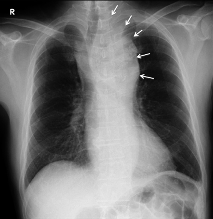
Chest X‐ray on admission. The aortic arch shadow is remarkably enlarged, resulting in tracheal deviation to the right side (arrows)

Computed tomography on admission reveals a specific and massive air‐filled collection, indicating emphysema, around the infected aortic arch aneurysm (arrows)

Computed tomography reveals a ruptured but shielded aortic arch aneurysm at 11 d after admission (arrow)

Intraoperative view. Infected aortic arch aneurysm involving the left common carotid and left subclavian arteries (arrows)

A, Computed tomography reveals the delayed esophageal perforation after surgery (white arrow); B, After 2 mo and a half, upper gastrointestinal endoscopy reveals the closure of esophageal perforation such as a small diverticulum without inflammation (black arrows)

3D computed tomography reveals the endovascular stent‐graft indwelling at the distal anastomosis
References
-
- Tokuno O, Kagawa D, Uchida D, Kinoshita S, Iwata K. A case of Salmonella‐infected thoracoabdominal aortic aneurysm making final diagnosis difficult. Kansenshogaku Zasshi. 2011;85:280‐283. - PubMed
-
- Kan CD, Lee HL, Yang YJ. Role of endovascular aortic repair in the treatment of infected aortic aneurysms complicated by aortoenteric or aortobronchial fistulae. Thorac Cardiovasc Surg. 2018;66(3):240‐247. - PubMed
-
- Sörelius K, Wanhainen A, Wahlgren CM, et al. Nationwide study on treatment of mycotic thoracic aortic aneurysms. Eur J Vasc Endovasc Surg. 2019;57(2):239‐246. - PubMed
Publication types
LinkOut - more resources
Full Text Sources
