. 2020 Nov;10(11):465.
doi: 10.1007/s13205-020-02457-1. Epub 2020 Oct 8.

Evaluation of the anticancer activity of enzymatically synthesized Baccatin III: an intermediate precursor of Taxol®

Affiliations

Evaluation of the anticancer activity of enzymatically synthesized Baccatin III: an intermediate precursor of Taxol®

Balendra Sah et al. 3 Biotech. 2020 Nov.

Abstract

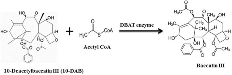
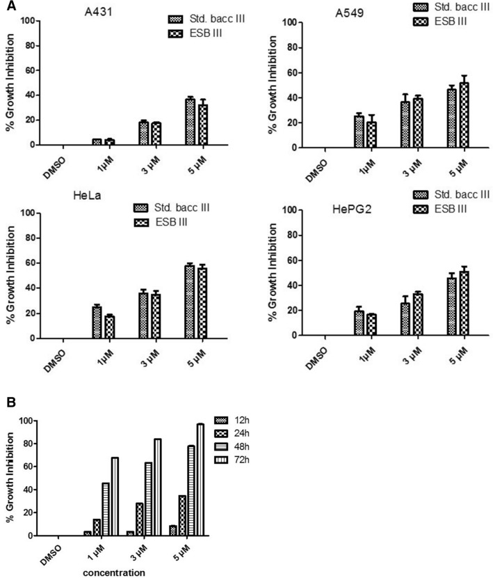
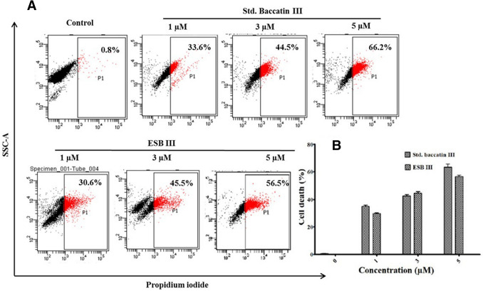
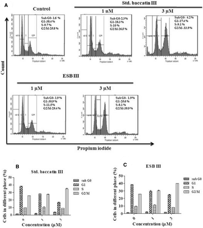
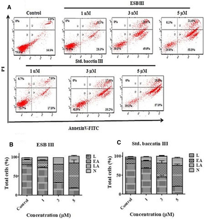
Baccatin III is an important precursor for the synthesis of clinically important anticancer drug Taxol. Previously, we have characterized a key enzyme of 10-deacetylbaccatin III-10-β-O-acetyltransferase (DBAT) which catalyses the 10-deacetylbaccatin III into baccatin III in taxol biosynthesis. Here, in the present study, we have evaluated and compared the cytotoxic properties of the enzymatically synthesized baccatin III (ESB III) with standard baccatin III in different human cancer cell lines, namely human cervical cancer (HeLa), human lung cancer (A549), human skin cancer (A431) and human liver cancer cells (HepG2). Among the various cancer lines tested, HeLa was more susceptible to ESB III with IC50 of 4.30 µM while IC50 values for A549, A431 and HepG2 ranged from 4 to 7.81 µM. Further, it showed G2/M phase cell cycle arrest, production of reactive oxygen species and depolarised mitochondrial membrane potential. In addition, annexin V-FITC staining was performed which showed the apoptotic cell death of HeLa cells, when treated with ESB III. Hence, ESB III was capable to show anticancer activities by inducing apoptotic cell death which could further be used for the semisynthesis of taxol in future.

Keywords: 10-Deacetylbaccatin III-10-β-O-acetyltransferase (DBAT); Apoptotic activity; Baccatin III; Cytotoxic; Paclitaxel.


Conflict of interest statement

Conflict of interestThe authors declare no conflict of interest.

Figures

Fig. 1
Fig. 1
Schematic diagram representing the enzymatic conversion of 10-deacetylbaccatin III (10-DAB) into baccatin III in the presence of acetyl coenzyme A and 10-deacetylbaccatin III-10-β-O-acetyltransferase (DBAT) enzyme (Sah 2018)
Fig. 2
Fig. 2
a Cytotoxic activity of enzymatically synthesized baccatin III (ESB III) and standard baccatin III against human skin cancer (A431), human lung cancer (A549), human liver cancer (HepG2) and human cervical cancer (HeLa) cell lines. (A431, A549, HepG2, and HeLa). Different concentrations of ESB III and standard baccatin III were treated to cells for 24 h and cytotoxicity was determined by MTT assay. b Cytotoxic activity of enzymatically synthesized baccatin III (ESB III) in human cervical cancer (HeLa) cell line at different time intervals. Cytotoxicity was determined by MTT assay with different concentrations of ESB III and standard baccatin III were treated to cells for 12, 24, 48 and 72 h
Fig. 3
Fig. 3
a Live and dead analyses of HeLa cells by PI staining while treated with different concentrations of enzymatically synthesized baccatin III (ESB III) and standard baccatin III. b Bar diagram showing the percentage of dead cells observed during PI live/dead staining. PI-based live/dead staining was performed by flow cytometry analysis upon treatment with different concentrations of ESB III and standard baccatin III
Fig. 4
Fig. 4
Effect of enzymatically synthesized baccatin III (ESB III) and standard baccatin III on cell cycle phase distribution. a Percent of HeLa cells in different phases of cell cycle after treatment with ESB III and standard baccatin III at different concentrations. b The bar diagram represents the distribution of cells at different phases of the cell cycle following treatment with standard baccatin III and c ESB III. The percentage of cells in each cell cycle phase was evaluated by propidium iodide-based staining by flow cytometry in the HeLa cells after treatment with ESB III and standard baccatin III
Fig. 5
Fig. 5
Effect of enzymatically synthesized baccatin III (ESB III) on mitochondrial membrane potential (MMP). a Scatter plot showing HeLa cells treated with different concentrations of ESB III and std. baccatin III. b Bar diagram representing the ratio of red to green fluorescence. Cells treated with different concentrations of ESB III and std. baccatin III, and were stained with JC-1 to detect depolarization of mitochondrial membrane using flow cytometry
Fig. 6
Fig. 6
Annexin V-FITC and PI staining to evaluate the different stages of apoptosis in HeLa cells following enzymatically synthesized baccatin III (ESB III) and standard baccatin III treatment. a In each panel, the lower left quadrant shows cells, which are negative for both PI and annexin V-FITC, while upper left quadrant shows only PI-positive cells, which are necrotic (N-necrotic). The lower right quadrant shows annexin positive cells (EA-early apoptotic) and the upper right quadrant shows annexin and PI-positive cells (LA-late apoptotic cells). b The bar diagram representing the comparison percentage of early and late apoptotic cells for ESB III and c standard baccatin III. All data of result from three independent experiments
Fig. 7
Fig. 7
Effect of enzymatically synthesized baccatin III (ESB III) and std. baccatin III on the generation of ROS in HeLa cells. a HeLa cells were treated with different concentrations of ESB III and standard baccatin III. b Bar diagram representing the fold increase in ROS after different treatment in comparison to control. Intracellular ROS production was observed by DCFH-DA dye after cells treated with ESB III and std. baccatin III by flow cytometry

References

    1. Alexandre J, Batteux F, Nicco C, Chéreau C, Laurent A, Guillevin L, Weill B, Goldwasser F. Accumulation of hydrogen peroxide is an early and crucial step for paclitaxel-induced cancer cell death both in vitro and in vivo. Int J Cancer. 2006;119(1):41–48. doi: 10.1002/ijc.21685. - DOI - PubMed
    1. Chakravarthi BV, Sujay R, Kuriakose GC, Karande AA, Jayabaskaran C. Inhibition of cancer cell proliferation and apoptosis-inducing activity of fungal taxol and its precursor baccatin III purified from endophytic Fusarium solani. Cancer Cell Int. 2013;13(1):105. doi: 10.1186/1475-2867-13-105. - DOI - PMC - PubMed
    1. Chow SY, Williams HJ, Pennington JD, Nanda S, Reibenspies JH, Scott AI. Studies on taxadiene synthase: interception of the cyclization cascade at the verticillene stage and rearrangement to phomactatriene. Tetrahedron. 2007;63(27):6204–6209. doi: 10.1016/j.tet.2007.03.029. - DOI
    1. Cossarizza A, Salvioli S. Flow cytometric analysis of mitochondrial membrane potential using JC-1. Curr Protocols Cytom. 2000;13(1):9–14. - PubMed
    1. Eruslanov E, Kusmartsev S. Identification of ROS using oxidized DCFDA and flow-cytometry. In: Armstrong D, editor. Advanced protocols in oxidative stress II. Methods in molecular biology (methods and protocols) 594. Totowa: Humana Press; 2010. - PubMed

LinkOut - more resources