. 2020 Sep 23;23(10):101600.
doi: 10.1016/j.isci.2020.101600. eCollection 2020 Oct 23.

Matrix Metalloprotease-7 Mediates Nucleolar Assembly and Intra-nucleolar Cleaving p53 in Gefitinib-Resistant Cancer Stem Cells

Affiliations

Matrix Metalloprotease-7 Mediates Nucleolar Assembly and Intra-nucleolar Cleaving p53 in Gefitinib-Resistant Cancer Stem Cells

Wei-Hsuan Yu et al. iScience. .

Abstract

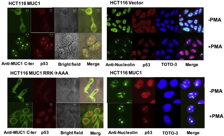
The enlarged distinct bulky-ball-like nucleolus matrix assembly is observed in most cancer stem cells (CSCs); however, the underlying mechanism is largely unknown. We show that matrix metalloproteinase-7 (MMP-7) shedding MUC-1 SEA domain releases MUC-1 C-ter, facilitating the nucleolus trafficking of p53 in gefitinib-resistant lung CSCs. The nucleolus colocalizations of p53, MUC-1 C-ter, MMP-7 and nucleolin were observed in the CD34+ CXADR+ CD44v3 + gefitinib-resistant EGFRL858R/T790M CSC colonies. MUC-1 C-ter induced a unique porous bulky-ball-shaped, cagelike nucleolus that functions as a nucleus molecular "garage" for potent tumor suppressor, p53. Nucleolus could also facilitate the novel sub-nucleus compartment for proteolytic processing p53 by MMP-7 to generate a 35 kDa fragment. Moreover, we show that salinomycin, an anti-CSC agent, disrupts nucleolus by inducing nucleoplasm translocation of p53 and sensitizing CSC to chemotherapy drugs. Thus, this study highlights the MMP-7-MUC-1-p53 axis in nucleolus as a potential therapeutic target for anti-CSCs to resolve the chemotherapy-resistance dilemma.

Keywords: Cancer; Cell Biology; Molecular Biology.


Conflict of interest statement

The authors declare no competing interests.

Figures

None
Graphical abstract
Figure 1
Figure 1
HRG and PMA Induce MUC-1 Shedding by MMP-7 (A) ZR-75-1 cells were treated with HRG or PMA for 30 min and subjected to immunoprecipitation with anti-MUC-1 N-ter Ab. The precipitates were analyzed by immunoblotting with anti-MMP-7 (RM7C) polyclonal Ab and anti-MUC-1 N-ter. Bottom panel: the total cell lysates were also immunoblotted with anti-MMP-7 (RM7C) polyclonal Ab. (B) Anti-MMP-7 immunoprecipitates from HRG- or PMA-treated cells were analyzed by immunoblotting with anti-MUC-1 C-ter. (C) ZR-75-1 cells were transfected to express an empty vector or MMP-7 and selected for 5 days in the presence of blasticidin-S. Anti-MMP-7 immunoprecipitates were analyzed by immunoblotting with anti-MUC-1 C-ter. (D) MMP-7 functions as an MUC-1 sheddase by cleaving MUC 1-ECD-Fc. Anti-MUC-1 N-ter immune precipitates from ZR-75-1 cells were incubated with MMP-7 alone and in the presence of SC44463. The proteins were subjected to an immunoblot analysis (non-denaturing conditions) with anti-MUC-1 C-ter. (E) An MUC-1 extracellular domain (ECD)-Fc fusion protein was incubated with 20 ng MMP-7 (alone and in the presence of SC44463), 40 ng MMP-2, or 40 ng MMP-9. The products were analyzed by SDS-PAGE and Coomassie Blue staining (upper panel). The N-terminal amino acid sequencing of the approximately 20-kDa band (lane 2) revealed cleavage at the PF site (lower panel). (F) Upper panel: lysates from untreated, HRG- and PMA-treated ZR-75-1 cells were subjected to immunoprecipitation with anti-MMP-7. MMP-7 activity was assessed by CM-transferin zymography. Pro-MMP-7 and MMP-7 were included as controls (lane 1). Lower panel: MCF-10A cells were stimulated with PMA, fixed, and stained with anti-MUC-1 C-ter and anti-MMP-7.
Figure 2
Figure 2
MUC-1 SEA Domain Complexes with Active MMP-7 in Silicodocking Model (A) Comparison of the docking models for the MUC-1 SEA domain/mature MMP-7 complexes upon cleavage of the MUC-1 SEA domain (based on PDB code: 2ACM). The enlarged window shows a clearer image of the loop near the cleavage site (G1097-S1098 (SEA domain: G115-S116)) snaking into the active site of mature MMP-7 (based on PDB code: 2Y6D; H120, H124, H130 E121 (pro-MMP-7: H219, H223, H229, E220)). The cleaved and uncleaved MUC-1 SEA domain molecules are indicated by tan and orchid ribbons. The mature MMP-7 molecule is presented as a spring green ribbon. (B and C) The binding complexes are shown as a spring green surface in mature MMP-7 and cleaved (C) and uncleaved (B) MUC-1 SEA domain molecules as tan and orchid ribbons. The two loops of the MUC-1 SEA domain shrink after G1097-S1098 site cleavage. (D and E) The binding complexes are presented as a spring green surface in mature MMP-7 and cleaved (E) and uncleaved (D) MUC-1 SEA domain molecules as tan and orchid surfaces. (E) The contact area between the MUC-1 SEA domain and mature MMP-7 decreases after cleavage. (F) Blue arrows indicate the putative MMP-7 specific cleavage sites in the SEA domain sequence. Red arrows indicate the N-terminal sequencing for the MMP-7 cleaves MUC-1 (ECD)-Fc fusion proteins products.
Figure 3
Figure 3
MMP-7 Shedding MUC-1 Releases MUC-1 C-Ter Inducing Nucleolus Translocation and Distinct Enlarged Nucleolus (A) ZR-75-1 cells were treated with the indicated recombinant proteins for 4 h. The cells were then fixed and stained with anti-MUC-1 N-ter. The nuclei were stained with TOTO-3. (B) HCT116/MUC-1 and HCT116/MUC-1(RRK→AAA) cells were transfected to express the empty vector or MMP-7 and selected in blasticidin-S for 5 days. The cells were fixed and stained with anti-MUC-1 C-ter and anti-MMP-7. The nuclei were stained with TOTO-3. (C) MUC-1 C-ter induces distinct, enlarged nucleoli. ZR-75-1 cells were infected with a retrovirus expressing the empty vector or MUC-1 siRNA and grown for the indicated number of days. Lysates from the cells were analyzed by immunoblotting with anti-MUC-1 N-ter and anti-tubulin. ZR-75-1 cells expressing the empty retroviral vector (top two panels) of MUC-1 siRNA (middle two panels) were left untreated or exposed to PMA for 60 min. HCH116 cells were stained with anti-MUC-1 C-ter, anti-nucleolin, and TOTO-3. Bottom panels: HCT116/vector and HCT116/MUC-1 cells were treated with PMA and stained with anti-MUC-1 C-ter, anti-nucleolin, and TOTO-3.
Figure 4
Figure 4
Co-expression of MUC-1 and MMP-7 Confirms the Anchorage Independent Growth Phenotypes (A) Top panel: ZR-75-1 cells grown in flasks for 5 and 10 days exhibited foci of multilayered domes. Middle panel: cells grown for 10 days in the presence of SC44463 or TIMP-3 exhibited flat foci consisting of a single layer of cells. Lower Panel: the cells were grown for 10 days in the presence of MMP-7 or the dominant-negative MMP-7 E219Q. (B) Lysates from the cells were analyzed by immunoblotting with anti-MUC-1 N-ter and anti-α-tubulin (lower panels). (C) The cells were infected with a retrovirus expressing the empty vector or MUC-1 siRNA and grown for 5 or 10 days. (D) MMP-7→MUC-1 signaling induces the formation of complex multi-acinar structures and targets MUC-1 C-ter to the nucleolus in MCF-10A 3D culture assay: (Top panels) MCF-10A cells were cultured for 10 days in Matrigel containing PMA or MMP-7 in the absence and presence of TIMP-3 or SC44463. The cells were fixed and stained with anti-MUC-1 C-ter. The nuclei were stained with TOTO-3. (Middle panels) MCF-10A cells were cultured for 10 days in Matrigel containing PMA in the absence and presence of TIMP-3. The cells were fixed and stained with anti-MUC-1 C-ter. The nuclei were stained with TOTO-3. (Bottom panels) MCF-10A cells were cultured for 10 days in Matrigel containing MMP-7 in the absence or presence of SC44463. (E) Left panel: sections of normal mammary ductal epithelium and an invasive ductal carcinoma were assessed to visualize their reactivity with anti-MUC-1 C-ter and anti-MMP-7. Right panel: enlarged concentric nucleoli were observed in breast carcinoma cells but not normal mammary gland epithelial cells.
Figure 5
Figure 5
MMP-7-Mediated MUC-1-Dependent Nucleolar Assembly and p53 Nucleolus Trafficking The assembly of the enlarged concentric nucleolus is an MUC-1 dependent on the PMA-treated HCT116 cell line. P53 significantly translocates into the nucleolus in the PMA-treated MUC-1-transfected HCT116 cell line. The immunofluorescence double staining for MUC-1 C-ter and p53 nucleolin indicate that both molecules are well colocalized in the nucleolus compartments, which are identified by immunostaining for nucleolin. In the absence of MUC-1, nucleolin assembly is not active and the MUC-1 with NLS mutation (RRK→AAA) mutant can abolish p53 translocating into nucleolus.
Figure 6
Figure 6
MUC-1 C-Ter and p53 Were Co-localized in Nucleolus of the CD34+ CXDR+ EGFRL858R/T790M CL1-0 Derived Lung CSCs (A) Increasing the anchorage-independent growth capacity of the gefitinib-resistant EGFRL858R/T790M CL1-0 colonies in soft agar assay. (B) Salinomycin can abolish the anchorage-independent growth abilities of EGFRL858R/T790M transfected CL1-0 cells in the presence of 10-μM gefitinib. (C) The EGFRL858R/T790M CL1-0 small lung carcinoma cells selected colonies from soft agar assay were further transfected with plasmid nucleolin-YFP fusion constructs, and the stable EGFRL858R/T790M CL1-0/nucleolin-YFP cell colony was used to perform the immunostaining with anti-CXADR, and anti-CD34 Ab followed by labeling with anti-mouse-Texas Red fluorescence, anti-rabbit GFP, and nucleus was stained with DAPI. Both p53 and MUC-1 C-ter are localized in nucleolin by using anti-p53 polyclonal Ab, anti-MUC-1 C-ter (MUC-1-CD) Ab, anti-CXADR, and anti-CD34 Ab followed by labeling with the pair of anti-mouse GFP/anti-rabbit Texas Red, anti-mouse-Texas Red/anti-rabbit FITC fluorescence, and nucleus was stained with DAPI.
Figure 7
Figure 7
MUC-1 C-Ter Is Involved in Nucleolus Matrix Assembly and Regulates Nucleolar Structure in CD34+ EGFRL858R/T790M CL1-0-Derived CSCs The MMP-7 upregulated expression EGFR constitutively active EGFRL858R/T790M CL1-0 small lung carcinoma cells selected from colonies from soft agar assay were further transfected with plasmid nucleolin-YFP fusion constructs, and the stable EGFRL858R/T790M CL1-0/nucleolin-GFP cell colonies were used to perform the immunostaining with anti-MUC-1 C-ter (MUC-1-CD) Ab.(A1–A4): Confocal microscopic analysis for immune-fluorescence staining for anti-MUC1 C-ter (MUC-1-CD) (Texas Red), nucleolin-YFP fusion protein expression (Green), and co-staining DAPI for nucleus chromosome (Blue). The sub-nucleus compartment colocalization of MUC1 C-ter (Red) with nucleolin (Green) in the DNA(DAPI) exclusive sub-nucleus compartment, nucleolus, (A1 is zoom out image and A2, A3, and A4 are zoom in images). The size and shape of nucleolus appear to be dependent on the relative amount of MUC-1 C-ter (Red) and nucleolin (Green) in the nucleolus. The complex of MUC-1 C-ter and nucleolin seem to be DNA (DAPI) exclusive and induce dome-like structure in the nucleus (see images of the DAPI merge with bright field), particularly in panel A3. In the lowest level of MUC-1 C-ter and nucleolin complex zone, the DNA clear zone starts to appear and the mosaic-architecture between MUC-1 C-ter (net-like in Red) and nucleolin (tetra-pod-like in Green) can be visualized.
Figure 8
Figure 8
MMP-7 Mediates Proteolytic Processing p53 to 35 kDa Fragments in the Bulky-Ball-like Nucleolus of the CD34+ EGFRL858R/T790M CL1-0-Derived CSCs The pcDNA p53 desRED and pcDNA nucleolin-YFP were co-transfected into floating sphere EGFRL858R/T790M CL1-0 CSC-like clones. Posttransfection 48 h, the cells were stained with DAPI to label nucleus and followed by confocal microscopic analysis. (A1 and A2) Sub-nucleus compartment colocalization of p53 (Red) with nucleolin (green) in the DNA (DAPI) exclusive sub-nucleus compartment, nucleolus, (A1 zoom out and A2 zoom in). The enlarged nucleolus appears as a bulky-ball-shaped cage, nucleolin-YFP (in green). The structure is just like a “bulky-ball-cage” and the abundant p53 (Texas Red) sequestrates inside the nucleolin cage-like compartment. (B) Immune-precipitation was performed directly from nucleus isolation extracts by using anti-nucleolin monoclonal Abs or anti-p53 monoclonal antibodies followed by western blot followed by immunoblotting with anti-p53, anti-MMP-7 (RM7C), anti-nucleolin, anti-CD44v3 mAb (R&D Systems), or anti-MUC-1 CD. U: Unbound; B Bound; MB: MicroBead
Figure 9
Figure 9
Salinomycin Disrupts Nucleolus and Releases Nucleolar p53 into Nucleoplasm Sensitizing CD34+ EGFRL858R/T790M CL1-0 Derived CSCs to Gefitinib Treatment (A) The localization of MMP7-CFP appears not only in the cell surface but also in the nucleolus compartment. Nucleolus localization of MMP-7 and proteolytic processing p53 by MMP-7 in the nucleolus of EGFRL858R/T790M CL1-0 CSC-like clones. (top panel) The fluorescent tag fusion proteins mammalian expression constructs, pcDNA MMP-7 CFP and pcDNA nucleolin YFP, were co-transfected into the floating sphere EGFRL858R/T790M CL1-0 CSC-like clones. Posttransfection 48 h, the cells were stained with DAPI to label nucleus and followed by confocal microscopic analysis, the colocalization of MMP-7 CFP and nucleolin YFP in porous-ring-like nucleolus were observed. The colocalization of MMP-7 CFP and nucleolin YFP were observed in the nucleolus compartments. The porous-ring-like nucleolus co-localized with MMP-7-CFP were observed in higher magnification. The major signal of nucleolin-YFP appears in the bulky-ball ring-like nucleolus is where the MMP-7 CFP also localized (bottom panel). (B) Top panel: well colocalization of anti-p53 polyclonal-Ab-desRed and anti-nucleolin monoclonal Ab-GFP in the absence of salinomycin treatment. Middle and bottom panels: in the presence of salinomycin treatment, the enlarged nucleolin is disrupted and the nucleoplasm redistribution of p53 from nucleolus is observed. EGFRL858R/T790M CL1-0 cancer-stem-cell like cells are sensitive to the nucleolin-targeting drug salinomycin. (C–E) Salinomycin promotes antitumor ability of gefitinib in vivo. Six-week-old BALB/c/nu mice were subcutaneously injected with 2 × 106 cells CL1-0 overexpressed double mutant EGFR single stable clone cells for 28 days then treated with DMSO (D), 5 mg/kg/day of salinomycin (S), 60 mg/kg/day of gefitinib (G), and combine treatment (G + S) for 28 days. (C) The body weight of experimental mice. (D) and (E) The mice with tumor were displayed and the quantitative results were shown. p < 0.05 v.s. DMSO group; #p < 0.05 v.s. G group.

Similar articles

Cited by

References

    1. Behrendtsen O., Werb Z. Metalloproteinases regulate parietal endoderm differentiating and migrating in cultured mouse embryos. Dev. Dyn. 1997;208:255–265. - PubMed
    1. Bergers G., Brekken R., McMahon G., Vu T.H., Itoh T., Tamaki K., Tanzawa K., Thorpe P., Itohara S., Werb Z., Hanahan D. Matrix metalloproteinase-9 triggers the angiogenic switch during carcinogenesis. Nat. Cell Biol. 2000;2:737–744. - PMC - PubMed
    1. Bergers G., Coussens L.M. Extrinsic regulators of epithelial tumor progression: metalloproteinases. Curr. Opin. Genet. Dev. 2000;10:120–127. - PubMed
    1. Bharti A.K., Olson M.O., Kufe D.W., Rubin E.H. Identification of a nucleolin binding site in human topoisomerase I. J. Biol. Chem. 1996;271:1993–1997. - PubMed
    1. Bitler B.G., Goverdhan A., Schroeder J.A. MUC1 regulates nuclear localization and function of the epidermal growth factor receptor. J. Cell Sci. 2010;123:1716–1723. - PMC - PubMed