. 2020 Nov;206(6):921-938.
doi: 10.1007/s00359-020-01448-0. Epub 2020 Oct 21.

Modular timer networks: abdominal interneurons controlling the chirp and pulse pattern in a cricket calling song

Affiliations

Modular timer networks: abdominal interneurons controlling the chirp and pulse pattern in a cricket calling song

Pedro F Jacob et al. J Comp Physiol A Neuroethol Sens Neural Behav Physiol. 2020 Nov.

Abstract

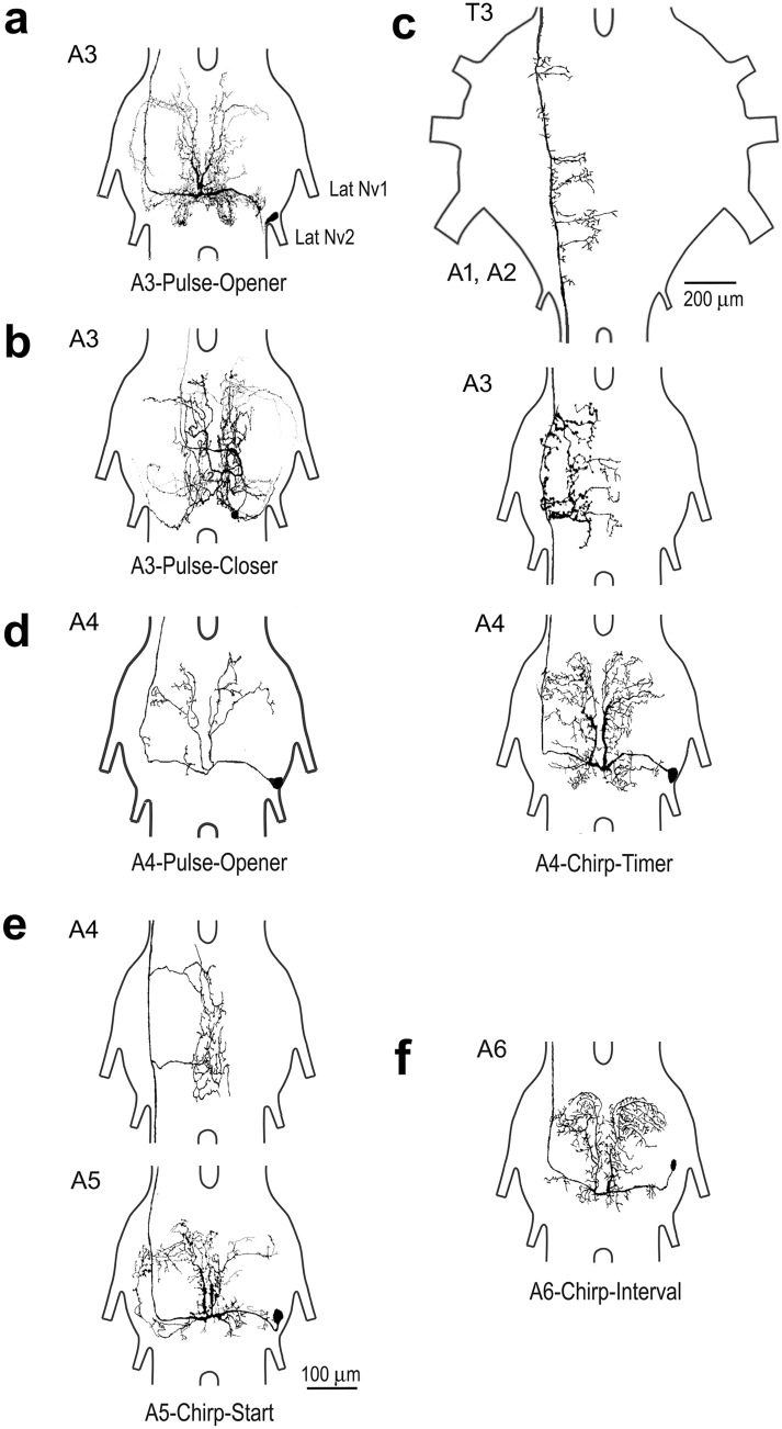
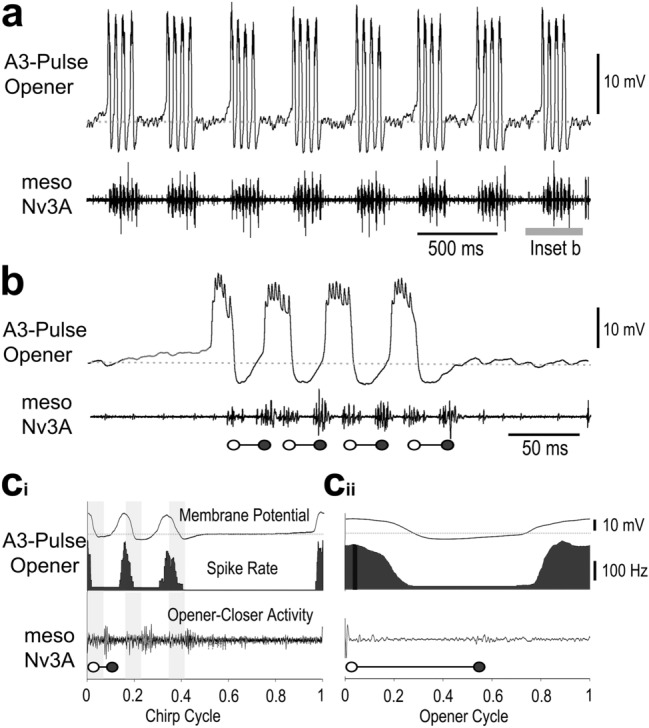
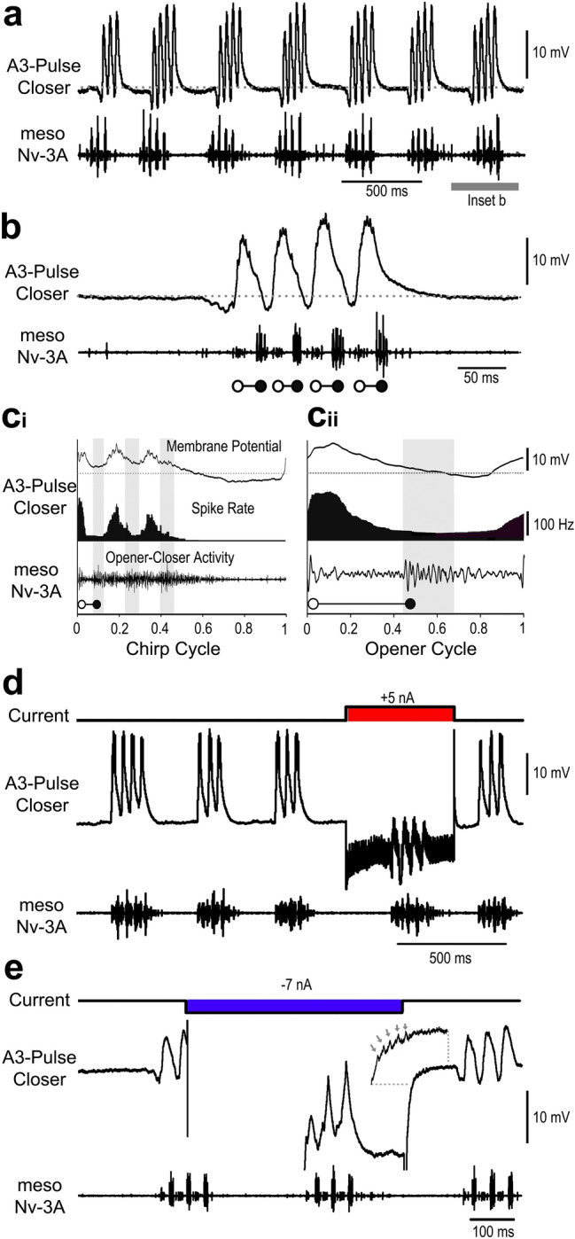
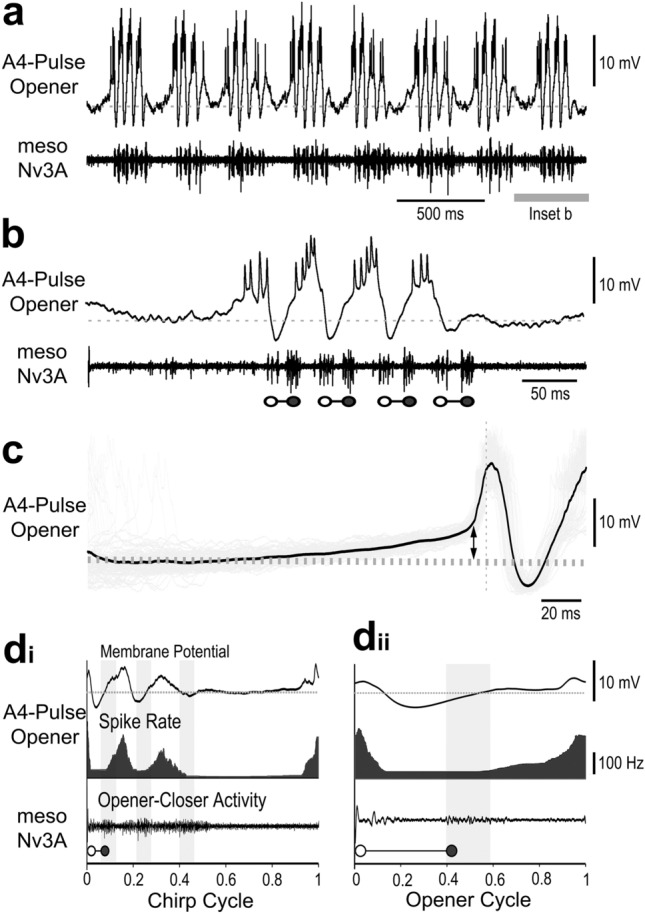
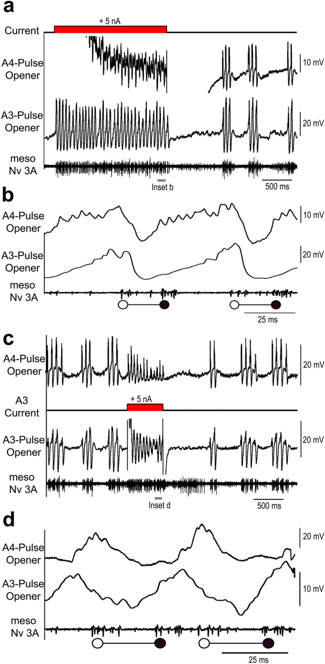
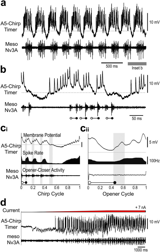
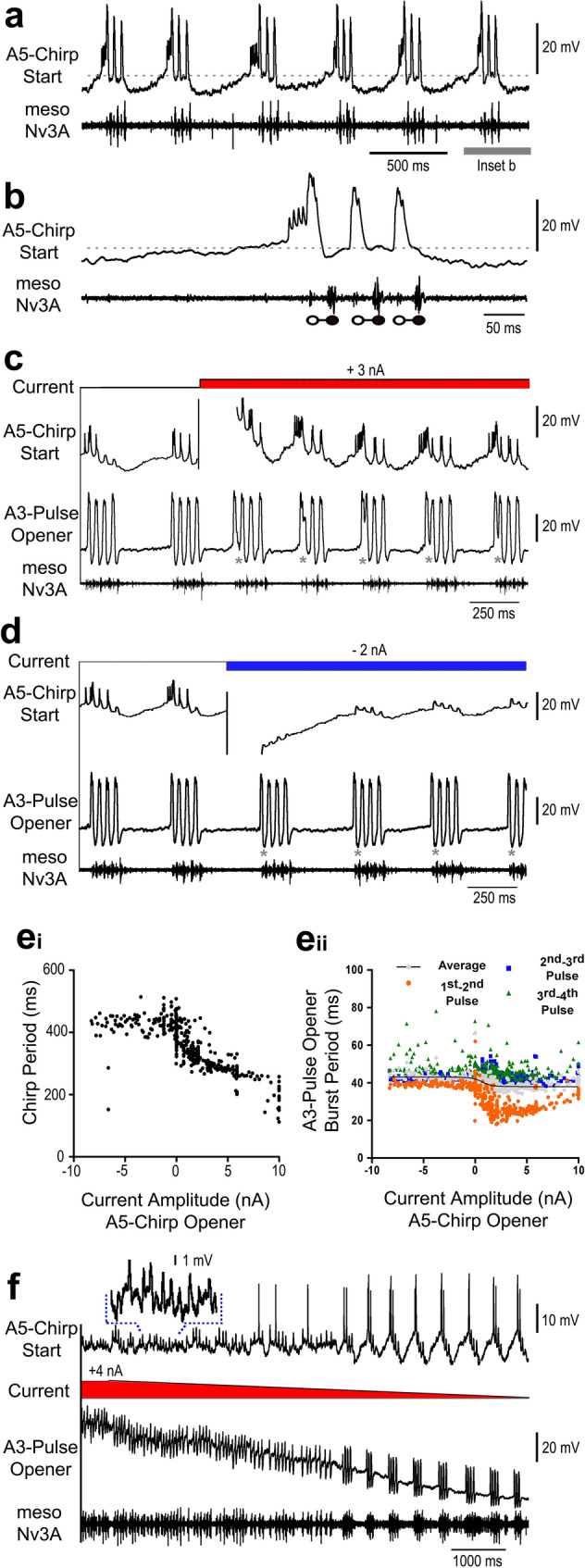
Chirping male crickets combine a 30 Hz pulse pattern with a 3 Hz chirp pattern to drive the rhythmic opening-closing movements of the front wings for sound production. Lesion experiments suggest two coupled modular timer-networks located along the chain of abdominal ganglia, a network in A3 and A4 generating the pulse pattern, and a network organized along with ganglia A4-A6 controlling the generation of the chirp rhythm. We analyzed neurons of the timer-networks and their synaptic connections by intracellular recordings and staining. We identified neurons spiking in phase with the chirps and pulses, or that are inhibited during the chirps. Neurons share a similar "gestalt", regarding the position of the cell body, the dendritic arborizations and the contralateral ascending axon. Activating neurons of the pulse-timer network elicits ongoing motor activity driving the generation of pulses; this activity is not structured in the chirp pattern. Activating neurons of the chirp-timer network excites pulse-timer neurons; it drives the generation of chirps and during the chirps the pulse pattern is produced. Our results support the hypothesis that two modular networks along the abdominal ganglion chain control the cricket calling song, a pattern generating network in the mesothoracic ganglion may not be required.

Keywords: Acoustic communication; Central pattern generator; Identified interneurons; Modular network; Timing of rhythms.


Conflict of interest statement

The authors declare no competing interest for this work.

Figures

Fig. 1
Fig. 1
Structure of singing-interneurons with cell bodies located in the abdominal ganglia. All neurons show a lateral cell body, extensive symmetrical dendrites in the dorsal neuropil and a contralateral axon projecting anteriorly, with medial collaterals, if labelled. a A3-Pulse-Opener interneuron, with cell body and dendrites located in A3 and axon projecting towards the T3 ganglion, dye coupling between bilateral neurons. b The A3-Pulse-Closer neuron, with complex dendrites in A3 and an ascending axon. c The A4-Chirp-Timer neuron in ganglion A4 with an axon projecting through A3 and T3, dye-coupling occurred between bilateral neurons. d A4-Pulse-Opener interneuron, with typically sparse dendrites. e A5-Chirp-Start interneuron in A5, the axon projects at least through A4. f A6-Chirp-Interval neuron with cell body and dendrites stained
Fig. 2
Fig. 2
Activity of the A3-Pulse-Opener neuron. a, b Rhythmic membrane potential depolarisations occur coupled to singing activity. High frequency bursts of spikes precede the opener MN activity (open circles), and hyperpolarisation precedes closer activity (closed circles) recorded from meso-Nv3A. Right after a chirp, activity terminates with a hyperpolarisation, followed by a ramp depolarisation but no spiking activity in the interchirp intervals. ci, cii Phase diagrams with the average membrane potential (top), the average spike rate (middle) and the average meso-Nv3A activity (bottom) for the chirp cycle (ci) and opener cycle (cii). Grey shaded bars indicate opener MN activity, grey dashed line represents the resting membrane potential. N = 6 crickets, n = 60 chirps/180 pulses, AP = 595
Fig. 3
Fig. 3
Activity the A3-Pulse-Closer neuron. a, b Rhythmic membrane potential depolarisations are coupled to singing activity. Hyperpolarisation precedes the opener MN activity (open circles) and high frequency bursts of spikes precede the closer MN activity (closed circles). At the end of a chirp activity terminates with a gradual decline to the resting membrane potential. No spiking activity occurs in the interchirp intervals. ci, cii Phase diagrams with the average membrane potential (top), the average spike rate (middle) and the average meso-Nv3A activity (bottom) for the chirp cycle (ci) and opener cycle (cii). Grey shaded bars indicate closer MN activity, grey dashed line represents the resting membrane potential. N = 5 crickets, n = 50 chirps/200 pulses, AP = 800. d Current injection into the A3-Pulse-Closer neuron with 5nA for 500 ms extends the ongoing chirp period. e Upon release from inhibition, a postinhibitory rebound generates spike activity. Inset shows spikes (arrows) in the rising phase of the rebound
Fig. 4
Fig. 4
Activity of the A4-Pulse-Opener. a During fictive singing the A4-Pulse-Opener is rhythmically activated during the chirps. b Recording shows a gradual ramp-depolarisation at the start of a chirp, bursts of spikes precede and the opener MN activity (open circles) and a pronounced hyperpolarisation precedes the closer MN activity (close circles). Grey dashed line indicates the resting membrane potential. c Ramp depolarisation at the start of a chirp leading to the first A4-Pulse-Opener spike, indicated by vertical dashed line. Double arrow indicates the peak amplitude of the ramp depolarisation. The black line represents the average and the grey shades the raw signals, respectively. N = 14 crickets, n = 140 chirps/105 pulses. di, dii Phase diagrams with the average membrane potential of the A4-Pulse-Opener (top), its average spike rate (middle) and the averaged meso-Nv3A activity (bottom) for the chirp cycle (di) and opener cycle (dii). Vertical grey bars indicate timing of closer MN activity, and grey dashed line the resting membrane potential. N = 14 crickets, n = 140 chirps/420 pulses, AP = 1540
Fig. 5
Fig. 5
Paired intracellular recordings of the A4-Pulse-Opener and the A3-Pulse-Opener. a Depolarising current injection with 5 nA for 1.8 s in the A4-Pulse-Opener drives rhythmic oscillations in in the A3-Pulse-Opener and the opener and closer MNs for the duration of the pulse. The chirp pattern resumes after current injection. b The recording at high resolution does not reveal synaptic coupling between the A3-Pulse-Opener and the A4-Pulse-Opener. c Depolarising current injection with 5 nA for 600 ms in the A3-Pulse-Opener drives rhythmic oscillations in the A4-Pulse-Opener and the opener-closer cycles for the duration of the pulse. The chirp pattern resumes after current injection. d The recording at high resolution does not reveal synaptic coupling between the interneurons. It reveals a shift in the timing of the depolarisations of both interneurons
Fig. 6
Fig. 6
Activity of the A4-Chirp-Timer. a The interneuron spike activity is rhythmically coupled to the chirp pattern. b In the interchirp interval the neuron shows a gradual increase in its membrane potential and starts spiking well before the start of a chirp. It is depolarised throughout the chirp, spikes occur before the opener MN burst (open circles), and it is hyperpolarised before the closer activity (close circles). Grey dashed line represents the resting membrane potential. ci, cii Phase diagrams with the average membrane potential of the A4-Chirp-Timer (top), its average spike rate (middle) and the averaged meso-Nv3A activity (bottom) for the chirp cycle (ci) and opener cycle (cii). Vertical grey bars indicate closer MN activity, and grey dashed line the resting membrane potential. N  = 6 crickets, n  = 60 chirps/240 pulses, AP = 970. d Paired recordings of the A4-Chirp-Timer and the A3-Pulse-Opener. Depolarising current injection in the A4-Chirp-Timer with 4 nA for 5 s shortens the chirp period of fictive singing and increases the A3-Pulse-Opener membrane potential oscillations. ei,ii Synaptic coupling between the A4-Chirp-Timer and the A3-Pulse-Opener, demonstrated during low spike activity by averaging the A3-Pulse-Opener membrane potential triggered by spikes of the A4-Chirp-Timer (N  = 2, n   = 82). f A ramp of depolarising current with 3 nA injected in the A3-Pulse-Opener terminates the chirp pattern; it elicits rhythmic opener-closer cycles in the A3 neuron and a barrage of EPSPs in the A4-Chirp-Timer
Fig. 7
Fig. 7
Activity of the A5-Chirp-Timer. a Spike activity is coupled to the generation of chirps. b The neuron is depolarised and starts spiking before the start of a chirp, a burst of spikes occurs before the opener activity (open circles), and the neuron repolarises before the closer activity (close circles). Grey dashed line indicates the resting membrane potential. ci, cii Phase diagrams with the average membrane potential (top), the average spike rate (middle) and the average meso-Nv3A activity (bottom) for the chirp cycle (ci) and opener cycle (cii). Grey shaded bars indicate closer MN activity, grey dashed line represents the resting membrane potential. N = 2 crickets, n = 35 chirps/140 pulses, AP = 455. d Increasing current injection in the A5-Chirp-Timer with a ramp of 7 nA induced singing activity, with a transition between subthreshold membrane potential oscillations (left) to the production of chirps (right)
Fig. 8
Fig. 8
Activity of A5-Chirp-Start and impact on A3-Pulse-Opener activity. a,b During fictive singing the A5-Chirp-Start neuron shows a high frequency burst of spikes right before a chirp. c,d Double intracellular recordings of the A5-Chirp-Start and the A3-Pulse-Opener, depolarisation of the A5-Chirp-Start neuron increases the chirp period and alters the activity pattern of the A3-Pulse-Opener neuron, which prematurely terminates the first depolarisation and starts the second one. Asterisks indicate change in time course (c) of the first depolarisation and the amplitude of the first hyperpolarisation (d). ei Effect of depolarising and hyperpolarising the A5-Chirp-Start neuron on the chirp period. eii Effect of depolarising the A5-Chirp-Start neuron on the A3-Pulse-Opener burst period. f An A3-Pulse-Opener ramp depolarisation decreasing from 4 nA is accompanied by the opener-closer MN activity in the pulse pattern and a transition to the chirp pattern as the current decreases. Activity in the A5-Chirp-Start neuron is coupled to the changing motor pattern. Inset shows section of the recording with higher resolution
Fig. 9
Fig. 9
Activity of A5-Chirp-Interval and A6-Chirp-Interval neurons. a,b During fictive singing the neurons spike in the interchirp intervals and are rhythmically inhibited during the chirps. ci, cii Phase diagrams with the averaged membrane potential (top), the average spike rate (middle) and the average meso-Nv3A activity (bottom) for the chirp cycle (ci) and opener cycle (cii). N = 2 crickets, n = 35 chirps/105 pulses, AP = 235. Grey shaded sections indicate closer MN activity, grey dashed line indicates the resting membrane potential. d Depolarisation of the A5-Chirp-Interval neuron increases the chirp period. e Hyperpolarisation of the A5-Chirp-Interval neuron reduces background activity in the meso-Nv3A recording and decreases the chirp period

References

    1. Aghajanian GK. Modulation of a transient outward current in serotonergic neurones by α1-adrenoceptors. Nature. 1985;315:501–503. - PubMed
    1. Alexander RD. Evolutionary change in cricket acoustical communication. Evolution. 1962;16(4):443–467.
    1. ASAB Ethics Committee Guidelines for the treatment of animals in behavioural research and teaching. Anim Behav. 1997;53:229–234.
    1. Bentley DR. Intracellular activity in cricket neurons during generation of song patterns. Z vgl Physiol. 1969;283:267–283. - PubMed
    1. Bidaye SS, Bockemühl T, Büschges A. Six-legged walking in insects: how CPGs, peripheral feedback, and descending signals generate coordinated and adaptive motor rhythms. J Neurophysiol. 2018;119:459–475. - PubMed

Publication types

LinkOut - more resources