

Diaphragmatic myoclonus due to SARS-CoV-2 infection
- PMID: 33090303
- PMCID: PMC7579554
- DOI: 10.1007/s10072-020-04766-y
Diaphragmatic myoclonus due to SARS-CoV-2 infection
Abstract
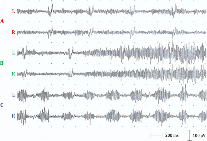
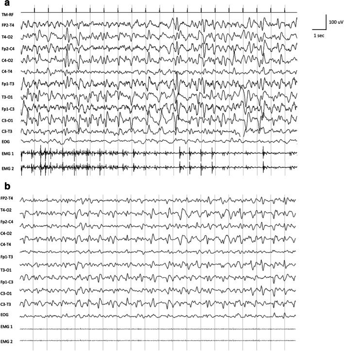
A wide range of neurological signs and symptoms have been associated with SARS-CoV-2 infection. In the present report, we described two Italian patients diagnosed with diaphragmatic myoclonus after COVID-19. In both cases, mild lymphocytosis at cerebrospinal fluid analysis and no structural brain changes were reported. The pathophysiological origin of the myoclonus in the two cases was different. In case 1, electroencephalogram did not reveal any cortical correlates and brain imaging of the spine was unremarkable, while in case 2, cortical origin of myoclonus was demonstrated. With the present two cases, we confirm and extend the neurological manifestations of SARS-CoV-2 infection.
Keywords: COVID-19; Diaphragmatic; Myoclonus; Neurology; SARS-CoV-2.
Conflict of interest statement
The authors declare that they have no conflict of interest.
Figures
Similar articles
-
Myoclonus status revealing COVID 19 infection.Seizure. 2023 Jan;104:12-14. doi: 10.1016/j.seizure.2022.11.010. Epub 2022 Nov 22. Seizure. 2023. PMID: 36446232 Free PMC article.
-
First case of SARS-COV-2 sequencing in cerebrospinal fluid of a patient with suspected demyelinating disease.J Neurol. 2020 Nov;267(11):3154-3156. doi: 10.1007/s00415-020-09996-w. Epub 2020 Jun 20. J Neurol. 2020. PMID: 32564153 Free PMC article.
-
Generalized myoclonus in COVID-19.Neurology. 2020 Aug 11;95(6):e767-e772. doi: 10.1212/WNL.0000000000009829. Epub 2020 May 21. Neurology. 2020. PMID: 32439821 Free PMC article.
-
Update on cerebrovascular manifestations of COVID-19.Neurol Sci. 2020 Dec;41(12):3423-3435. doi: 10.1007/s10072-020-04837-0. Epub 2020 Oct 20. Neurol Sci. 2020. PMID: 33083934 Free PMC article. Review.
-
A First Case of Acute Cerebellitis Associated with Coronavirus Disease (COVID-19): a Case Report and Literature Review.Cerebellum. 2020 Dec;19(6):911-914. doi: 10.1007/s12311-020-01177-9. Cerebellum. 2020. PMID: 32737799 Free PMC article. Review.
Cited by
-
Movement Disorders Associated with COVID-19.Parkinsons Dis. 2021 Nov 8;2021:3227753. doi: 10.1155/2021/3227753. eCollection 2021. Parkinsons Dis. 2021. PMID: 34790346 Free PMC article. Review.
-
Cerebrospinal fluid in COVID-19: A systematic review of the literature.J Neurol Sci. 2021 Feb 15;421:117316. doi: 10.1016/j.jns.2021.117316. Epub 2021 Jan 10. J Neurol Sci. 2021. PMID: 33561753 Free PMC article.
-
De Novo Movement Disorders Associated with COVID-19- A Systematic Review of Individual Patients.Ann Indian Acad Neurol. 2023 Sep-Oct;26(5):702-707. doi: 10.4103/aian.aian_572_23. Epub 2023 Oct 18. Ann Indian Acad Neurol. 2023. PMID: 38022478 Free PMC article.
-
Movement Disorders and SARS-CoV-2.Mov Disord Clin Pract. 2022 Dec 7;10(Suppl 2):S9-S17. doi: 10.1002/mdc3.13615. eCollection 2023 Aug. Mov Disord Clin Pract. 2022. PMID: 37637980 Free PMC article. No abstract available.
-
Diaphragm Muscle Atrophy Contributes to Low Physical Capacity in COVID-19 Survivors.Life (Basel). 2024 Sep 5;14(9):1117. doi: 10.3390/life14091117. Life (Basel). 2024. PMID: 39337900 Free PMC article.
References
-
- Benussi A, Pilotto A, Premi E et al. (2020) Clinical characteristics and outcomes of inpatients with neurologic disease and COVID-19 in Brescia, Lombardy, Italy. Neurology in press - PubMed
Publication types
MeSH terms
LinkOut - more resources
Full Text Sources
Miscellaneous
