Review
. 2020 Oct 22;16(10):e1008876.
doi: 10.1371/journal.ppat.1008876. eCollection 2020 Oct.

A molecular understanding of alphavirus entry

Affiliations
Review

A molecular understanding of alphavirus entry

Autumn C Holmes et al. PLoS Pathog. .

Abstract

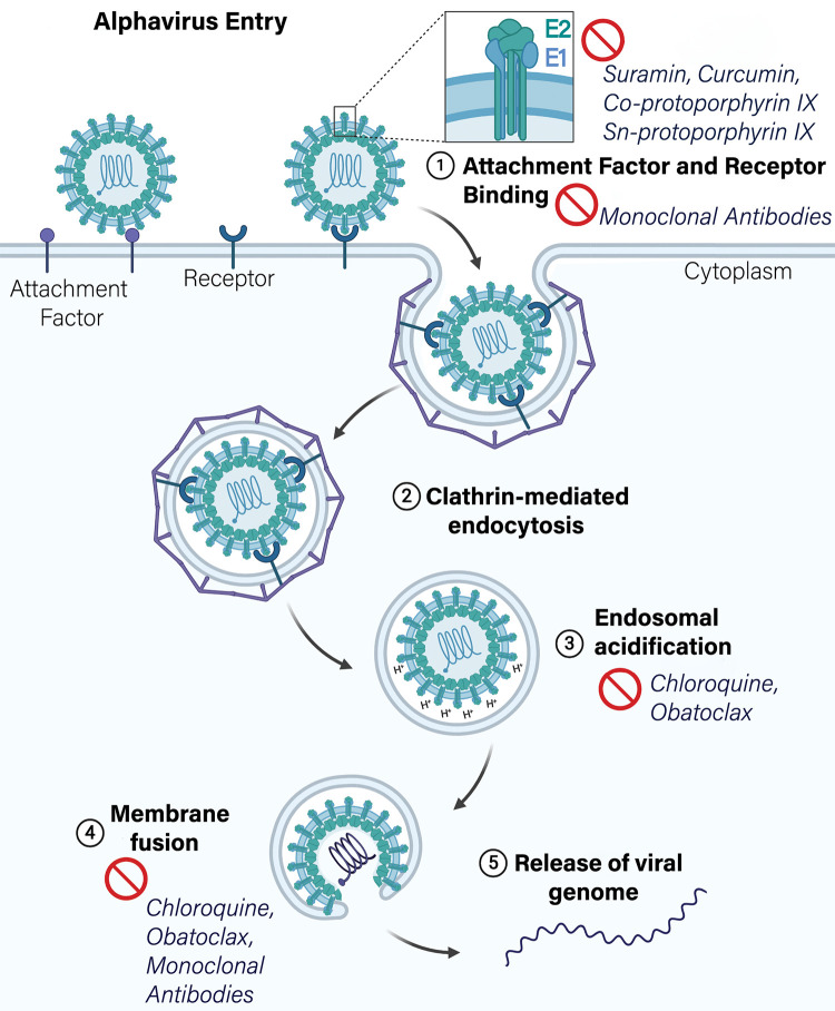
Alphaviruses cause severe human illnesses including persistent arthritis and fatal encephalitis. As alphavirus entry into target cells is the first step in infection, intensive research efforts have focused on elucidating aspects of this pathway, including attachment, internalization, and fusion. Herein, we review recent developments in the molecular understanding of alphavirus entry both in vitro and in vivo and how these advances might enable the design of therapeutics targeting this critical step in the alphavirus life cycle.


Conflict of interest statement

M.S.D. is a consultant for Inbios, Vir Biotechnology, NGM Biopharmaceuticals, and on the Scientific Advisory Board of Moderna. The Diamond laboratory has received unrelated funding under sponsored research agreements from Vir Biotechnology, Moderna, and Emergent BioSolutions.

Figures

Fig 1
Fig 1. Alphavirus genome organization and molecular structure of the virion.
(A) Cryo-EM reconstruction of CHIKV VLP (EMDB: 9393) colored by radial distance and depicted from the surface (left half) and an equatorial cross section (right half). The white triangle indicates 1 icosahedral asymmetric unit, with the 5-fold (i5), 3-fold (i3), and 2-fold (i2) icosahedral axes of symmetry labeled with a pentagon, triangles, and an oval, respectively. Trimeric spikes are labeled “i3” if coincident with the i3 axes and “q3” if on a quasi-3-fold axis. The black arrows indicate the directions of the icosahedral symmetry axes. Radial distance color scheme: red, electron dense core and RNA; yellow, capsid; green, membrane lipid; cyan, E1; and dark blue, E2 spike. Scale bar: 100 A°. E1 and E2 are embedded in the viral membrane and assemble into a heterotrimer: E1 is responsible for membrane fusion, while E2 facilitates receptor engagement. E3 is also bound in some alphaviruses including CHIKV, SFV, and VEEV, but the significance of this is not fully understood. (B) The alphavirus genome is a single-stranded, plus-sense RNA molecule of approximately 11 kb and encodes 4 nonstructural proteins, nsP1–4 and 5 structural proteins, capsid, E3, E2, 6k/TF, and E1. NsP4 forms the primary RNA-dependent RNA polymerase, but the synthesis of the genome requires all 4 nonstructural proteins. The RNA is capped at the 5′ end and polyadenylated at the 3′ end. (C) The alphavirus structural proteins E2 and E3 are produced as a polyprotein termed p62 (left). P62 acts as a chaperone to ensure proper folding of E1 (right) in the ER and is proteolytically processed into the mature E2 and E3 proteins by host furin-like proteases. E3 binds E2 during transport of the protein complex to the cell surface and also remains bound in the mature virion for some alphaviruses. Structural image generated with Chimera software using structural data reported in [11] (PDB:3N40). Both glycoproteins are colored by domain. E3, yellow. E2: domain A, light blue; β-ribbon connector, light cyan; domain B, dark cyan; domain C, dark blue; cartoon of subdomain D/stem region, black. E1: DI, light gray; DII, medium gray; DIII, dark gray; FL, orange. (D) Upon exposure to low pH, E2 dissociates from E1, which drives E1 homotrimer formation (structural representation pictured). The FLs are exposed, which insert into the target membrane and facilitate membrane fusion in the early endosome. Image generated with Chimera software and structural data reported in [34] (PDB:1RER). One of the 3 E1 monomers is colored by domain. CHIKV, Chikungunya; cryo-EM, cryo-electron microscopy; ER, endoplasmic reticulum; FL, fusion loop; SFV, Semliki Forest; TF, transframe; VEEV, Venezuelan equine encephalitis virus; VLP, virus-like particle.
Fig 2
Fig 2. Alphavirus attachment factors entry receptors.
Alphaviruses utilize several cell surface molecules including HS, DC-SIGN/L-SIGN, and PS receptors to carry out an initial attachment to target cells. These molecules increase infectivity of multiple alphaviruses and may also enhance virus internalization. Alphavirus receptors that satisfy all criteria to describe a protein as a virus receptor include NRAMP2 and Mxra8. Other putative receptors requiring further corroboration include laminin receptor and PHB1. CHIKV, Chikungunya; DC-SIGN, dendritic cell-specific intercellular adhesion molecule-3-grabbing non-integrin; EEEV, Eastern equine encephalitis virus; HS, heparan sulfate; L-SIGN, liver-specific SIGN; MAYV, Mayaro; Mxra8, matrix remodeling associated protein 8; NRAMP2, natural resistance-associated macrophage protein 2; ONNV, O'nyong-nyong; PHB1, prohibitin1; PS, phosphatidylserine; RRV, Ross River; SFV, Semliki Forest; SINV, Sindbis; TIM-1, T cell immunoglobulin mucin domain 1.
Fig 3
Fig 3. Mxra8 binding to CHIKV E1 and E2.
Side view (A) and top view (B and C) of Mxra8 bound to E1 and E2. Mxra8 (purple) wraps around 1 E1 (gray)-E2 (blue) heterodimer, contacts a second heterodimer within the same spike, and makes contacts with a third heterodimer on the adjacent spike. Capsid proteins are pictured in red. Mxra8 engages the CHIKV spike structure in a complex quaternary epitope. Image generated with Chimera software using [21] as a reference (PDB: 6NK6). CHIKV, Chikungunya virus; Mxra8, matrix remodeling associated protein 8.
Fig 4
Fig 4. Schematic diagram of alphavirus entry pathway and inhibitors targeting each step.
Inhibitors that target alphavirus attachment and receptor binding, endosomal acidification, membrane fusion, and E1/E2 stability.

References

    1. Silva LA, Dermody TS. Chikungunya virus: epidemiology, replication, disease mechanisms, and prospective intervention strategies. J Clin Invest. 2017;127(3):737–749. 10.1172/JCI84417 - DOI - PMC - PubMed
    1. Fuller SD. The T = 4 envelope of Sindbis virus is organized by interactions with a complementary T = 3 capsid. Cell. 1987;48(6):923–934. 10.1016/0092-8674(87)90701-x - DOI - PubMed
    1. Garoff H, Sjoberg M, Cheng RH. Budding of alphaviruses. Virus Res. 2004;106(2):103–116. 10.1016/j.virusres.2004.08.008 - DOI - PubMed
    1. Rupp JC, Sokoloski KJ, Gebhart NN, Hardy RW. Alphavirus RNA synthesis and non-structural protein functions. J Gen Virol. 2015;96(9):2483–2500. 10.1099/jgv.0.000249 - DOI - PMC - PubMed
    1. Jose J, Snyder JE, Kuhn RJ. A structural and functional perspective of alphavirus replication and assembly. Future Microbiol. 2009;4(7):837–856. 10.2217/fmb.09.59 - DOI - PMC - PubMed

Publication types