

Inflammatory Variant of Atypical Lipomatous Tumor/Well-Differentiated Liposarcoma of the Buccal Mucosa: An Overview and Case Report with a 10-Year Follow-Up
- PMID: 33091145
- PMCID: PMC8384926
- DOI: 10.1007/s12105-020-01242-z
Inflammatory Variant of Atypical Lipomatous Tumor/Well-Differentiated Liposarcoma of the Buccal Mucosa: An Overview and Case Report with a 10-Year Follow-Up
Abstract
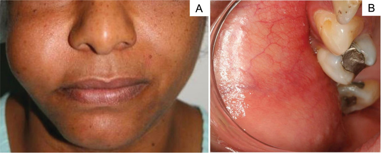
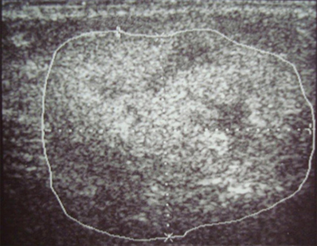
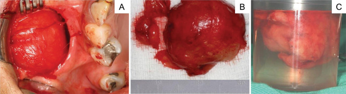
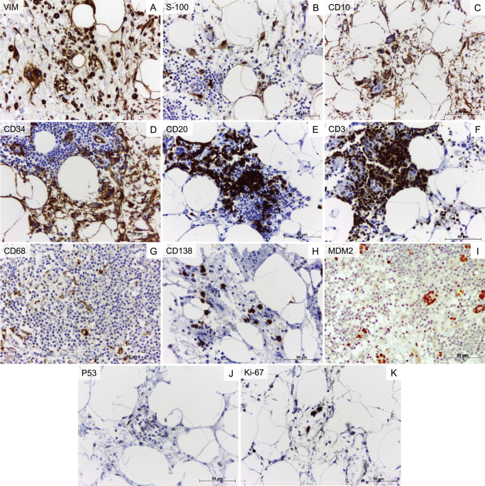
Liposarcomas of the oral cavity are rare. Those originating in the buccal mucosa cause challenging diagnostic and therapeutic issues since less than 40 cases of liposarcomas of the buccal mucosa and cheek have been reported in the worldwide literature. Herein, we present a case of atypical lipomatous tumor/well-differentiated liposarcoma affecting a 45-year-old female patient. Ultrasonography and magnetic resonance imaging confirmed a well-defined mass located in the right buccal mucosa, extending to the submucosal layers of the cheek. Histopathologically, a well-differentiated fatty neoplasm with presence of prominent stromal inflammatory cells was observed. Multifocally scattered bizarre hyperchromatic stromal cells, some of which multinucleated, were also observed. An immunohistochemical panel comprising vimentin, S-100, CD10, CD34, CD20, CD3, CD68, CD138, MDM2, Ki-67, and P53 was employed to better characterize the lesion. A local recurrence event occurred during a 10-year follow-up period. Surgical resection was performed during both episodes. We also provided an overview of demographic and clinicopathological characteristics, immunohistochemical features, imaging findings, and the differential diagnosis of liposarcoma of the oral cavity. Knowledge of the etiopathological and clinical aspects of this rare neoplasm is fundamental in order to rule out other conditions, including lipomatous lesions that affect the buccal mucosa.
Keywords: Atypical lipomatous tumor; Liposarcoma; Mouth mucosa; Oral cancer; Recurrence; Well differentiated liposarcoma.
© 2020. Springer Science+Business Media, LLC, part of Springer Nature.
Conflict of interest statement
No conflict of interest to disclose.
Figures


References
-
- Virchow R. Ein fall von Bosartigen zum Theil in der form des Neurons auftretenden Fettgeschwulsten. Arch A Pathol Anat Phys. 1857;11:281–288. doi: 10.1007/BF01995372. - DOI
Publication types
MeSH terms
Grants and funding
- 404710/2018-2/Conselho Nacional de Desenvolvimento Científico e Tecnológico
- 310797/2019-5/Conselho Nacional de Desenvolvimento Científico e Tecnológico
- 305493/2018-3/Conselho Nacional de Desenvolvimento Científico e Tecnológico
- 455644/2018-1/Conselho Nacional de Desenvolvimento Científico e Tecnológico
- 001/Coordenação de Aperfeiçoamento de Pessoal de Nível Superior
LinkOut - more resources
Full Text Sources
Medical
Research Materials
Miscellaneous
