. 2020 Oct 27;33(4):108322.
doi: 10.1016/j.celrep.2020.108322. Epub 2020 Oct 12.

Structure-Based Design with Tag-Based Purification and In-Process Biotinylation Enable Streamlined Development of SARS-CoV-2 Spike Molecular Probes

Affiliations

Structure-Based Design with Tag-Based Purification and In-Process Biotinylation Enable Streamlined Development of SARS-CoV-2 Spike Molecular Probes

Tongqing Zhou et al. Cell Rep. .

Abstract

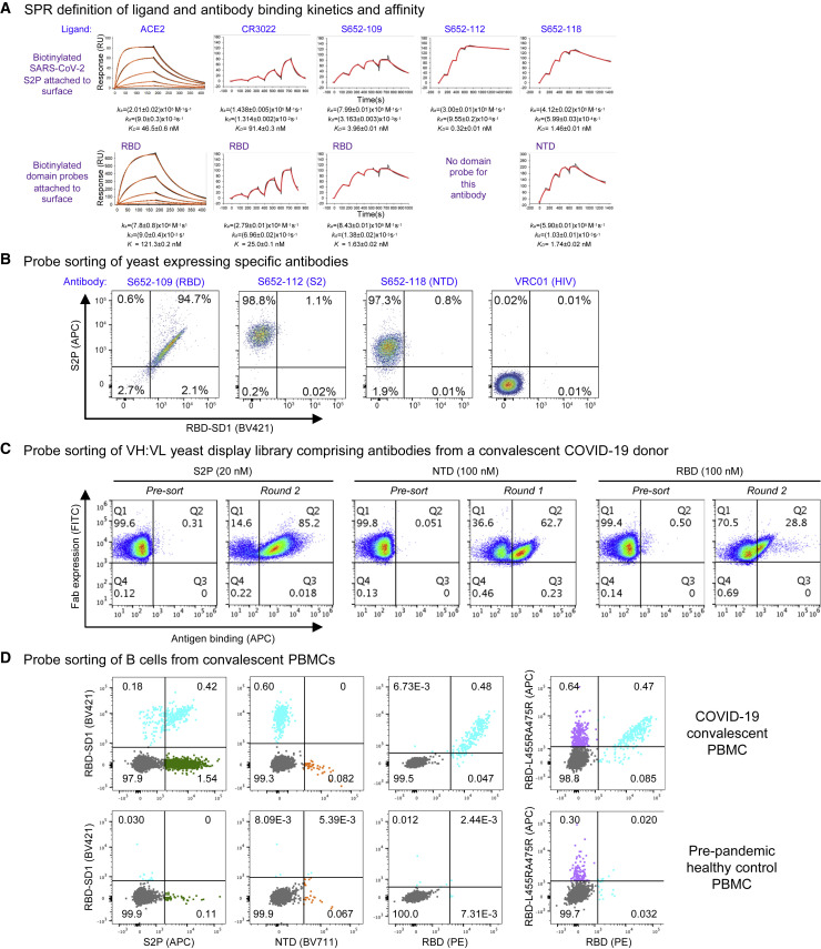
Biotin-labeled molecular probes, comprising specific regions of the severe acute respiratory syndrome coronavirus 2 (SARS-CoV-2) spike, would be helpful in the isolation and characterization of antibodies targeting this recently emerged pathogen. Here, we design constructs incorporating an N-terminal purification tag, a site-specific protease-cleavage site, the probe region of interest, and a C-terminal sequence targeted by biotin ligase. Probe regions include full-length spike ectodomain as well as various subregions, and we also design mutants that eliminate recognition of the angiotensin-converting enzyme 2 (ACE2) receptor. Yields of biotin-labeled probes from transient transfection range from ∼0.5 mg/L for the complete ectodomain to >5 mg/L for several subregions. Probes are characterized for antigenicity and ACE2 recognition, and the structure of the spike ectodomain probe is determined by cryoelectron microscopy. We also characterize antibody-binding specificities and cell-sorting capabilities of the biotinylated probes. Altogether, structure-based design coupled to efficient purification and biotinylation processes can thus enable streamlined development of SARS-CoV-2 spike ectodomain probes.

Keywords: COVID-19; HRV3C protease; antibody; biotinylated probe; coronavirus disease 2019; human rhinovirus 3C; single-chain Fc; structure-based design.


Conflict of interest statement

Declaration of Interests The authors declare no competing interest.

Figures

None
Graphical abstract
Figure 1
Figure 1
Strategy for Tag-Based Purification with On-Column Biotinylation (A) Schematic design of the expression construct of SARS-CoV-2 molecular probes. A single-chain human Ig constant domain (scFc) was added at the N terminus to facilitate expression and purification. The AVI tag was placed at the C terminus after a 10-amino-acid linker for biotinylation. The red arrows in the second and fourth Fc domains showed the “knob-in-hole” mutations to prevent dimerization of the scFc. (B) Biotinylation and HRV3C digestion. Cell culture supernatant from cells transiently transfected with plasmid was loaded onto protein A affinity column. Biotinylation and HRV3C cleavage reactions can be carried out in series or simultaneously, as buffers for both reactions are compatible.
Figure 2
Figure 2
Design of the SARS-CoV-2 Spike S2P Molecular Probe (A) Schematic of construction of the SARS-CoV-2 spike probe containing the ectodomain of the spike protein, the GSAS mutations replacing the RRAR furin cleavage site, and the K986PV987P (2P) mutation stabilizing the spike in pre-fusion conformation, a foldon domain, and a 10-amino-acid linker (10lnQQ), followed by the AVI tag. (B) Structural model of a SARS-CoV-2 spike protomer with the NTD, RBD, and SD1 domains highlighted. (C) Sequence of the SARS-CoV-2 spike probe with key regions annotated. See also Table S1.
Figure 3
Figure 3
Physical Properties, Antigenic Characteristics, and Cryo-EM Structure of a Biotinylated SARS-CoV-2 S2P Probe (A) Size-exclusion chromatography of the biotinylated SARS-CoV-2 S2P probe in PBS buffer. (B) SDS-PAGE of the SARS-CoV-2 S2P probe with and without the reducing agent DTT. Molecular weight marker (MWM) was run alongside the probe. (C) Negative-stain EM of the SARS-CoV-2 S2P probe. The 2D-class averages were shown below the wide-field raw EM image in 5-fold magnification. Scale bar: 10 nm. (D) Biotinylation, receptor recognition, and antigenicity of the SARS-CoV-2 S2P probe. The level of biotinylation was evaluated by capture of probes at 40 μg/mL onto the streptavidin biosensors. A biotinylated HIV-1 Env containing the same 10lnQQ-AVI tag at the C terminus was used for comparison (top left). Binding to the ACE2 receptor (top right) and binding to SARS-CoV (middle) and SARS-CoV-2 (bottom) antibodies were assessed using 80 μg/mL S2P or 20 μg/mL HIV Env probes, respectively. Receptors and antibodies were set at 500 nM. Error bars represent standard deviation of triplicate measurements. HIV-1 receptor CD4 and antibody VRC01 were used as controls. (E) Cryo-EM structures of biotinylated SARS-CoV-2 S2P probe. Domains are colored as in Figure 2C sequence. The C-terminal residues 1153-1208 plus the foldon, 10lnQQ-AVI tag, and biotin were not visible in the electron density. See also Figures S2 and S3 and Table S2.
Figure 4
Figure 4
Structure-Based Definition of SARS-CoV-2 Molecular Probes Comprising the NTD, RBD, and RBD-SD1 Domains (A) Cryo-EM structure of the NTD domain in the S2P probe determined in this study (Figure 3E), with reconstruction density shown in orange for NTD domain and in gray otherwise. First ordered residue with density (A27) is highlighted with a blue sphere; last residue of NTD domain (S305) is highlighted with a red sphere. (B) Close-up view of the NTD termini. (C) Sequence of NTD domain probe. The sequence is highlighted in orange except for residues 14-26, which are disordered in the cryo-EM structures. (D) Cryo-EM structure of the RBD domain in spike (Figure 3E), with reconstruction density shown in cyan for RBD domain and in gray otherwise. First residue with density (L329) is highlighted with a blue sphere; last ordered residue of RBD domain (G526) is highlighted with a red sphere. (E) Close-up view of the spike RBD termini. (F) Sequence of RBD domain probe highlighted in cyan. (G) Cryo-EM structure of the RBD-SD1 domains in spike (Figure 3E), with reconstruction density shown in green for RBD-SD1 domain and in gray otherwise. First residue with density (R319) is highlighted with a blue sphere; last ordered residue of RBD-SD1 domain (S591) is highlighted with a red sphere. (H) Close-up view of the spike RBD-SD1 termini. (I) Sequence of the RBD-SD1 domain probe highlighted in green. See also Figures S1 and S3 and Tables S1 and S2.
Figure 5
Figure 5
Physical Properties and Antigenic Characteristics of Molecular Probes Comprising SARS-CoV-2 NTD, RBD, and RBD-SD1 Domains (A) Size-exclusion chromatography of biotinylated NTD, RBD, and RBD-SD1 molecular probes. (B) SDS-PAGE of NTD, RBD, and RBD-SD1 molecular probes. RBD-SD1 peak 1 appeared to be partially disulfide linked and was therefore removed from further analysis; RBD-SD1 thus refers to the monomeric peak 2. (C) Binding of receptor ACE2 to both RBD and RBD-SD1 probes. (D) Binding of SARS-CoV antibodies to NTD (left) and RBD (right) probes. Both RBD and RBD-SD1 probes bound to antibodies CR3022 and S652-109, while the NTD probe specifically interacted with antibody S652-118. HIV-1 antibody VRC01 was used as control. (E) Binding of SARS-CoV-2 antibodies to NTD (left) and RBD (right) probes. Antibodies 4-8 and 2-51 showed specific binding to the NTD probe. Antibodies 2-4 and B38 showed specific binding to RBD and RBD-SD1 probes. Error bars represent standard deviation of triplicate measurements. See also Figure S2.
Figure 6
Figure 6
Molecular Probes Comprising Specific Mutations for Knocking Out ACE2 Interaction with RBD (A) Structural model of the RBD-ACE2 complex, with inset panels highlighting RBD mutations designed to reduce ACE2 recognition. ACE2 and RBD are shown in cartoon representation and colored blue and cyan, respectively. Arg mutations are shown in stick representation, with potential clashes with ACE2 highlighted with red discs. (B) Schematic and sequence of mutant RBD probes with sites mutated to Arg highlighted in magenta. (C) Size-exclusion chromatography of RBDs with mutations altering ACE2 binding. (D) SDS-PAGE of mutant RBD probes with and without reducing agent as indicated. (E) Binding to receptor ACE2. All the knockout mutations abolished RBD binding to the ACE2 receptor. (F) Binding to antibodies. Both SARS-CoV (top) and SARS-CoV-2 (bottom) antibodies were tested. Binding to all the SARS-CoV-2 antibodies and to SARS-CoV antibody S652-109 was reduced or abolished by the knockout mutations. Binding to CR3022, whose epitope is not on the ACE2-binding site, was not significantly affected. All binding was assessed using 7.5 μg/mL RBD-L455RA475R, 80 μg/mL RBD-L455RG496R, or 15 μg/mL RBD-L455RA475RG502R to account for varying biotinylation levels among the mutants. Receptor or antibodies were at 500 nM. HIV-1 antibody VRC01 was used as a negative control. Error bars represent standard deviation of triplicate measurements. See also Figure S2 and Table S1.
Figure 7
Figure 7
Validation of Biotinylated Probes (A) SPR standard kinetic experiments for ACE2 and single-cycle kinetic experiments for Fabs CR3022, S652-109, S652-112, and S652-118 binding over biotinylated spike (top row) and biotinylated RBD or biotinylated NTD (bottom row) loaded onto a streptavidin sensor chip. Black traces represent the experimental data, and the red traces represent the fit to a 1:1 interaction model. The error in each measurement represents the error of the fit. (B) Binding of yeast expressing SARS-CoV cross-reactive Fabs to SARS-CoV-2 antigenic probes. HIV envelope-targeting VRC01 Fab was used as a negative control. (C) Sorting of yeast cells encoding antibody transcripts from a convalescent COVID-19 donor with S2P-, NTD-, and RBD-based probes. (D) Sorting of B cells from COVID-19 convalescent PBMCs. (Top row) PBMCs obtained from a SARS-CoV-2-infected subject 75 days after symptom onset were stained with surface antibodies and probes as outlined in the STAR Methods to allow for identification and specificity-determination of SARS-CoV-2 spike-specific IgG+ and IgA+ B cells. (Bottom row) PBMCs obtained pre-pandemic from a healthy donor serve as a control for background binding of the probes. See also Figures S4–S7.

Update of

References

    1. Adams P.D., Gopal K., Grosse-Kunstleve R.W., Hung L.W., Ioerger T.R., McCoy A.J., Moriarty N.W., Pai R.K., Read R.J., Romo T.D. Recent developments in the PHENIX software for automated crystallographic structure determination. J. Synchrotron Radiat. 2004;11:53–55. - PubMed
    1. Antoniou G., Papakyriacou I., Papaneophytou C. Optimization of Soluble Expression and Purification of Recombinant Human Rhinovirus Type-14 3C Protease Using Statistically Designed Experiments: Isolation and Characterization of the Enzyme. Mol. Biotechnol. 2017;59:407–424. - PubMed
    1. Barad B.A., Echols N., Wang R.Y., Cheng Y., DiMaio F., Adams P.D., Fraser J.S. EMRinger: side chain-directed model and map validation for 3D cryo-electron microscopy. Nat. Methods. 2015;12:943–946. - PMC - PubMed
    1. Brouwer P.J.M., Caniels T.G., van der Straten K., Snitselaar J.L., Aldon Y., Bangaru S., Torres J.L., Okba N.M.A., Claireaux M., Kerster G. Potent neutralizing antibodies from COVID-19 patients define multiple targets of vulnerability. Science. 2020;369:643–650. - PMC - PubMed
    1. Callaway E. The race for coronavirus vaccines: a graphical guide. Nature. 2020;580:576–577. - PubMed

Publication types

MeSH terms