. 2020 Dec:184:104955.
doi: 10.1016/j.antiviral.2020.104955. Epub 2020 Oct 19.

Antiviral activity and safety of remdesivir against SARS-CoV-2 infection in human pluripotent stem cell-derived cardiomyocytes

Affiliations

Antiviral activity and safety of remdesivir against SARS-CoV-2 infection in human pluripotent stem cell-derived cardiomyocytes

Seong Woo Choi et al. Antiviral Res. 2020 Dec.

Abstract

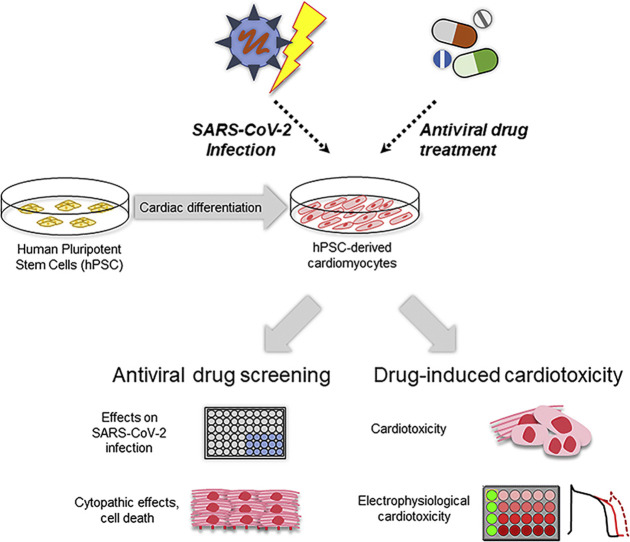
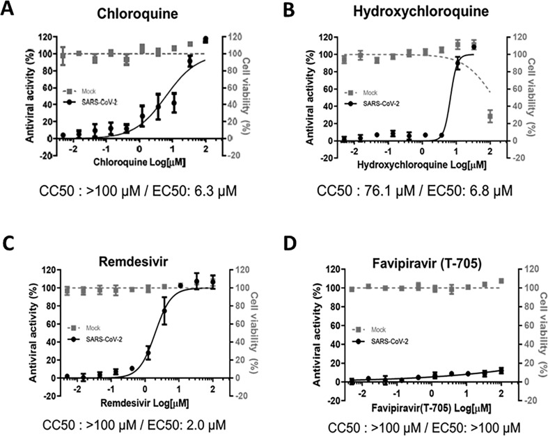
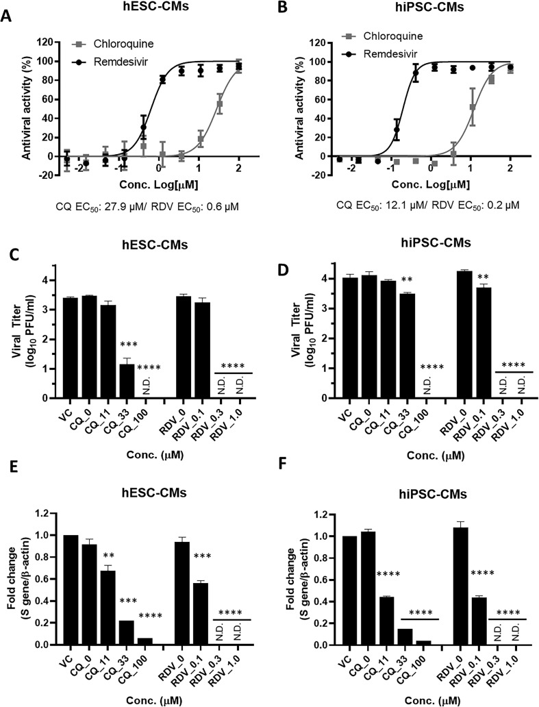
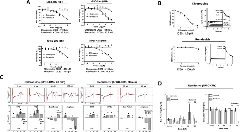
Coronavirus disease 2019 (COVID-19), caused by severe acute respiratory syndrome coronavirus-2 (SARS-CoV-2), is considered as the most significant global public health crisis of the century. Several drug candidates have been suggested as potential therapeutic options for COVID-19, including remdesivir, currently the only authorized drug for use under an Emergency Use Authorization. However, there is only limited information regarding the safety profiles of the proposed drugs, in particular drug-induced cardiotoxicity. Here, we evaluated the antiviral activity and cardiotoxicity of remdesivir using cardiomyocytes-derived from human pluripotent stem cells (hPSC-CMs) as an alternative source of human primary cardiomyocytes (CMs). In this study, remdesivir exhibited up to 60-fold higher antiviral activity in hPSC-CMs compared to Vero E6 cells; however, it also induced moderate cardiotoxicity in these cells. To gain further insight into the drug-induced arrhythmogenic risk, we assessed QT interval prolongation and automaticity of remdesivir-treated hPSC-CMs using a multielectrode array (MEA). As a result, the data indicated a potential risk of QT prolongation when remdesivir is used at concentrations higher than the estimated peak plasma concentration. Therefore, we conclude that close monitoring of the electrocardiographic/QT interval should be advised in SARS-CoV-2-infected patients under remdesivir medication, in particular individuals with pre-existing heart conditions.

Keywords: COVID-19; Human cardiomyocytes; Pluripotent stem cells; Remdesivir; SARS-CoV-2.


Conflict of interest statement

The authors declare that they have no competing interests.

Figures

Image 1
Graphical abstract
Fig. 1
Fig. 1
Characterization of cardiomyocytes-derived from human pluripotent stem cells (hPSC-CMs). (A) Schematic diagram of hPSC differentiation into cardiomyocytes. (B) The morphology of differentiated hPSC-CMs derived from two hPSC cell lines (hESCs: H9 and hiPSCs: CMC-11; 10 × magnification). (C) The RT-qPCR was performed to measure the expression of cardiac sarcomeric markers, cTnT, and MYH6 genes, in hPSC-CMs. The data represent the mean (±SD) of at least two independent experiments performed in triplicate. Statistical analyses were determined using paired student's t-tests, and P < 0.05 was considered as significant (*). (D) Expression of TNNT2 and ACTN2 in hPSC-CMs (hESC-CMs [upper panel] and hiPSC-CMs [lower panel]) at day 15 of differentiation were determined by flow cytometry and immunofluorescence analyses. Scale bar, 100 μm. (E) Beating cardiomyocytes were prepared for action potential (AP) recording using the patch-clamp technique in hESC-CMs and field potential (FP) recording using a multielectrode array (MEA) system in hiPSC-CMs. The hPSC-CMs were treated with 100 nM isoprenaline (β2-adrenergic receptor agonist), 0.3 μM and 1 μM nifedipine (L-type Ca2+ channel blocker), and 10 nM dofetilide and 30 nM E4031 (hERG K+ channel blockers).
Fig. 2
Fig. 2
SARS-CoV-2 infects hPSC-CMs. (A) The mRNA expression levels of ACE2 and TMPRSS2 genes in hPSC-CMs (hESCs: H9 and hiPSCs: CMC-11) were measured by RT-qPCR. The data represent the mean (±SD) of at least two independent experiments performed in triplicate. Statistical analyses were determined using paired student's t-tests, and P < 0.05 was considered as significant (*). (B) Immunofluorescence assay was performed to evaluate the expression of ACE2 and TMPRSS2 proteins in cTnT-positive hPSC-CMs. ACE2 (red) or TMPRSS2 (red) was co-expressed with cTnT (green) positive hESC-CMs and hiPSC-CMs. (C) SARS-CoV-2 infection of hPSC-CMs. The hiPSC-CMs were infected with SARS-CoV-2 (betaCoV/Korea/KCDC) at the indicated multiplicity of infection (MOI) for 48 h. Immunofluorescence image of SARS-CoV-2 infected hiPSC-CMs positively stained for the viral spike (S) protein (green) is shown. The percentage of SARS-CoV-2 infected hiPSC-CMs at various MOI was determined by the quantification of S protein-positive cells. The data represent the mean (±SD) of at least two independent experiments performed in duplicate.
Fig. 3
Fig. 3
Antiviral efficacy of chloroquine, hydroxychloroquine, remdesivir, and favipiravir in Vero E6 cells. (A to D) Dose-response curve analyses were performed in Vero E6 cells infected with SARS-CoV-2 at a multiplicity of infection (MOI) of 0.1 in the presence of various concentrations of chloroquine (A), hydroxychloroquine (B), remdesivir (C), or favipiravir (T-705) (D). At 72 h post-infection, cell viability was measured using the CellTiter 96® AQueous One Solution Cell Proliferation Assay. The data represent the mean (±SD) of at least three independent experiments performed in duplicate. Mock-infected (grey square) or infected with SARS-CoV-2 (black circle). CC50, 50% cytotoxic concentrations; EC50, 50% effective concentration.
Fig. 4
Fig. 4
Remdesivir potently inhibits SARS-CoV-2 infection in hPSC-CMs. (A and B) Dose-response curve (DRC) analyses of SARS-CoV-2 inhibition by chloroquine and remdesivir in hPSC-CMs. The hESC-CMs (A) and hiPSC-CMs (B) were infected with SARS-CoV-2 at an MOI 2.5 in the presence of various concentrations of chloroquine (grey square) or remdesivir (black circle). At 48 h p.i., the percentage of infected cells was visualized and measured using an automated high-throughput confocal fluorescence imaging system. The data represent the mean (±SD) of at least three independent experiments performed in duplicate. (C to F) Reduction of SARS-CoV-2 replication by chloroquine and remdesivir in hPSC-CMs as determined by infectious viral titer and RT-qPCR. The hESC-CMs (C and E) and hiPSC-CMs (D and F) were infected with SARS-CoV-2 at an MOI of 2.5 in the presence of the indicated concentrations of chloroquine or remdesivir. (C and D) Viral titers from hPSC-CM supernatants were determined with plaque assay in Vero E6 cells. (E and F) Quantification of intracellular SARS-CoV-2 genome RNA by RT-qPCR. Total RNA was isolated from lysates of infected cells for quantification of intracellular SARS-CoV-2 RNA levels (S gene), and results were normalized to β-actin mRNA. Data represent means (±SD) of at least two independent experiments performed in duplicate. Statistical analyses were determined using paired student's t-tests, and significant differences are indicated by **P < 0.01, ***P < 0.005, and ****P < 0.0001. CQ, chloroquine; RDV, remdesivir; N.D, not detected.
Fig. 5
Fig. 5
Remdesivir induces cardiotoxicity in hPSC-CMs. (A) Cardiomyocyte toxicity of chloroquine and remdesivir in hPSC-CMs. Dose-response curve analyses of drug-induced cardiotoxicity in hPSC-CMs (hESC-CMs and hiPSC-CMs) were performed in the presence of various concentrations of each drug. At 24 h and 48 h post-treatment, cell viability was measured using the CellTiter 96® AQueous One Solution Cell Proliferation Assay (MTS, Promega). The data represent the mean (±SD) of at least two independent experiments performed in triplicate. CC50, 50% cytotoxic concentration. (B) The inhibitory effects of chloroquine and remdesivir on hERG currents in stably overexpressing hERG HEK-293 cell line. The hERG currents were recorded with the conventional whole-cell voltage-clamp configuration. The peak hERG current was activated at −50 mV, followed by a 50 mV depolarization from −80 mV of holding voltage. The data represent the mean (±SD) of at least two independent experiments performed in triplicate. (C, D) Arrhythmogenic effects of chloroquine and remdesivir in hiPSC-CMs. The FP signals were analyzed with FP duration (FPD), corrected FPD (FPDc; field potential duration corrected by Fredericia's formula), beat period, the amplitude of Na+ Peak (amplitude), beat period irregularity (coefficient of variation of the beat period), and conduction velocity using Cardiac Analysis Tool 2.2.7 (Axion BioSystems). All data are expressed as mean ± SD (n = 5, exclusion of quiescent and flat T). Statistical analyses were determined using paired student's t-tests, and P < 0.05 was considered as significant (*). N.D, not detected.

References

    1. Agency E.M. 2020. Summary on Compassionate Use: Remdesivir Gilead.https://www.ema.europa.eu/en/documents/other/summary-compassionate-use-r...
    1. Aggarwal G., Cheruiyot I., Aggarwal S., Wong J., Lippi G., Lavie C.J., Henry B.M., Sanchis-Gomar F. Association of cardiovascular disease with coronavirus disease 2019 (COVID-19) severity: a meta-analysis. Curr. Probl. Cardiol. 2020;45:100617. - PMC - PubMed
    1. Aggarwal G., Henry B.M., Aggarwal S., Bangalore S. Cardiovascular safety of potential drugs for the treatment of coronavirus disease 2019. Am. J. Cardiol. 2020;128:147–150. - PMC - PubMed
    1. Bansal M. Cardiovascular disease and COVID-19. Diabetes Metabol. Syndr. 2020;14:247–250. - PMC - PubMed
    1. Bojkova D., Wagner J.U.G., Shumliakivska M., Aslan G.S., Saleem U., Hansen A., Luxan G., Gunther S., Pham M.D., Krishnan J., Harter P.N., Ermel U.H., Frangakis A.S., Milting H., Zeiher A.M., Klingel K., Cinatl J., Dendorfer A., Eschenhagen T., Tschope C., Ciesek S., Dimmeler S. SARS-CoV-2 infects and induces cytotoxic effects in human cardiomyocytes. Cardiovasc. Res. 2020 - PMC - PubMed

Publication types

MeSH terms