Review
. 2021 Feb;1868(2):118893.
doi: 10.1016/j.bbamcr.2020.118893. Epub 2020 Oct 20.

Copper metabolism as a unique vulnerability in cancer

Affiliations
Review

Copper metabolism as a unique vulnerability in cancer

Vinit C Shanbhag et al. Biochim Biophys Acta Mol Cell Res. 2021 Feb.

Abstract

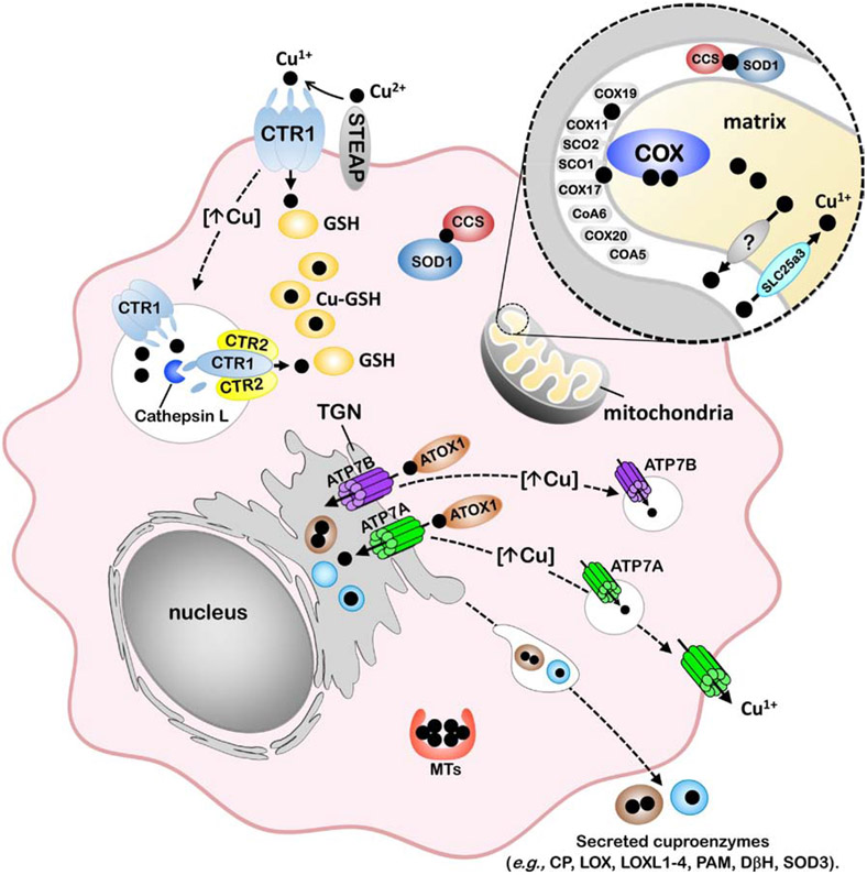
The last 25 years have witnessed tremendous progress in identifying and characterizing proteins that regulate the uptake, intracellular trafficking and export of copper. Although dietary copper is required in trace amounts, sufficient quantities of this metal are needed to sustain growth and development in humans and other mammals. However, copper is also a rate-limiting nutrient for the growth and proliferation of cancer cells. Oral copper chelators taken with food have been shown to confer anti-neoplastic and anti-metastatic benefits in animals and humans. Recent studies have begun to identify specific roles for copper in pathways of oncogenic signaling and resistance to anti-neoplastic drugs. Here, we review the general mechanisms of cellular copper homeostasis and discuss roles of copper in cancer progression, highlighting metabolic vulnerabilities that may be targetable in the development of anticancer therapies.

Keywords: Cancer; Cisplatin chemotherapy; Copper chelation; Copper homeostasis; Metallochaperone; Nutrition.


Conflict of interest statement

Declaration of competing interest

The authors declare that they have no known competing financial interests or personal relationships that could have appeared to influence the work reported in this paper.

Figures

Figure 1.
Figure 1.. Model of cellular copper homeostasis in mammals.
The transport of Cu(I) (black circles) into the cytoplasm occurs via the CTR1 copper importer, which is located at the plasma membrane and within endosomal compartments. The uptake of Cu(I) via CTR1 is thought to depend on the reduction of Cu(II) to Cu(I) by members of the STEAP family of metalloreductases. The oligomerization of CTR1 and another SLC31 protein, CTR2, facilitates the cleavage of the CTR1 ectodomain by cathepsin L to enhance Cu(I) transport from endosomal compartments into the cytoplasm. Cytoplasmic Cu(I) is bound to glutathione (GSH) and can be transferred to metallochaperones for targeted trafficking to proteins. The CCS metallochaperone is required for insertion of Cu(I) into apo-SOD1 in both the cytoplasm and the mitochondrial intermembrane space. The metalation of cuproenzymes within the secretory pathway occurs via ATOX1-mediated Cu(I) delivery to the P-type ATPases, ATP7A or ATP7B, which are located in the trans-Golgi network (TGN) and facilitate Cu(I) loading into this organelle. Cu(I) transport into the mitochondrial matrix is facilitated by SLC25A3 located within the inner membrane. The metalation of COX occurs within the intermembrane space of the mitochondria and requires copper to be exported from the matrix via an unknown transporter. COX metalation and maturation requires the metallochaperones COX17, SCO1 and COX11, and other assembly / redox control factors. Protective mechanisms of Cu tolerance include sequestration by metallothioneins (MTs) as well as Cu(I)-stimulated trafficking of copper transporters, including the endocytosis and degradation of CTR1 which reduces Cu(I) import and exocytic trafficking of ATP7A and ATP7B to post-Golgi vesicles and the plasma membrane to facilitate copper export from the cell.
Figure 2.
Figure 2.. Structure models of the human CTR1 copper importer.
A) Homology-based model of the human CTR1 homotrimer derived from the crystal structure of sCtr1 from Atlantic salmon. Each hCTR1 monomer is depicted with a different color for each monomer. The copper-binding MXXM motif within the second transmembrane domain of each monomer forms two methionine rings that are thought to function as a Cu(I) selective filter at the extracellular entrance. B) A top view highlighting copper co-ordination within the methionine rings.
Figure 3.
Figure 3.. Schematic model of ATP7A/B structure and predicted Cu entry site.
A) A schematic illustration of ATP7A/B highlighting domains that are important for transport and trafficking function. The copper-binding MXCXXC motifs in the N-terminal domain are required for copper-induced trafficking from the TGN and to receive copper from the ATOX1 metallochaperone. Canonical sequences conserved in all P-type ATPases include the GDGIND required for ATP binding, DKTGT which contains the aspartic acid that is phosphorylated during catalysis, and TGE within the phosphatase domain required for removing the aspartyl phosphate. A conserved Met-Asp-Glu triad at the channel entrance and an intramembranous Cys-Pr-Cys motif are thought to coordinate copper during transport. The C-terminal tail harbors a di-leucine (ATP7A) or trileucine (ATP7B) required for endocytosis. A C-terminal DTAL motif is required for basolateral targeting of ATP7A in polarized epithelial cells, whereas an N-terminal FAFDNVGYE motif is required for apical targeting of ATP7B. B and C) Homology-derived structure of human ATP7A based on the crystal structure of LCopA from Legionella pneumophila. A bend in TM2 domain (helical ribbon) is predicted to form a platform at the cytosolic-membrane interface adjacent to the Met-Glu-Asp triad that may serve as a metallochaperone docking site for copper delivery.
Figure 4.
Figure 4.. Copper-dependent mechanisms of oncogenic signaling.
Copper uptake via CTR1 allosterically activates MEK1 to potentiate oncogenic signaling via the MAP kinase pathway. Copper is also an allosteric activator of the ULK1/2 kinases, which, under amino acid starvation conditions, stimulate formation of the autophagosome for the degradative recycling of biomolecules.
Figure 5.
Figure 5.. Copper-dependent mechanisms of cancer cell migration and metastasis.
The copper-binding protein MEMO1 is required for motility and metastasis of cancer cells in response to receptor tyrosine kinase signaling. MEMO1 is postulated to produce ROS at the leading edge of cancer cells which and activates pathways that influence cancer cell migration and invasion. Copper uptake via CTR1 facilitates ATOX1-mediated copper delivery to the ATP7A copper pump, which transports copper to the lysyl oxidase family of enzymes (LOX) within the secretory pathway. The LOX family of enzymes mediate the cross linking of collagen fibers generating H2O2 which facilitates activation of integrin-associated focal adhesion kinase (FAK1) and proto-oncogene tyrosine-protein kinase (SRC) which promote subsequent migration and metastasis. LOX enzymes also facilitate the formation of pre-metastatic niches at distant metastatic sites.

References

    1. Nevitt T, Ohrvik H, Thiele DJ, Charting the travels of copper in eukaryotes from yeast to mammals, Biochim Biophys Acta, 1823 (2012) 1580–1593. - PMC - PubMed
    1. Ochiai EI, Copper and the biological evolution, Biosystems, 16 (1983) 81–86. - PubMed
    1. Chi Fru E, Rodriguez NP, Partin CA, Lalonde SV, Andersson P, Weiss DJ, El Albani A, Rodushkin I, Konhauser KO, Cu isotopes in marine black shales record the Great Oxidation Event, Proc Natl Acad Sci U S A, 113 (2016) 4941–4946. - PMC - PubMed
    1. Macomber L, Imlay JA, The iron-sulfur clusters of dehydratases are primary intracellular targets of copper toxicity, Proc Natl Acad Sci U S A, 106 (2009) 8344–8349. - PMC - PubMed
    1. Pierson H, Yang H, Lutsenko S, Copper Transport and Disease: What Can We Learn from Organoids?, Annu Rev Nutr, 39 (2019) 75–94. - PMC - PubMed

Publication types