Review
. 2020 Oct 20;10(10):1461.
doi: 10.3390/biom10101461.

Understanding the Pathogenesis of Spondyloarthritis

Affiliations
Review

Understanding the Pathogenesis of Spondyloarthritis

Aigul Sharip et al. Biomolecules. .

Abstract

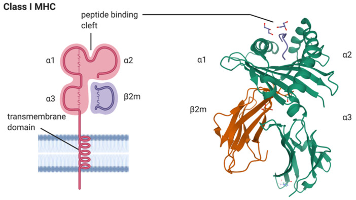
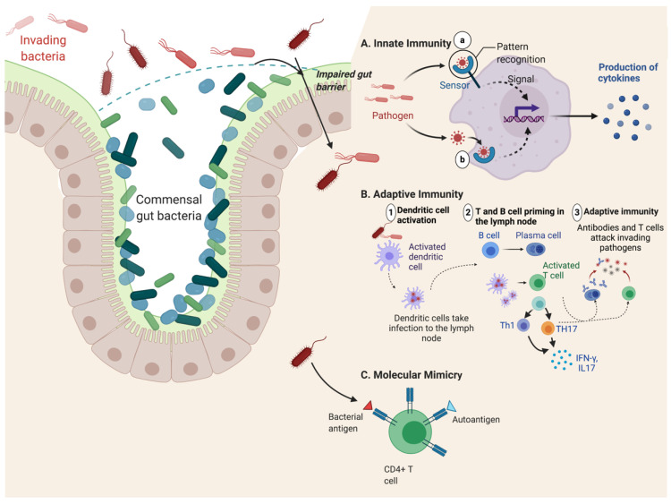
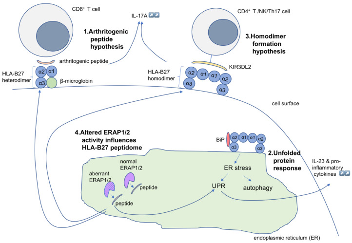
Spondyloarthritis comprises a group of inflammatory diseases of the joints and spine, with various clinical manifestations. The group includes ankylosing spondylitis, reactive arthritis, psoriatic arthritis, arthritis associated with inflammatory bowel disease, and undifferentiated spondyloarthritis. The exact etiology and pathogenesis of spondyloarthritis are still unknown, but five hypotheses explaining the pathogenesis exist. These hypotheses suggest that spondyloarthritis is caused by arthritogenic peptides, an unfolded protein response, HLA-B*27 homodimer formation, malfunctioning endoplasmic reticulum aminopeptidases, and, last but not least, gut inflammation and dysbiosis. Here we discuss the five hypotheses and the evidence supporting each. In all of these hypotheses, HLA-B*27 plays a central role. It is likely that a combination of these hypotheses, with HLA-B*27 taking center stage, will eventually explain the development of spondyloarthritis in predisposed individuals.

Keywords: ERAP1; HLA-B*27; arthritogenic peptides; gut dysbiosis; inflammation; pathogenesis; spondyloarthritis; unfolded protein response.


Conflict of interest statement

The authors declare no conflict of interest.

Figures

Figure 1
Figure 1
A model depicting the major histocompatibility complex (MHC) class I HLA-B*27 heavy chain in complex with beta 2-macroglobulin (β2m) is shown on the left (created with BioRender.com). The alpha 1 (α1), alpha 2 (α2), and alpha 3 (α3) domains of the HLA-B*27 molecule and the peptide binding cleft composed of α1 and α2 domains of HLA-B*27 are indicated, as is the transmembrane domain inserted into the lipid bilayer of the plasma membrane. A ribbon model of the crystal structure of the human class I MHC molecule HLA-B*2705 heavy chain (green) bound to nona-peptide m9 (blue), and in complex with β2m (orange), is shown on the right. Orientation: cell surface at top of picture. The image was sourced from the Research Collaboratory for Structural Bioinformatics (RCSB; rcsb.org) Protein Data Bank (PDB) entry ID 1JGE (DOI:10.2210/pdb1JGE/pdb; [9]) generated by PISA [10].
Figure 2
Figure 2
Contribution of gut dysbiosis to autoimmune disease. Due to impaired gut barrier function, pathogenic bacteria can invade the gut lumen and promote overactivation of innate and adaptive immune responses. (A) Innate immunity is activated by exposure to bacteria present in the gut tissue and circulation, resulting in excess production of proinflammatory cytokines. (B) Bacterial antigens can be presented to CD4+ cells by dendritic cells, leading to the activation of different T cell subtypes (Th1 and Th17) and production of cytokines. B cells are also activated either directly, or through interaction with antigen-presenting dendritic cells, leading to their differentiation into plasma cells and the production of anti-microbial antibodies. (C) Bacterial antigens can also provoke autoimmunity by molecular mimicry. Shared sequence similarity between foreign- and self-peptides results in the cross-activation of autoreactive T or B cells by a pathogen-derived antigen. The figure was adapted from “Keystone Gut Microbiota Species Provide Colonization Resistance to Invading Bacteria”, by BioRender.com (2020), retrieved from https://app.biorender.com/biorender-templates. Abbreviations: IL, interleukin; TH-1, T helper type 1; TH-17 T helper type 17.
Figure 3
Figure 3
Illustration of the hypothesis for the pathogenetic role of HLA-B*2705 molecules in spondyloarthritis. (1) Arthritogenic peptides displayed by properly folded HLA-B*27 can be recognized by autoreactive CD8+ T cells, resulting in inflammation. (2) Misfolded HLA-B*27 chains and binding of BiP causes ER stress and activation of UPR, leading to increased production of IL-23 and other proinflammatory cytokines. (3) Cell surface HLA homodimers interact with CD4+ T cells through innate immune receptors, such as KIR3DL2, and facilitate cell-mediated autoimmune responses. (4) Altered ERAP1 activity can result in changes in peptide processing, with pathological consequences. ER, endoplasmic reticulum; ERAP1, ER aminopeptidases (ERAP)1; KIR3DL2, killer immunoglobulin-like receptor; UPR, unfolded protein response.

Similar articles

Cited by

References

    1. Zochling J., Brandt J., Braun J. The current concept of spondyloarthritis with special emphasis on undifferentiated spondyloarthritis. Rheumatology. 2005;44:1483–1491. doi: 10.1093/rheumatology/kei047. - DOI - PubMed
    1. Khan M.A. Polymorphism of HLA-B27: 105 subtypes currently known. Curr. Rheumatol. Rep. 2013;15:362. doi: 10.1007/s11926-013-0362-y. - DOI - PubMed
    1. Stolwijk C., Boonen A., van Tubergen A., Reveille J.D. Epidemiology of spondyloarthritis. Rheum. Dis. Clin. N. Am. 2012;38:441–476. doi: 10.1016/j.rdc.2012.09.003. - DOI - PMC - PubMed
    1. Bakland G., Nossent H.C. Epidemiology of Spondyloarthritis: A Review. Curr. Rheumatol. Rep. 2013;15:351. doi: 10.1007/s11926-013-0351-1. - DOI - PubMed
    1. Braun J., Sieper J. Ankylosing spondylitis. Lancet. 2007;369:1379–1390. doi: 10.1016/S0140-6736(07)60635-7. - DOI - PubMed

Publication types

MeSH terms

Substances