. 2020 Nov 10;117(45):28046-28055.
doi: 10.1073/pnas.2016093117. Epub 2020 Oct 22.

Engineered ACE2 receptor traps potently neutralize SARS-CoV-2

Affiliations

Engineered ACE2 receptor traps potently neutralize SARS-CoV-2

Anum Glasgow et al. Proc Natl Acad Sci U S A. .

Abstract

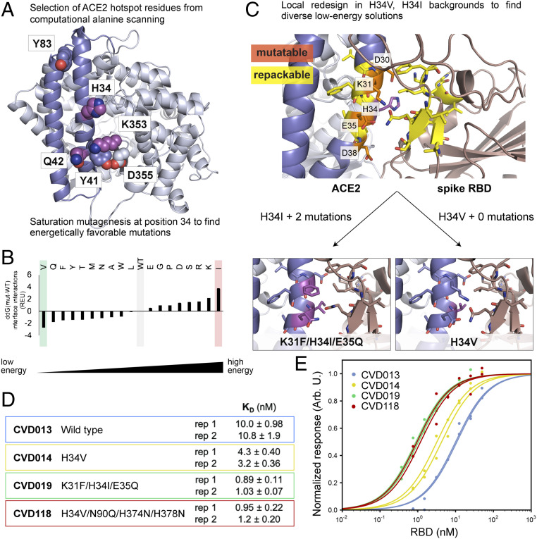
An essential mechanism for severe acute respiratory syndrome coronavirus 1 (SARS-CoV-1) and severe acute respiratory syndrome coronavirus 2 (SARS-CoV-2) infection begins with the viral spike protein binding to the human receptor protein angiotensin-converting enzyme II (ACE2). Here, we describe a stepwise engineering approach to generate a set of affinity optimized, enzymatically inactivated ACE2 variants that potently block SARS-CoV-2 infection of cells. These optimized receptor traps tightly bind the receptor binding domain (RBD) of the viral spike protein and prevent entry into host cells. We first computationally designed the ACE2-RBD interface using a two-stage flexible protein backbone design process that improved affinity for the RBD by up to 12-fold. These designed receptor variants were affinity matured an additional 14-fold by random mutagenesis and selection using yeast surface display. The highest-affinity variant contained seven amino acid changes and bound to the RBD 170-fold more tightly than wild-type ACE2. With the addition of the natural ACE2 collectrin domain and fusion to a human immunoglobulin crystallizable fragment (Fc) domain for increased stabilization and avidity, the most optimal ACE2 receptor traps neutralized SARS-CoV-2-pseudotyped lentivirus and authentic SARS-CoV-2 virus with half-maximal inhibitory concentrations (IC50s) in the 10- to 100-ng/mL range. Engineered ACE2 receptor traps offer a promising route to fighting infections by SARS-CoV-2 and other ACE2-using coronaviruses, with the key advantage that viral resistance would also likely impair viral entry. Moreover, such traps can be predesigned for viruses with known entry receptors for faster therapeutic response without the need for neutralizing antibodies isolated from convalescent patients.

Keywords: SARS-CoV-2; antiviral therapeutics; computational design; receptor trap; yeast display.


Conflict of interest statement

Competing interest statement: A.G., J.G., I.L., X.X.Z., T.K., and J.A.W. have filed a provisional patent related to this work.

Figures

Fig. 1.
Fig. 1.
ACE2 receptor trap strategy. Two computational design strategies were used to predict mutations to ACE2(614) (light blue shape) that enhance its affinity for the SARS-CoV-2 spike RBD: 1) saturation mutagenesis at computational alanine scanning hot spots, followed by local ACE2 redesign (blue star mutations) and 2) combining mutations from computational saturation mutagenesis and experimental DMS data (green star mutations). Designed ACE2 variants were mutagenized and screened for binding to the RBD, and additional mutations that improved the binding affinity were isolated (red star mutations). Engineered ACE2(614) variants were fused to the ACE2 collectrin domain (residues 615 to 740, yellow ovals) and expressed as Fc fusions (purple shapes) with an additional mutation to inactivate ACE2 peptidase activity (white star mutations). KD,app values represent apparent binding affinities measured between yeast surface-displayed ACE2(614) variants and the monomeric RBD. IC50 values represent those measured for the four most potent neutralizing variants in SARS-CoV-2 pseudotyped virus assays.
Fig. 2.
Fig. 2.
Computational design of ACE2 for improved binding affinity to the spike RBD. (A) Computational alanine scanning identified ACE2 hot spot residues (shown as spheres) that contribute strongly to binding the spike RBD. Residues 18 to 90 are shown in blue, and residues 91 to 614 are shown in light blue. H34, Q42, and K353, shown as magenta spheres, were selected for computational saturation mutagenesis. (B) Computational saturation mutagenesis predicted several stabilizing mutations to H34. H34V and H34I were selected for further design to generate diverse ACE2 receptor trap models. (C) Flexible backbone design was performed around V34 ACE2 and I34 ACE2 (WT residue H34 shown in magenta). ACE2 residues that were permitted to change amino acid identity (“mutable” residues) are labeled and shown in orange; ACE2 and RBD residues that were allowed to change rotameric conformations and/or backbone atom positions (“repackable” residues) are shown in yellow. Design around H34I resulted in two additional mutations, shown in magenta. (D and E) In vitro BLI measurements show that designed ACE2(614)-Fc binding affinities to the RBD are improved 2- to 11-fold over WT ACE2(614)-Fc. To generate CVD118, the N90Q glycan knockout mutation from DMS (24) was added to V34 ACE2, as well as two histidine mutations to inactivate ACE2 peptidase activity (25). Each “rep” is a separate biological replicate. The table in D lists the KD values for the designed ACE2 variants, with errors of the fits for titration curves shown in E.
Fig. 3.
Fig. 3.
Affinity maturation by yeast surface display of pooled error-prone PCR libraries on designed ACE2 variants and DMS-guided design. (A) Monomeric ACE2(614) variants were expressed as Aga2p-GFP fusions on the surface of yeast cells. Binding of ACE2(614) to the spike RBD was quantified by incubation of the cells in a solution of biotinylated RBD, followed by staining the cells with streptavidin Alexa Fluor 647, washing the cells, and measuring fluorescent populations by flow cytometry. (B) Stringency was increased in each round of yeast display to enrich the cell population for ACE2(614) variants that bound the RBD tightly. Equilibrium sorts with decreasing RBD concentrations were used for sorts 1 to 3, and off-rate sorts with increasing dissociation times and decreasing gate size were used for the last two sorts. (C) Representative cell populations by flow cytometry for yeast expressing a tight binding ACE2(614) variant from sort 4 compared with yeast expressing the computationally designed parent, K31F/H34I/E35Q ACE2(614), incubated with 0.1 nM RBD monomer. The additional mutations in the sort 4 variant shift the cell population higher on the y axis due to tighter RBD binding. (D) On-yeast titration curves for cells expressing ACE2(614) variants chosen from sorts 3 (Y313), 4 (Y353), and 5 (Y373) that bind the RBD most tightly, compared with WT ACE2(614) (Y208) and K31F/H34I/E35Q ACE2(614) (Y293). (E) On-yeast titration curves for cells expressing the ACE2(614) variants generated by DMS-guided design, compared with WT ACE2(614) (Y208). Titration curves in D and E are fit to biological duplicates, shown as points. Variant names, mutations, and KD,app values are in SI Appendix, Table S3. MFI, median fluorescence intensity.
Fig. 4.
Fig. 4.
Increased stability, affinity, and avidity effects result in tighter binding between ACE2(740)-Fc and FL spike and potent viral neutralization. (A) Plasmid constructs for expression of ACE2(614) and ACE2(740) (Left) and spike in the monomeric RBD and full-length forms (Right). Avi tag, target sequence for intracellular biotinylation by BirA; hIgG1-Fc, human immunoglobulin 1 Fc domain; interleukin-2 (IL2) SS, IL2 signal sequence (cleaved); native SS, native spike signal sequence; thrombin site, thrombin cleavage site; T4 trimer, T4 bacteriophage fibritin trimerization motif; tobacco etch virus (TEV)-linker, TEV protease cleavage sequence and glycine-serine linker; 8X- or 6XHis tag, polyhistidine tag. (B) BLI measurements show decreased KD for ACE2(740)-Fc compared with ACE2(614)-Fc for the interaction with the spike RBD. Binding between ACE2-Fc and the FL spike protein results in aKD less than 100 pM due to very low off rates. Solid lines show response curves for twofold dilution titration spanning 0.37 to 50 nM RBD. Dotted lines show calculated fits. (C) Table of all ACE2 variants with scaffolds and mutations tested in pseudotyped and authentic SARS-CoV-2 viral neutralization assays, listed with their origin. (D) Fc fusion, inclusion of the collectrin domain, and affinity-enhancing mutations improve neutralization of ACE2 constructs against pseudotyped SARS-CoV-2 virus, except for misfolded variant 353. Error bars represent SDs over all technical replicates from two to four biological replicates. Biological replicates were separate experiments using different preparations of the ACE2 variant and pseudovirus, each with two or four technical replicates. Statistical significance with P < 0.01 was determined using a homoscedastic two-tailed t test. Authentic SARS-CoV-2 viral neutralization experiments with ACE2 variants in VeroE6 cells showed that (E) Fc fusion (CVD013) and addition of computationally predicted mutations (CVD118) enhance neutralization by greater than 50,000-fold over a control anti-GFP IgG antibody sample and that (F) inclusion of the collectrin domain, computationally predicted mutations (CVD293), DMS-guided mutations (CVD310), and mutations from affinity maturation in yeast (CVD313) enhance neutralization potency over a control anti-GFP IgG antibody sample to IC50 values of 73 to 136 ng/mL. E and F show results from different experiments in biological duplicate. Error bars represent the SEM.
Fig. 5.
Fig. 5.
WT and engineered ACE2(740)-Fc bind the SARS-CoV-1 RBD and the HCoV-NL63 RBD. Representative BLI measurements for (A, D, and G) WT ACE2(740)-Fc, (B, E, and H) K31F/H34I/E35Q ACE2(740)-Fc, and (C, F, and I) K31F/N33D/H34S/E35Q/H345L ACE2(740)-Fc interactions with the SARS-CoV-2 RBD (A–C), the SARS-CoV-1 RBD (D–F), and the HCoV-NL63 RBD (G–I) at concentrations of 0.375 to 100 nM RBD, with the highest RBD concentration tested and KD values as indicated. Dotted lines show calculated fits. The extremely slow off rate observed in C precluded KD determination.

Update of

Comment in

References

    1. Beigel J. H., et al. , Remdesivir for the treatment of Covid-19: Preliminary report. N. Engl. J. Med. 383, 994 (2020). - PubMed
    1. Zhou P., et al. , A pneumonia outbreak associated with a new coronavirus of probable bat origin. Nature 579, 270–273 (2020). - PMC - PubMed
    1. Shi R., et al. , A human neutralizing antibody targets the receptor-binding site of SARS-CoV-2. Nature 584, 120–124 (2020). - PubMed
    1. Wu Y., et al. , A noncompeting pair of human neutralizing antibodies block COVID-19 virus binding to its receptor ACE2. Science 368, 1274–1278 (2020). - PMC - PubMed
    1. Baum A., et al. , Antibody cocktail to SARS-CoV-2 spike protein prevents rapid mutational escape seen with individual antibodies. Science 369, 1014–1018 (2020). - PMC - PubMed

Publication types

MeSH terms