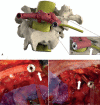
Accuracy of Patient-Specific 3D-Printed Drill Guides for Pedicle and Lateral Mass Screw Insertion: An Analysis of 76 Cervical and Thoracic Screw Trajectories
- PMID: 33093310
- PMCID: PMC7787187
- DOI: 10.1097/BRS.0000000000003747
Accuracy of Patient-Specific 3D-Printed Drill Guides for Pedicle and Lateral Mass Screw Insertion: An Analysis of 76 Cervical and Thoracic Screw Trajectories
Abstract
Study design: Single-center retrospective case series.
Objective: The purpose of this study was to assess the safety and accuracy of three-dimensional (3D)-printed individualized drill guides for pedicle and lateral mass screw insertion in the cervical and upper-thoracic region, by comparing the preoperative 3D surgical plan with the postoperative results.
Summary of background data: Posterior spinal fusion surgery can provide rigid intervertebral fixation but screw misplacement involves a high risk of neurovascular injury. However, modern spine surgeons now have tools such as virtual surgical planning and 3D-printed drill guides to facilitate spinal screw insertion.
Methods: A total of 15 patients who underwent posterior spinal fusion surgery involving patient-specific 3D-printed drill guides were included in this study. After segmentation of bone and screws, the postoperative models were superimposed onto the preoperative surgical plan. The accuracy of the realized screw trajectories was quantified by measuring the entry point and angular deviation.
Results: The 3D deviation analysis showed that the entry point and angular deviation over all 76 screw trajectories were 1.40 ± 0.81 mm and 6.70 ± 3.77°, respectively. Angular deviation was significantly higher in the sagittal plane than in the axial plane (P = 0.02). All screw positions were classified as "safe" (100%), showing no neurovascular injury, facet joint violation, or violation of the pedicle wall.
Conclusions: 3D virtual planning and 3D-printed patient-specific drill guides appear to be safe and accurate for pedicle and lateral mass screw insertion in the cervical and upper-thoracic spine. The quantitative 3D deviation analyses confirmed that screw positions were accurate with respect to the 3D-surgical plan.Level of Evidence: 4.
Copyright © 2020 The Author(s). Published by Wolters Kluwer Health, Inc.
Figures


References
-
- Esses SI, Sachs BL, Dreyzin V. Complications associated with the technique of pedicle screw fixation. A selected survey of ABS members. Spine (Phila Pa 1976) 1993; 18:2231–2238. - PubMed
-
- Gertzbein SD, Robbins SE. Accuracy of pedicular screw placement in vivo. Spine (Phila Pa 1976) 1990; 15:11–14. - PubMed
-
- Gaines RW., Jr The use of pedicle-screw internal fixation for the operative treatment of spinal disorders. J Bone Joint Surg Am 2000; 82:1458–1476. - PubMed
-
- Blumenthal S, Gill K. Complications of the wiltse pedicle screw fixation system. Spine (Phila Pa 1976) 1993; 18:1867–1871. - PubMed
-
- Liljenqvist UR, Halm HFH, Link TM. Pedicle screw instrumentation of the thoracic spine in idiopathic scoliosis. Spine (Phila Pa 1976) 1997; 22:2239–2245. - PubMed
Publication types
MeSH terms
LinkOut - more resources
Full Text Sources
Research Materials

