. 2020 Oct 22;9(10):94.
doi: 10.1038/s41389-020-00278-8.

Antileukemic activity of the VPS34-IN1 inhibitor in acute myeloid leukemia

Affiliations

Antileukemic activity of the VPS34-IN1 inhibitor in acute myeloid leukemia

Godelieve Meunier et al. Oncogenesis. .

Abstract

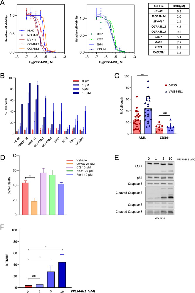
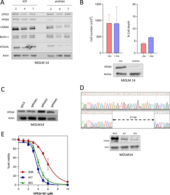
Acute myeloid leukemia (AML) is an aggressive disease with a poor prognosis. Vacuolar protein sorting 34 (VPS34) is a member of the phosphatidylinositol-3-kinase lipid kinase family that controls the canonical autophagy pathway and vesicular trafficking. Using a recently developed specific inhibitor (VPS34-IN1), we found that VPS34 inhibition induces apoptosis in AML cells but not in normal CD34+ hematopoietic cells. Complete and acute inhibition of VPS34 was required for the antileukemic activity of VPS34-IN1. This inhibitor also has pleiotropic effects against various cellular functions related to class III PI3K in AML cells that may explain their survival impairment. VPS34-IN1 inhibits basal and L-asparaginase-induced autophagy in AML cells. A synergistic cell death activity of this drug was also demonstrated. VPS34-IN1 was additionally found to impair vesicular trafficking and mTORC1 signaling. From an unbiased approach based on phosphoproteomic analysis, we identified that VPS34-IN1 specifically inhibits STAT5 phosphorylation downstream of FLT3-ITD signaling in AML. The identification of the mechanisms controlling FLT3-ITD signaling by VPS34 represents an important insight into the oncogenesis of AML and could lead to new therapeutic strategies.


Conflict of interest statement

The authors declare that they have no conflict of interest.

Figures

Fig. 1
Fig. 1. The VPS34 IN1 inhibitor has antileukemic activity against AML.
A AML cell lines were cultured for 48 h in the presence of VPS34-IN1 over a large concentration range and viability was quantified using the fluorescence-based Uptiblue assay. This enabled a determination of the IC50 values for VPS34-IN1; n = 3, error bars represent the standard deviation. B AML cell lines were cultured for 48 h with vehicle or 1, 5, or 10 µM of VPS34-IN1. Cell death was quantified by flow cytometry analysis of the percentage of annexin-V positive cells; n = 3, bars represent the standard error of the mean. C Primary AML cells from 23 newly diagnosed AML patients (Supplemental Table 1) were treated for 48 h with vehicle or 5 µM VPS34-IN1. Normal CD34+ hematopoietic progenitors from 6 allogenic bone marrow donors were cultured under similar conditions after positive sorting. Cell death was quantified by the percentage of annexin-V positive cells. Bars represent standard error of the mean. D Cell death (%) was quantified by flow cytometry analysis of the percentage of annexin-V positive cells for MOLM14 cells at 24 h post-VPS34-IN1 treatment (5 µM) with vehicle or ferrostatine-1 (10 µM), necrostatine-1 (20 µM), chloroquine (20 µM) or QVD-oph (25 µM); n = 3, errors bars represent the standard deviation. All other compounds were added to the medium 2 h before VPS34-IN1. E MOLM-14 cells were cultured for 48 h with vehicle or VPS34-IN1 at 1, 5, or 10 µM and western blot analysis was performed using antibodies directed against PARP, caspase-3, cleaved caspase-3, caspase-8, cleaved caspase-8, or p85. F MOLM-14 cells were cultured for 48 h with vehicle or VPS34-IN1 at 1, 5, or 10 µM and TMRE staining was performed to assess mitochondrial depolarization; n = 3, bars represent the standard error of the mean.
Fig. 2
Fig. 2. The complete and acute inhibition of VPS34 is required for VPS34-IN1 anti-leukemic activity.
A MOLM-14 cells engineered to express a doxycycline-inducible VPS34 shRNA were cultured with vehicle or doxycycline for 2, 4, and 7 days. The expression of β-actin, VPS15, VPS34, UVRAG, beclin-1, and ATG14L was assayed by western blotting. B MOLM-14 cells expressing a VPS34 shRNA construct were cultured with vehicle or doxycycline and the cell number and cell death were quantified at 48 h. C VSP34 expression in MOLM-14 scrambled (SCR) cells or MOLM-14 cells with a disrupted expression of VPS34 induced using CRISPR/CAS9 technology (bulk) was assessed by western blot. D Upper panel: representative Sanger sequencing of VPS34 exon 1 in MOLM-14 SCR cells or MOLM-14 cells with disrupted VPS34 expression (subclone 17). Lower panel: western blot analysis of VSP34 expression in MOLM-14 SCR cells or MOLM-14 cells with disrupted VPS34 expression (subclones 17 and 23). E MOLM14 SCR and MOLM-14 cells with disrupted VPS34 expression (subclones 17 and 23) were cultured for 48 h in the presence of VPS34-IN1 over a large concentration range. Viability was quantified using the fluorescence-based Uptiblue assay; n = 3, bars represent standard error of the mean.
Fig. 3
Fig. 3. VPS34-IN1 inhibits intracellular vesicle trafficking and basal autophagy in AML cells.
A MOLM-14 cells were cultured for 48 h with vehicle (left panel) or VPS34-IN1 (5 µM, right panel) and observed by optical microscopy (×100) after May Gruenwald Giemsa staining. B MOLM-14 cells were cultured for 72 h with vehicle (upper panel) or VPS34-IN1 (5 µM, lower panel) and observed by electron microscopy. Large-sized empty vacuoles were observed in VPS34-IN1 treated cells representing single-membraned swollen endosomes/lysosomes (*). Lipid droplets (arrowheads) were also observed in VPS34-IN1 treated cells. C MOLM-14 cells were cultured for 24 h with vehicle (left) or VPS34-IN1 (5 µM, right) and observed by immunofluorescence microscopy (×100) following Lysotracker deep red staining. MFI was also measured by FCM. D MOLM 14 cells were cultured for 6 h with vehicle or chloroquine (10 µM) and various doses of VPS34-IN1. Western blot analysis was then performed using antibodies directed against LC3-II and actin. E Representative immunofluorescence results of MOLM-14 GFP LC3 cells treated with vehicle, chloroquine alone (10 µM), or chloroquine with VPS34-IN1 (5 µM). F MOLM-14 cells were cultured for 24 h with vehicle, chloroquine (10 µM) or VPS34-IN (5 µM) and p62 accumulation was assessed by western blotting.
Fig. 4
Fig. 4. VPS34-IN1 inhibits l-asparaginase induced autophagy and bot drugs act synergistically.
A MOLM-14 cells were cultured for 6 h with l-asparaginase and/or chloroquine (10 µM) as indicated. Autophagy induction was assessed by analyzing both LC3-I and LC3-II expression by western blot. Upon autophagic induction, LC3-I is transformed to LC3-II which is then degraded in autophagolysosome. Chloroquine is able to inhibit acidification of autophagolysosome, allowing the evaluation of LC3-II formation which is an indirect marker of autophagy induction B Number of LC3B puncta per cells (right) and immunofluorescence analysis of GFP LC3 (left) in vehicle-treated or l-Asparaginase-treated (10 UI/ml) MOLM-14 cells were determined; n = 3, bars represent standard error of the mean. C MOLM-14 cells were cultured for 24 h with vehicle, chloroquine (10 µM) or l-asparaginase (10 UI/ml) and p62 accumulation was assessed by western blot. D MOLM-14 cells were cultured with increasing concentrations of VPS34-IN1 and in the presence of both l-asparaginase (10 UI/ml) and chloroquine (10 µM). Western blotting analysis of the LC3-I and LC3-II bands was performed at 6 h. E Synergy map (left) and viability matrix (right) of l-asparaginase with VPS34-IN1 for the MOLM-14 cell line. The mean viability measurement from three independent experiments was used. F Summary of synergy score from 48 h co-treatments of seven primary AML samples with l-Asparaginase and VPS34-IN1.
Fig. 5
Fig. 5. VPS34-IN1 modulates mTORC1 and FLT3-ITD signaling in AML cells.
A MOLM-14 cells were cultured for 6 h with vehicle or VPS34-IN1 (5 µM). Western blot analysis was used to evaluate the activation of the mTORC1 pathway using Thr 37/46 p4EBP1 and Thr 389 pP70S6K antibodies. B MOLM-14 cells were cultured for 0–4 h with VPS34-IN1 (5 µM). Western blot analysis was used to study the activation of the mTORC1 pathway using a Thr389 pP70S6K antibody and PI3K/AKT pathway activation was assessed using antibodies directed against phosphorylated AKT Thr308 or AKT S473 residues. AZD8055, a TOR kinase inhibitor, was used as a control for the inhibition of the mTORC1 and AKT Ser473 signaling pathways. C Percentage inhibition of phosphotyrosine in the 11 most downregulated proteins identified by phosphoproteomic analysis. D BA/F3 WT and BA/F3 cells with FLT3-ITD expression, with or without IL-3, were cultured for 48 h in the presence of VPS34-IN1 over a large concentration range. Viability was quantified using the fluorescence based Uptiblue assay; n = 3, bars represent standard error of the mean. E Comparison of VPS34 expression (normalized RPKM) in AML cells from patients with or without FLT3 mutation. Data are from Vizome database, a part of the Beat AML project. F MOLM-14 cells were cultured with vehicle or 5 µM VPS34-IN1 for 1 h. Western blotting was then used to analyze FLT3 expression, and STAT5 and P42/44MAPK phosphorylation. G Ba/F3 cells were cultured in the presence of IL-3 with vehicle or VPS34-IN1 (5 µM) for 3 h. Ba/F3 cells engineered to harbor either the FLT3-ITD, FLT3-ITD-TKD D835V or FLT3-ITD-TKD F691L mutations, which render them IL-3 independent for growth, were cultured under similar conditions. Western blot analysis was used to study the activation of the STAT5 pathway using a pSTAT5 antibody and MAPK1/3 pathway using a p42/44MAPK antibody. H MOLM-14 cells and BA/F3 cells expressing FLT3-ITD were cultured without or with 5 µM VPS34-IN1 for 1 h. The FLT3-ITD inhibitor AC220 was used as a positive control for the inhibition of FLT3 phosphorylation. Western blotting was used to analyze FLT3 expression, FLT3 phosphorylation and STAT5 pathway activation. I Ba/F3 cells were cultured in the presence of IL-3 with vehicle or various dose of VPS34-IN1, PIK-III, autophinib, sorafenib, AC220, and crenolanib for 48 h. Ba/F3 cells engineered to harbor either the FLT3-ITD, FLT3-ITD-TKD D835V or FLT3-ITD-TKD F691L mutations, which render them IL-3 independent for growth, were cultured under similar conditions. Viability was quantified using the fluorescence based Uptiblue assay; n = 3, bars represent standard error of the mean.

References

    1. Patel JP, et al. Prognostic relevance of integrated genetic profiling in acute myeloid leukemia. N. Engl. J. Med. 2012;366:1079–1089. doi: 10.1056/NEJMoa1112304. - DOI - PMC - PubMed
    1. Cerrano M, Itzykson R. New treatment options for acute myeloid leukemia in 2019. Curr. Oncol. Rep. 2019;21:16. doi: 10.1007/s11912-019-0764-8. - DOI - PubMed
    1. Backer JM. The intricate regulation and complex functions of the Class III phosphoinositide 3-kinase Vps34. Biochem. J. 2016;473:2251–2271. doi: 10.1042/BCJ20160170. - DOI - PubMed
    1. Ohashi Y, Tremel S, Williams RL. VPS34 complexes from a structural perspective. J. Lipid Res. 2019;60:229–241. doi: 10.1194/jlr.R089490. - DOI - PMC - PubMed
    1. Bilanges B, Posor Y, Vanhaesebroeck B. PI3K isoforms in cell signalling and vesicle trafficking. Nat. Rev. Mol. Cell Biol. 2019;20:515–534. doi: 10.1038/s41580-019-0129-z. - DOI - PubMed