. 2020 Oct 22;11(10):900.
doi: 10.1038/s41419-020-03117-1.

BET degrader inhibits tumor progression and stem-like cell growth via Wnt/β-catenin signaling repression in glioma cells

Affiliations

BET degrader inhibits tumor progression and stem-like cell growth via Wnt/β-catenin signaling repression in glioma cells

Tao Tian et al. Cell Death Dis. .

Abstract

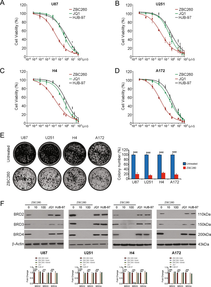
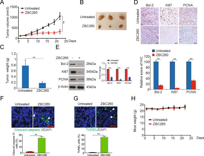
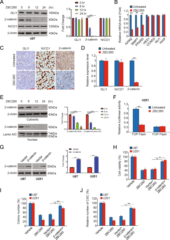
Based on their histological appearance, gliomas are a very common primary tumor type of the brain and are classified into grades, Grade I to Grade IV, of the World Health Organization. Treatment failure is due to the cancer stem cells (CSC) phenotype maintenance and self-renewal. BET degraders such as ZBC260 represents a novel class of BET inhibitors that act by inducing BET proteins degradation. This study explores the mode of action and effects of ZBC260 in vivo and in vitro against glioma. By inhibiting cell proliferation and inducting cell cycle arrest, the fact that glioma cell lines show sensitivity to ZBC260. Notably, ZBC260 targeted glioma without side effects in vivo. In addition, the stem cell-like properties of glioma cells were inhibited upon ZBC260 treatment. When the mechanism was examined, our findings indicated that Wnt/β-catenin pathway repression is required for ZBC260-induced stem cell-like properties and tumor growth suppression. In conclusion, the growth of tumors and stem cell-like properties were inhibited by ZBC260 via Wnt/β-catenin repression, which suggests ZBC260 as a potential therapeutic agent for glioma.


Conflict of interest statement

The authors declare that they have no conflicts of interest.

Figures

Fig. 1
Fig. 1. ZBC260 inhibits proliferation and induces cell cycle arrest in glioma cells.
A–D Indicated glioma cells were treated with increasing dose of ZBC260, JQ1 or HJB-97 for 72 h. The viability of cells was analyzed by the MTT assay. E Indicated cells were treated with 100 nM ZBC260 for 24 h. Colony formation assay was done by seeding an equal number of treated cells in 6-well plates, and then staining attached cells with crystal violet 14 days later. F Indicated cells were treated ZBC260 at indicated concentration, 10 μM JQ1 or 10 μM HJB-97 for 24 h. Indicated proteins level were analyzed by western blotting and normalized to β-actin. Results were expressed as means ± SD of three independent experiments. ***P < 0.001.
Fig. 2
Fig. 2. ZBC260 induces cell cycle arrest and apoptosis in glioma cells.
A Indicated cell lines were treated with 100 nM ZBC260 for 24 h. Cell cycle was analyzed by flow cytometry. B Indicated cell lines were treated with 100 nM ZBC260 for 24 h. Indicated protein level was analyzed by western blotting and normalized to β-actin. C Indicated cell lines were treated with 100 nM ZBC260 for 24 h. Indicated protein level was analyzed by western blotting and normalized to β-actin. D Indicated cell lines were treated with 100 nM ZBC260 for 24 h. Apoptosis was analyzed by flow cytometry. E U87 cells were treated with 100 nM ZBC260 for 24 h with or without z-VAD-fmk pretreatment. Caspase 3/7 activity was analyzed by the caspase 3/7 activation kit. F U87 cells were treated with 100 nM ZBC260 for 24 h. Indicated protein level was analyzed by western blotting and normalized to β-actin. Results were expressed as means ± SD of three independent experiments. *P < 0.05; **P < 0.01; ***P < 0.001.
Fig. 3
Fig. 3. ZBC260 regulates migration, invasion, and EMT in glioma cells.
A Representative results of wound healing assay showing the effect of ZBC260 (100 nM) treatment on the migration ability in U87 cells. B Representative results of wound healing assay showing the effect of ZBC260 (100 nM) treatment on the migration ability in U251 cells. C Representative results of Transwell assay showing the effect of ZBC260 treatment invasive ability in U87 cells. D Representative results of Transwell assay showing the effect of ZBC260 treatment invasive ability in U251 cells. E U87 cells were treated with 100 nM ZBC260 for 24 h. Indicated protein level was analyzed by western blotting and normalized to β-actin. Results were expressed as means ± SD of three independent experiments. **P < 0.01; ***P < 0.001.
Fig. 4
Fig. 4. ZBC260 inhibits the growth of xenograft in mouse model.
A Nude mice were injected s.c. with U251 cells. After one week, mice were treated with ZBC260 or vehicle control. Tumor volume at indicated time points post treatment was calculated and plotted with P-values, n = 6. B Representative tumors at the end of the experiment in A. C Tumor weight at the end of the experiment in A. D Paraffin-embedded sections of tumor tissues from mice were analyzed by IHC for Bcl2, Ki67, and PCNA. Lower, The score of IHC staining. E Western blotting of Bcl2, Ki67, and PCNA in tumor tissues from the experiment in A. The western blotting was normalized to β-actin (F) Paraffin-embedded sections of tumor tissues from mice were analyzed by cleaved caspase 3 staining. G Paraffin-embedded sections of tumor tissues from mice were analyzed by TUNEL staining. H Effect of ZBC260 on the body weight of mice over the treatment time. Results were expressed as means ± SD of three independent experiments. **P < 0.01; ***P < 0.001.
Fig. 5
Fig. 5. ZBC260 inhibits stem-cell-like properties of glioma cells.
A, B Sarcosphere-formation capacity of indicated cells was analyzed post 100 nM ZBC260 treatment. C, D Secondary sarcosphere formation capacity of U251 cells was analyzed without further treatment after 100 nM ZBC260 treatment in primary sarcosphere-formation assay. E mRNA expression of stem cell markers in U251 cells treated with 100 nM ZBC260 for 24 h was examined by Real-time RT-PCR. F U251 cells were treated with 100 nM ZBC260 for 24 h. The protein level of stem cell markers was analyzed by western blotting and normalized to β-actin. Results were expressed as means ± SD of three independent experiments. **P < 0.01; ***P < 0.001.
Fig. 6
Fig. 6. ZBC260 regulates glioma-cell-like properties via Wnt/β-catenin pathway.
A U87 cells were treated with 100 nM ZBC260 at indicated time point. The expression of β-catenin, NICD1, and GLI1 involved in CSC pathways was analyzed by western blotting and normalized to β-actin. B U251 cells were treated with 100 nM ZBC260 for 24 h. mRNA level of target genes was analyzed by Real-time RT-PCR. C Paraffin-embedded sections of tumor tissues from mice were analyzed by IHC for β-catenin, NICD1 and GLI1. D The score of IHC staining. E U251 cells were treated with 100 nM ZBC260 at indicated time point. The level of cytosolic and nuclear β-catenin was analyzed by western blotting and normalized to β-actin or Lamin A/C as indicated. F The activity of TCF/β-catenin reporter (TOP/FOP Flash) in 100 nM ZBC260-treated U251 cells. G The efficiency of β-catenin overexpression was analyzed by western blotting and normalized to β-actin. H Indicated cell lines transfected with β-catenin were treated with 100 nM ZBC260 for 72 h. The viability of cells was analyzed by the MTT assay. I Indicated cell lines transfected with β-catenin were treated with 100 nM ZBC260 for 24 h. Colony formation assay was done by seeding anequal number of treated cells in 6-well plates, and then staining attached cells with crystal violet 14 days later. J Indicated cell lines transfected with β-catenin were treated with 100 nM ZBC260 for 24 h. Sarcosphere-formation capacity was analyzed. Results were expressed as means ± SD of three independent experiments. *P < 0.05; **P < 0.01; ***P < 0.001.

References

    1. Hanif F, Muzaffar K, Perveen K, Malhi SM, Simjee SU. Glioblastoma multiforme: a review of its epidemiology and pathogenesis through clinical presentation and treatment. Asian Pac. J. Cancer Prev. 2017;18:3–9. - PMC - PubMed
    1. Gupta M, et al. Prognostic factors in elderly patients with high-grade gliomas: a retrospective analysis of 24 cases. J. Neurosci. Rural Pr. 2018;9:312–316. doi: 10.4103/jnrp.jnrp_576_17. - DOI - PMC - PubMed
    1. Kern M, Auer TA, Picht T, Misch M, Wiener E. T2 mapping of molecular subtypes of WHO grade II/III gliomas. BMC Neurol. 2020;20:8. doi: 10.1186/s12883-019-1590-1. - DOI - PMC - PubMed
    1. Liang J, et al. Prognostic factors of patients with gliomas-an analysis on 335 patients with glioblastoma and other forms of gliomas. BMC Cancer. 2020;20:35. doi: 10.1186/s12885-019-6511-6. - DOI - PMC - PubMed
    1. Hayashi S, Kitamura Y, Hirose Y, Yoshida K, Sasaki H. Molecular-genetic and clinicopathological prognostic factors in patients with gliomas showing total 1p19q loss: gain of chromosome 19p and histological grade III negatively correlate with patient’s prognosis. J. Neurooncol. 2017;132:119–126. doi: 10.1007/s11060-016-2344-1. - DOI - PubMed

MeSH terms