

Retinal vascular development in an immature retina at 33-34 weeks postmenstrual age predicts retinopathy of prematurity
- PMID: 33093504
- PMCID: PMC7582165
- DOI: 10.1038/s41598-020-75151-0
Retinal vascular development in an immature retina at 33-34 weeks postmenstrual age predicts retinopathy of prematurity
Abstract
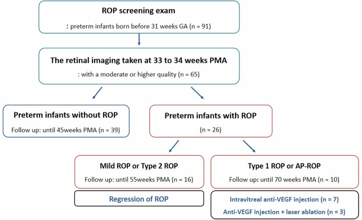
In preterm birth, the immature retina can develop a potentially blinding disorder of the eye known as retinopathy of prematurity (ROP). The vaso-proliferative phase of ROP begins at an approximate postmenstrual age (PMA) of 32 weeks. There is little or no evidence of an association between ROP development and retinal status in the early vaso-proliferative phase. We aimed to evaluate the retinal vascular findings of infants at 33-34 weeks PMA to determine their risk of ROP. We reviewed 130 serial wide-field retinal images from 65 preterm infants born before the gestational age of 31 weeks. ROP occurred more frequently in infants having a leading vascular edge within posterior Zone II. This was in contrast to normal infants, who are characterized by complete retinal vascularization up to Zone II at 34 weeks PMA. The probability of ROP development in preterm infants with retinal edge hemorrhage was 24.58 times higher than in preterm infants without retinal edge hemorrhage. Eyes with ROP that required treatment showed significantly delayed retinal vascularization accompanied by pre-plus disease. In conclusion, retinal status in the early vaso-proliferation phase might determine the risk of ROP.
Conflict of interest statement
J.H.J. reports an honorarium from Bayer and Novartis, outside the submitted work. Y.C.K. reports an honorarium from Allergan, Bayer, Novartis and Santen, and a research grant from Bayer, outside the submitted work.
Figures


Similar articles
-
Retinopathy of Prematurity.2025 Jun 2. In: StatPearls [Internet]. Treasure Island (FL): StatPearls Publishing; 2025 Jan–. 2025 Jun 2. In: StatPearls [Internet]. Treasure Island (FL): StatPearls Publishing; 2025 Jan–. PMID: 32965990 Free Books & Documents.
-
Maturation of rod function in preterm infants with and without retinopathy of prematurity.J Pediatr. 2008 Nov;153(5):605-11. doi: 10.1016/j.jpeds.2008.05.018. Epub 2008 Jul 14. J Pediatr. 2008. PMID: 18621392
-
RATE OF AND TIME TO COMPLETE RETINAL VASCULARIZATION IN PREMATURE INFANTS AND ASSOCIATED FACTORS.Retina. 2023 Jan 1;43(1):102-110. doi: 10.1097/IAE.0000000000003627. Retina. 2023. PMID: 36201755
-
Retinopathy of Prematurity: Therapeutic Strategies Based on Pathophysiology.Neonatology. 2016;109(4):369-76. doi: 10.1159/000444901. Epub 2016 Jun 3. Neonatology. 2016. PMID: 27251645 Review.
-
Retinopathy of prematurity in Indonesia: Incidence and risk factors.J Neonatal Perinatal Med. 2017;10(1):85-90. doi: 10.3233/NPM-915142. J Neonatal Perinatal Med. 2017. PMID: 28304327 Review.
Cited by
-
Systemic Cytokines in Retinopathy of Prematurity.J Pers Med. 2023 Feb 5;13(2):291. doi: 10.3390/jpm13020291. J Pers Med. 2023. PMID: 36836525 Free PMC article. Review.
-
Cord blood transforming growth factor-β-induced as predictive biomarker of retinopathy of prematurity in preterm infants.Graefes Arch Clin Exp Ophthalmol. 2023 Sep;261(9):2477-2488. doi: 10.1007/s00417-023-06056-7. Epub 2023 Apr 6. Graefes Arch Clin Exp Ophthalmol. 2023. PMID: 37022494
-
Retinopathy of prematurity: contribution of inflammatory and genetic factors.Mol Cell Biochem. 2022 Jun;477(6):1739-1763. doi: 10.1007/s11010-022-04394-4. Epub 2022 Mar 9. Mol Cell Biochem. 2022. PMID: 35262882 Review.
-
Evaluation of the Course and Outcome of Aggressive Retinopathy of Prematurity.Cureus. 2025 May 21;17(5):e84589. doi: 10.7759/cureus.84589. eCollection 2025 May. Cureus. 2025. PMID: 40546518 Free PMC article.
-
The Role of HIF-1α in Retinopathy of Prematurity: A Review of Current Literature.J Clin Med. 2024 Jul 10;13(14):4034. doi: 10.3390/jcm13144034. J Clin Med. 2024. PMID: 39064074 Free PMC article. Review.
References
Publication types
MeSH terms
LinkOut - more resources
Full Text Sources
Medical
