. 2020 Oct 22;10(1):17995.
doi: 10.1038/s41598-020-75057-x.

CELF2 regulates the species-specific alternative splicing of TREM2

Affiliations

CELF2 regulates the species-specific alternative splicing of TREM2

Motoaki Yanaizu et al. Sci Rep. .

Abstract

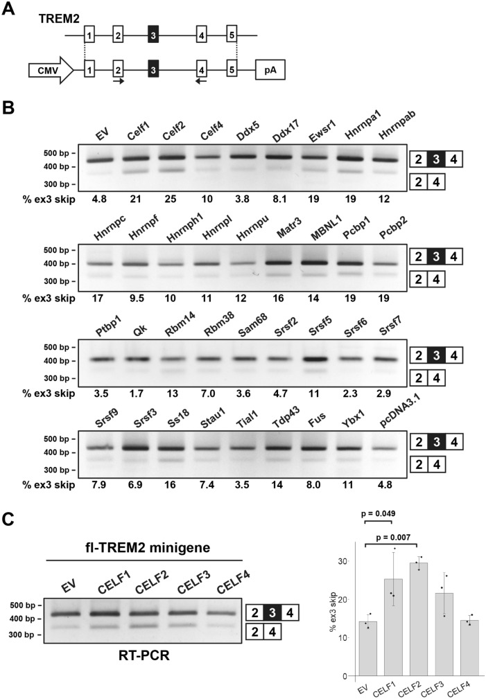
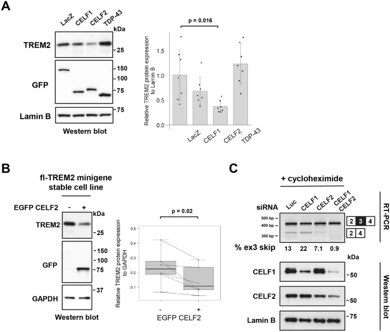
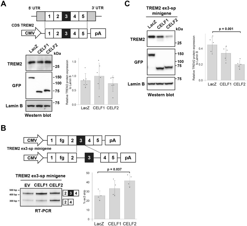
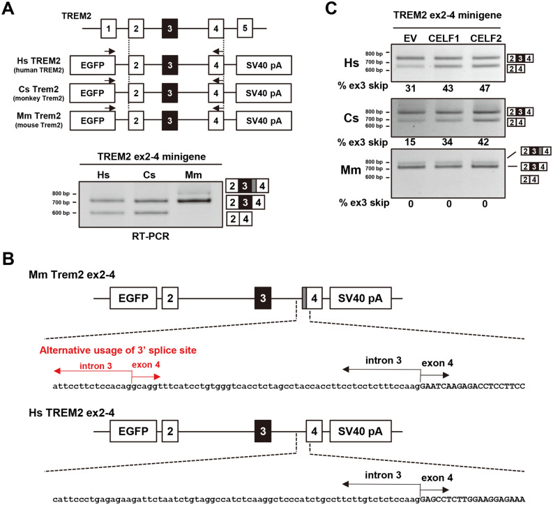
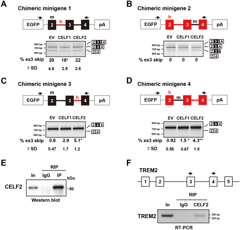
Genetic variations of TREM2 have been implicated as a risk factor of Alzheimer's disease (AD). Recent studies suggest that the loss of TREM2 function compromises microglial responses to the accumulation of amyloid beta. Previously, we found that exon 3 of TREM2 is an alternative exon whose skipping leads to a reduction in full-length TREM2 protein by inducing nonsense-mediated mRNA decay. Here, we aimed to identify factors regulating TREM2 splicing. Using a panel of RNA-binding proteins, we found that exon 3 skipping of TREM2 was promoted by two paralogous proteins, CELF1 and CELF2, which were both linked previously with risk loci of AD. Although the overexpression of both CELF1 and CELF2 enhanced exon 3 skipping, only CELF2 reduced the expression of full-length TREM2 protein. Notably, the TREM2 ortholog in the green monkey, but not in the mouse, showed alternative splicing of exon 3 like human TREM2. Similarly, splicing regulation of exon 3 by CELF1/2 was found to be common to humans and monkeys. Using chimeric minigenes of human and mouse TREM2, we mapped a CELF-responsive sequence within intron 3 of human TREM2. Collectively, our results revealed a novel regulatory factor of TREM2 expression and highlighted a species-dependent difference of its regulation.


Conflict of interest statement

The authors declare no competing interests.

Figures

Figure 1
Figure 1
Screening of RNA-binding proteins (RBPs) that regulate the alternative splicing of TREM2 exon 3. (A) Schematic diagram of the fl-TREM2 minigene. The arrows indicate the primer set to detect the splicing pattern of exon 3. (B) Results of the splicing assay using the fl-TREM2 minigene and a panel of RBPs. RBPs were expressed as a fusion with EGFP. Splice products were detected by RT-PCR using agarose gels. The proportion of exon 3 skipping is indicated at the bottom of each lane. EGFP (EV) and pcDNA3.1 were used as controls. (C) The effect of human CELF proteins (CELF1/2/3/4) on the splicing of TREM2 exon 3. The fl-TREM2 minigene was co-transfected with EGFP-fused CELF proteins into HEK cells. Bar chart shows the proportion of exon 3 skipping. Error bars represent SD (n = 3). Tukey’s test was used for statistical evaluation.
Figure 2
Figure 2
CELF2 promotes exon 3 skipping and decreases the protein expression of TREM2. (A) Western blot analysis of the fl-TREM2 minigene co-transfected with EGFP-fused CELF1 or CELF2 into HEK cells. Protein expression was detected using antibodies as indicated. EGFP-LacZ was used as a control. Bar chart shows TREM2 protein levels relative to Lamin B (arbitrary unit). Error bars represent the SD (n = 7). Tukey’s test was used for statistical evaluation. (B) HEK cells that stably express the fl-TREM2 minigene were transfected with EGFP-CELF2, and the EGFP-positive cells were separated from the EGFP-negative cells via fluorescence-activated cell sorting. The cells were subjected to western blot, using antibodies as indicated (lest panel). The box plot shows TREM2 protein expression levels relative to GAPDH (arbitrary units). The collected EGFP-positive and EGFP-negative cells were considered as a pair at each sorting, and the two-tailed paired t test was used for statistical analysis (n = 6). (C) RT-PCR products of TREM2 in THP-1 cells treated with siRNA and cycloheximide (CHX) were resolved by polyacrylamide gels (upper panel). The lower panel shows western blot results of CELF proteins in siRNA-treated THP-1 cells.
Figure 3
Figure 3
The reduction of TREM2 expression by CELF2 is coupled with exon 3 skipping. (A) Schematic diagram of the CDS TREM2 construct (upper panel). Western blot analysis of CDS TREM2 that was co-transfected into HEK cells with EGFP-fused CELF1 or CELF2 (lower left panel). Quantification of TREM2 expression levels (lower right panel). Bar chart shows TREM2 protein levels relative to Lamin B (arbitrary units). Error bars represent the SD (n = 6). No significant alteration of TREM2 protein expression was observed (Tukey’s test). Lamin B was used as a loading control as well as a reference for normalization. (B) Schematic diagram of the TREM2 ex3-sp minigene. The fragment of the genomic region of TREM2 intron 2 to intron 4 was inserted into TREM2 cDNA. fg represents the 3xDDDDK tag (upper panel). Splicing assay results of the TREM2 ex3-sp minigene co-expressed with CELF proteins using polyacrylamide gels (lower left panel). Bar chart shows the portion of exon 3 skipping (lower right panel). Error bars represent the SD (n = 4). Tukey’s test was used for statistical evaluation. (C) Western blot analysis of TREM2 ex3-sp co-transfected with EGFP-fused CELF1 or CELF2 into HEK cells (left panel). Bar chart shows TREM2 protein levels relative to Lamin B (right panel). Error bars represent the SD (n = 5). Tukey’s test was used for statistical evaluation.
Figure 4
Figure 4
Primate-specific alternative splicing of TREM2 exon 3 is regulated by CELF1 and CELF2. (A) Schematic diagram of minigenes covering a genomic region from exon 2 to exon 4 of Homo sapiens TREM2 (HsTREM2), Chlorocebus sabaeus Trem2 (CsTrem2), and Mus musculus Trem2 (MmTrem2). Each fragment was inserted into the pEGFP-C1 vector. The black arrows show the primer sets for RT-PCR to detect the splicing pattern of exon 3 in each species. A species-specific reverse primer was used in this experiment. RT-PCR products were resolved by polyacrylamide gels. (B) Schematic diagram of MmTrem2-specific splice products represented by a gray box. These splice products were confirmed by sequencing. (C) Each minigene was co-transfected with EGFP-fused cDNA into HEK cells to detect the alteration of exon 3 splicing. RT-PCR products were resolved by agarose gels.
Figure 5
Figure 5
Chimeric minigene assay and RNA immunoprecipitation to map the determinants of the alternative splicing of exon 3. (A–D) The results of the chimeric minigene assay. Chimeric minigenes of human and mouse TREM2 were inserted into the pEGFP-C1 vector. The black boxes and lines indicate mouse exons and introns, respectively. The red boxes and lines indicate human exons and introns, respectively. The arrows exhibit the primer set. RT-PCR products were resolved by polyacrylamide gels. Tukey’s test was adopted for statistical tests (n = 4). (E) Western blot analysis to confirm the immunoprecipitation of CELF2 using the anti-CELF2 antibody. In input fraction. (F) RT-PCR analysis of cDNA synthesized from CELF2-immunoprecipitated products. The primer set, indicated by the black arrows, was used to detect the binding of CELF2 to TREM2 RNA.

References

    1. Duyckaerts C, Delatour B, Potier M-C. Classification and basic pathology of Alzheimer disease. Acta Neuropathol. 2009;118:5–36. doi: 10.1007/s00401-009-0532-1. - DOI - PubMed
    1. Bertram L, Tanzi RE. Alzheimer disease risk genes: 29 and counting. Nat. Rev. Neurol. 2019;15:191–192. doi: 10.1038/s41582-019-0158-4. - DOI - PubMed
    1. Gosselin D, et al. An environment-dependent transcriptional network specifies human microglia identity. Science. 2017;356:eaal3222. doi: 10.1126/science.aal3222. - DOI - PMC - PubMed
    1. Yeh FL, Hansen DV, Sheng M. TREM2, microglia, and neurodegenerative diseases. Trends Mol. Med. 2017;23:512–533. doi: 10.1016/j.molmed.2017.03.008. - DOI - PubMed
    1. Paolicelli RC, et al. Synaptic pruning by microglia is necessary for normal brain development. Science. 2011;333:1456–1458. doi: 10.1126/science.1202529. - DOI - PubMed

Publication types