. 2017 Jan 6;3(1):005.
doi: 10.18063/IJB.2017.01.005. eCollection 2017.

3D bioprinting of stem cells and polymer/bioactive glass composite scaffolds for bone tissue engineering

Affiliations

3D bioprinting of stem cells and polymer/bioactive glass composite scaffolds for bone tissue engineering

Caroline Murphy et al. Int J Bioprint. .

Abstract

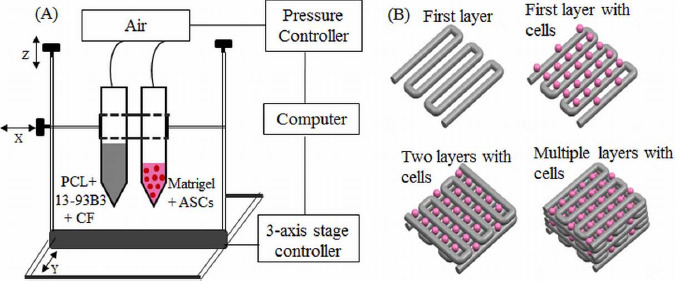
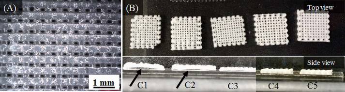
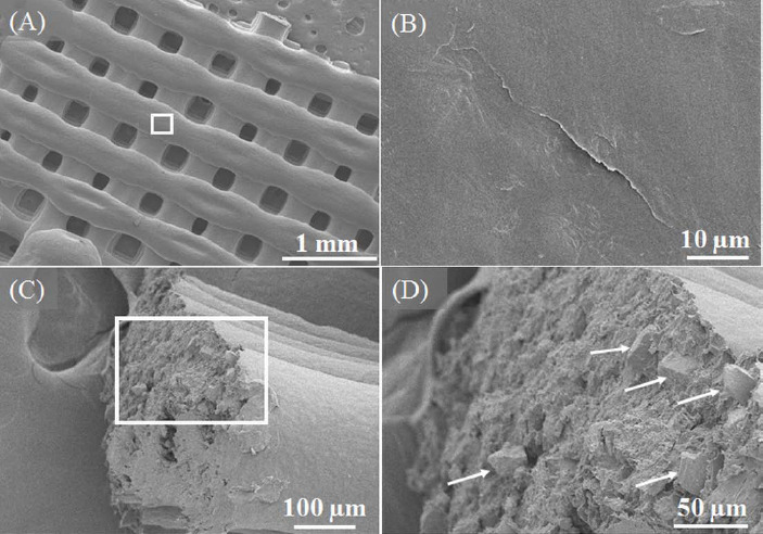
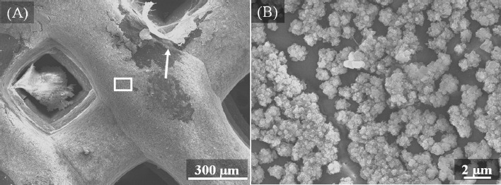
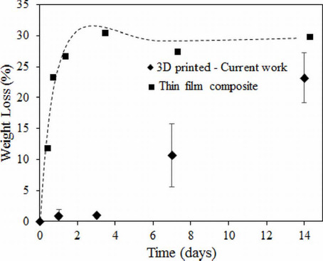
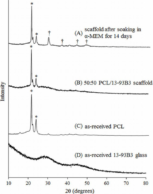
A major limitation of using synthetic scaffolds in tissue engineering applications is insufficient angiogenesis in scaffold interior. Bioactive borate glasses have been shown to promote angiogenesis. There is a need to investigate the biofabrication of polymer composites by incorporating borate glass to increase the angiogenic capacity of the fabricated scaffolds. In this study, we investigated the bioprinting of human adipose stem cells (ASCs) with a polycaprolactone (PCL)/bioactive borate glass composite. Borate glass at the concentration of 10 to 50 weight %, was added to a mixture of PCL and organic solvent to make an extrudable paste. ASCs suspended in Matrigel were ejected as droplets using a second syringe. Scaffolds measuring 10 x 10 x 1 mm3 in overall dimensions with pore sizes ranging from 100 - 300 μm were fabricated. Degradation of the scaffolds in cell culture medium showed a controlled release of bioactive glass for up to two weeks. The viability of ASCs printed on the scaffold was investigated during the same time period. This 3D bioprinting method shows a high potential to create a bioactive, highly angiogenic three-dimensional environment required for complex and dynamic interactions that govern the cell's behavior in vivo.

Keywords: MSCs; bioactive glass; biofabrication; bioprinting; human adipose-derived stem cell; polycaprolactone; scaffold; tissue engineering.


Conflict of interest statement

No conflict of interest was reported by the authors.

Figures

Figure 1
Figure 1
(A) Schematic of the printing set-up. One syringe contained PCL, 13-93B3 glass, and chloroform, while the other syringe contained ASCs suspended in Matrigel. (B) The composite layers are printed in 0°-90° pattern using one syringe while a second syringe prints the bio-ink droplets on top of every other layer.
Figure 2
Figure 2
(A) Optical microscopic image showing the pores (~160 μm) in a composite scaffold fabricated with C5 paste. (B) Scaffolds fabricated with different composite pastes (C1 to C5). The bottom panel shows scaffold warpage with an arrow indicating space between scaffold and slide. Warpage was minimal in C3/C4 scaffolds and completely absent in C5 scaffolds.
Figure 3
Figure 3
SEM images of the 50:50 PCL/13-93B3 glass scaffold. (A) Low magnification (30x) image of scaffold surface showing filaments and pores, (B) smooth surface morphology of filament (2000x magnified image of the region marked in (A), (C) fractured surface of a broken filament with PCL matrix and glass particles, (D) magnified image of the region marked in (C) with arrows indicating glass particles present a few microns beneath the surface.
Figure 4
Figure 4
SEM images of the 50:50 PCL/13-93B3 glass scaffold after immersion in α-MEM for 14 days. (A) ~1 μm thick layer was formed on the filament surface (a piece of the reacted layer indicated by arrow raised to expose the polymer beneath), (B) magnified image (8000x) of the area marked in (A) showing the formation of HA-like florets on the filament.
Figure 5
Figure 5
EDX analysis on the surface of the 50:50 PCL/13-93B3 glass scaffold soaked in α-MEM. (A) Graph of line scan data showing the variation in Ca, P, O, and C in atomic weight percentages; presence of Ca, P, and O on the reacted surface confirms the glass reaction and formation of HA-like material, (B) SEM image with the arrow line indicating the scanned area for EDX analysis.
Figure 6
Figure 6
Live/Dead images of ASCs suspended in Matrigel and printed on the 50:50 PCL/13-93B3 glass composite scaffold. Imaged after (A-B) 24 hours, and (C-D) 1 week. The dotted lines indicate the outline of the filament and dark space indicates the pore.
Figure 7
Figure 7
Schematic highlighting the difference in two methods of extrusion printing. (A) Melt-deposition of polymer-glass composite resulting in a dense filament and low bioactivity, (B) solvent-based extrusion printed composite resulting in a porous filament with high bioactivity, (C) SEM images showing surface cracks on the filament indicated by arrows in (i) and (ii), and pores inside the filament measuring less than 10 μm are also indicated by arrows in (iii).
Figure 8
Figure 8
Weight loss percentage comparison of 3D printed 50:50 PCL/13-93B3 glass composite scaffolds vs. thin film composite made using PCL, CF, and 50% 13-93B3 glass[35].
Figure 9
Figure 9
XRD patterns of (A) 50:50 PCL/13-93B3 glass composite scaffold soaked in α-MEM for 14 days, (B) PCL/13-93B3 glass scaffold, (C) as-received PCL showing a semi-crystalline nature with characteristic peaks marked by *, and (D) as-received 13-93B3 glass with characteristic amorphous hump (25° to 35° and 40° to 50°).

References

    1. Zhang P, Zhang X, Brown J B, et al. Economic impact of diabetes. IDF Diabetes Atlas 2010
    1. Banwart J C, Asher M A, Hassanein R S. Iliac crest bone graft harvest donor site morbidity. A statistical evaluation. Spine (Phila. Pa. 1976) 1995;20(9):10551060. https://doi.org/10.1097/00007632-199505000-00012. - PubMed
    1. Goulet J A, Senunas L E, DeSilva G L, et al. Autogenous iliac crest bone graft. Complications and functional assessment. Clinical Orthophopedics and Related Research. 1997;(339):76–81. https://doi.org/10.1097/00003086-199706000-00011. - PubMed
    1. Giannoudis P V, Dinopoulos H, Tsiridis E. Bone substitutes: an update. Injury. 2005;36(3):S20–S27. http://dx.doi.org/10.1016/j.injury.2005.07.029. - PubMed
    1. Doiphode N D, Huang T, Leu M C, et al. Freeze extrusion fabrication of 13-93 bioactive glass scaffolds for bone repair. Journal of Materials Science:Materials in Medicine. 2011;22(3):515–523. http://dx.doi.org/10.1007/s10856-011-4236-4. - PubMed

LinkOut - more resources