

A Semi-supervised Joint Network for Simultaneous Left Ventricular Motion Tracking and Segmentation in 4D Echocardiography
- PMID: 33094292
- PMCID: PMC7576886
- DOI: 10.1007/978-3-030-59725-2_45
A Semi-supervised Joint Network for Simultaneous Left Ventricular Motion Tracking and Segmentation in 4D Echocardiography
Abstract
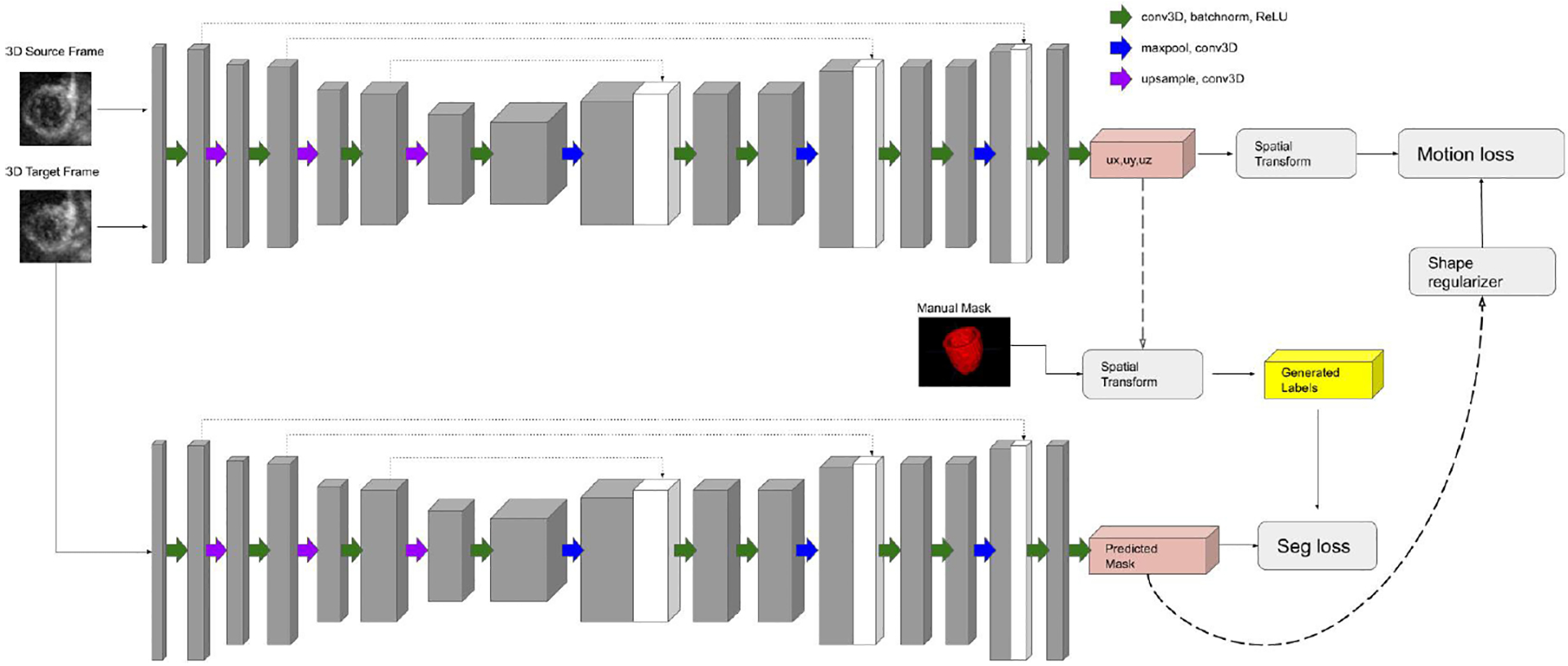
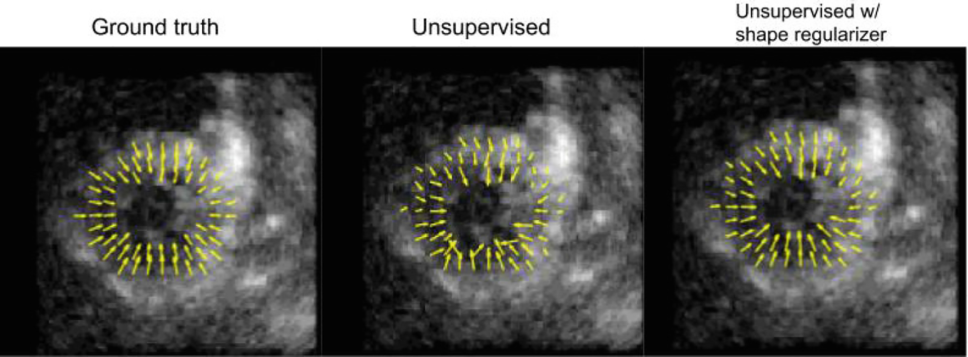
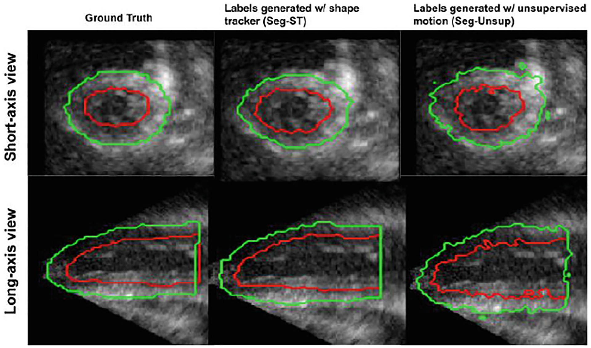
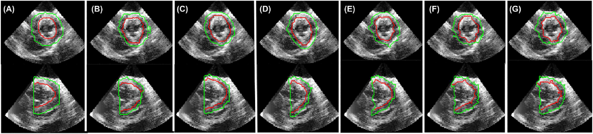
This work presents a novel deep learning method to combine segmentation and motion tracking in 4D echocardiography. The network iteratively trains a motion branch and a segmentation branch. The motion branch is initially trained entirely unsupervised and learns to roughly map the displacements between a source and a target frame. The estimated displacement maps are then used to generate pseudo-ground truth labels to train the segmentation branch. The labels predicted by the trained segmentation branch are fed back into the motion branch and act as landmarks to help retrain the branch to produce smoother displacement estimations. These smoothed out displacements are then used to obtain smoother pseudo-labels to retrain the segmentation branch. Additionally, a biomechanically-inspired incompressibility constraint is implemented in order to encourage more realistic cardiac motion. The proposed method is evaluated against other approaches using synthetic and in-vivo canine studies. Both the segmentation and motion tracking results of our model perform favorably against competing methods.
Keywords: Echocardiography; Motion tracking; Segmentation.
Figures

References
-
- Alessandrini M, et al.: A pipeline for the generation of realistic 3D synthetic echocardiographic sequences: Methodology and open-access database. IEEE Trans. Med. Imaging 34, 1436–1451 (2015) - PubMed
-
- Balakrishnan G, et al.: An unsupervised learning model for deformable medical image registration. In: The IEEE Conference on Computer Vision and Pattern Recognition (CVPR) (2018)
-
- Cheng J, et al.: Segflow: joint learning for video object segmentation and optical flow. In: IEEE International Conference on Computer Vision (ICCV) (2017)
Grants and funding
LinkOut - more resources
Full Text Sources