Review
. 2020 Dec;10(6):1229-1253.
doi: 10.1007/s13555-020-00458-9. Epub 2020 Oct 23.

The Pathogenic Role of Demodex Mites in Rosacea: A Potential Therapeutic Target Already in Erythematotelangiectatic Rosacea?

Affiliations
Review

The Pathogenic Role of Demodex Mites in Rosacea: A Potential Therapeutic Target Already in Erythematotelangiectatic Rosacea?

Fabienne M N Forton. Dermatol Ther (Heidelb). 2020 Dec.

Abstract

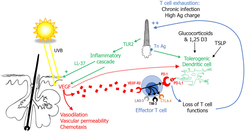
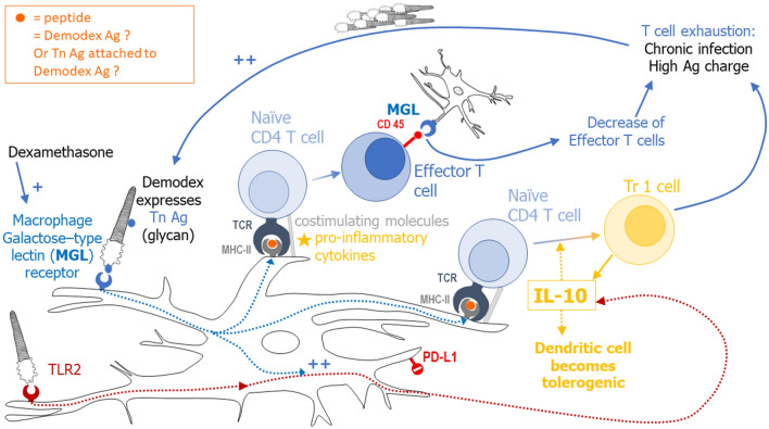
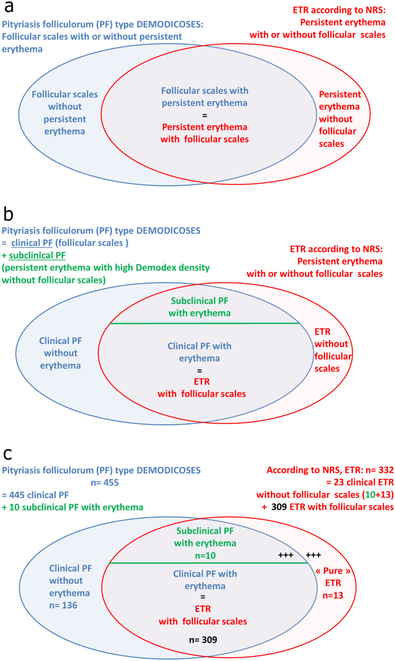
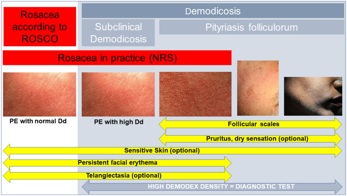
Rosacea is a common facial dermatosis but its definition and classification are still unclear, especially in terms of its links with demodicosis. Triggers of rosacea (ultraviolet light, heat, spicy foods, alcohol, stress, microbes) are currently considered to induce a cascading innate and then adaptive immune response that gets out of control. Recent histological and biochemical studies support the concept that this inflammatory response is a continuum, already present from the onset of the disease, even when no clinical signs of inflammation are visible. The Demodex mite is beginning to be accepted as one of the triggers of this inflammatory cascade, and its proliferation as a marker of rosacea; moreover, the papulopustules of rosacea can be effectively treated with topical acaricidal agents. Demodex proliferation appears to be a continuum process in rosacea, and may not be clinically visible at the onset of the disease. Molecular studies suggest that Demodex may induce tolerogenic dendritic cells and collaborate with vascular endothelial growth factor (VEGF) to induce T cell exhaustion and favor its own proliferation. These interactions among VEGF, Demodex, and immunity need to be explored further and the nosology of rosacea adapted accordingly. However, treating early rosacea, with only clinically visible vascular symptoms, with an acaricide may decrease early inflammation, limit potential flare-ups following laser treatment, and prevent the ultimate development of the papulopustules of rosacea. The effectiveness of this approach needs to be confirmed by prospective controlled clinical trials with long-term follow-up. Currently, the evidence suggests that patients with only vascular symptoms of rosacea should be carefully examined for the presence of follicular scales as signs of Demodex overgrowth or pityriasis folliculorum so that these patients, at least, can be treated early with an acaricidal cream.

Keywords: Benzyl benzoate; Demodicosis; Dendritic cell; Immunotolerance; Ivermectin; MGL; Rosacea; Standardized skin surface biopsy; Tn Ag; VEGF.


Figures

Fig. 1
Fig. 1
Erythema of rosacea and pityriasis folliculorum. a Erythema of rosacea on white skin, according to the consensus of the National Rosacea Society (NRS): original photograph published by the NRS [6]. b However, as shown on a zoom on the right cheek, this photo clearly reveals the presence of follicular scales, suggesting a diagnosis of pityriasis folliculorum. c Demodicosis associated with vascular symptoms of rosacea: discreet thin whitish follicular scales at the base of the hair give a frosted appearance and a rough texture, suggesting a diagnosis of pityriasis folliculorum; this was confirmed by the diagnostic test. Each follicular scale corresponds to the most superficial part of numerous Demodex mites agglutinated on a single follicle (blue box). d Subclinical demodicosis with vascular symptoms of rosacea: the follicular scales were not detected on close clinical examination, even after cleaning the skin with ether and using tangential illumination, leading to the clinical diagnosis of erythematotelangiectatic rosacea. However, this patient had a high Demodex density, suggesting a likely diagnosis of subclinical demodicosis. e, f Pityriasis folliculorum diagnosed as rosacea and treated with intense pulsed light (IPL): this 41-year-old woman complained of sensitive skin and redness of the whole face for 2 years. She consulted a dermatologist and was treated with isotretinoin for 8 months (30 mg/day for 6 months and 40 mg/day for 2 months) and then by IPL flash lamp (which emits simultaneous wavelengths between 530 and 1200 nm), with no resolution of her problems and even some aggravation. The dermatologist then sent the patient to our clinic for our advice. The patient had diffuse redness all over the face (not shown), more pronounced at the follicular orifices, with slight diffuse edema (visible on the lobule of the ear): the skin appeared irritated. On close examination, there was no vellus hair or follicular scales on the skin of the central face. After the skin was cleaned with ether, two standardized skin surface biopsies were consecutively performed on the right cheek and confirmed the absence of Demodex mite (0 + 0 D/cm2). Nevertheless, on small areas not treated by IPL, i.e., the lobule of the ear and the preauricular zone of the cheek, we discovered follicular scales suggesting Demodex mites. e On the preauricular zone, the mite density was very high, confirming the diagnosis of pityriasis folliculorum. The patient was instructed to apply an acaricidal cream (benzyl benzoate 12% and crotamiton 10% in Cetomacrogol cream) all over the face (not on the eyelashes or the lips) once daily for 1 week, then twice daily. f Two months later, facial signs and symptoms had cleared and the Demodex density was normalized on the preauricular zone (0 + 0 D/cm2). We concluded that the IPL may have killed the mites on the treated zones with release of their antigens and flare-up of the inflammation, or that the mites may still have persisted more deeply in the skin, in sufficient number to induce the inflammation. The standardized skin surface biopsy (SSSB1 + SSSB2) values are indicated on the figure. Part a, so also the zoom b, was reprinted from “Gallo RL et al. Standard classification and pathophysiology of rosacea: The 2017 update by the National Rosacea Society Expert Committee. J Am Acad Dermatol. 2018;78(1):148–55”, 2018, with permission from Elsevier. Parts c and d were reprinted from “Forton FMN, De Maertelaer V. Erythematotelangiectatic rosacea may be associated with a subclinical stage of demodicosis. A case control study. Br J Dermatol. 2019; 181: 818–25”, 2019, with permission from John Wiley and Sons
Fig. 2
Fig. 2
Schematic of hypothesis that vascular endothelial growth factor (VEGF) may promote T cell exhaustion and therefore Demodex proliferation, by analogy with what happens with tumors. This figure assembles information from tumor pathology, from studies on Demodex and rosacea, and from the hypothesis formulated in Fig. 3. In tumor pathology, it is known that tumors secrete VEGF, which favors their development through its pro-angiogenic properties, but also by favoring T cell exhaustion: when VEGF is bound by the VEGF receptor (VEGF-R2) present on CD8+ cytotoxic T lymphocytes, inhibitory receptors, such as programmed cell death 1 (PD-1), cytotoxic T lymphocyte antigen (CTLA-4), T cell immunoglobulin and mucin 3 domain (TIM-3), or lymphocyte activation gene 3 protein (LAG-3), are expressed on the lymphocyte surface [140]. When these receptors bind to their ligands, expressed by the tumors, this causes loss of lymphocyte function, with accumulation of regulatory T cells, reflecting T cell exhaustion [140]. In the skin, VEGF is produced by keratinocytes and fibroblasts under ultraviolet (UV) B induction [237, 238] and is increased in the dermis in rosacea, both in erythematotelangiectatic rosacea (ETR) and papulopustular rosacea (PPR) [26]. VEGF may play the same role in rosacea as in tumor pathology and collaborate with the tolerogenic dendritic cells to induce T cell exhaustion. The PD-1 receptor, induced on the effector T cell surface by its synapse with VEGF, binds to the programmed death ligand 1 (PD-L1), expressed on the surface of tolerogenic dendritic cells: this synapse then causes a loss of T cell function [145]. Tolerogenic dendritic cells may be induced by the mite (Fig. 3), thymic stromal lymphopoietin (TSLP) [36], vitamin D3 (1,25 D3) and/or glucocorticoids [145, 149], and production is also favored by VEGF [139]. The Demodex mite activates a Toll-like receptor 2 (TLR2) pathway immune response [58], which induces increased production of the cathelicidin peptide, LL-37, and subsequent angiogenesis and inflammation [15, 18]. As LL-37 stimulates the activity of endothelial cells after UV exposure and may lead to increased sensitivity to UVB radiation [20, 21], theoretically, Demodex mites may also contribute to a higher sensitivity of the skin to UVB. This suggests a vicious circle that includes mite proliferation, TLR2, LL-37, sensitivity to UVB, and VEGF, providing a physiopathogenic link between ETR and PPR
Fig. 3
Fig. 3
How Demodex may manipulate the host immune system via its Tn Ag to induce dendritic cell immunotolerance. This schematic figure assembles information from immunological studies on dendritic cells and from immunohistological studies on Demodex and rosacea. Immunological studies have shown that when dendritic cells connect with the Thomsen-nouveau antigen (Tn Ag), through its macrophage galactose-type lectin receptor (MGL), the cells migrate towards the draining lymph node, where they initiate adaptive immunity [149]. The dendritic cells interact with naïve T cells to induce immunotolerance: a peptide Ag (small orange circle) with the major histocompatibility complex (MHC) type II is presented to the T cell receptor (TCR) of the naïve T cell, together with co-stimulation molecules (gray bar). If the dendritic cell also secretes pro-inflammatory cytokines (yellow star), the Ag presentation transforms the naïve T cell into an effector T cell expressing CD45. This interacts again with the MGL receptor of the dendritic cell [150], inducing loss of the functions of the effector T cell (decreasing proliferation, reducing production of inflammatory cytokines, and increasing apoptosis) [151]. If, instead of pro-inflammatory cytokines, there is interleukin (IL-10), contact with the naïve T cell results in its transformation into a Tr1 lymphocyte (with immunosuppressive functions), which in turn produces more IL-10 [148, 152]. The production of IL-10 by the dendritic cell after stimulation of its TLR2 is strongly increased when the MGL receptor is also stimulated [151]. IL-10 is thought to induce tolerogenic transformation of the dendritic cell and to stimulate the expression of inhibitory receptors (including programmed death-ligand 1 (PD-L1)) [145]. As Demodex mites express the Tn Ag [147], these immune reactions may also occur after contact of mite Tn Ag with dendritic cells. The peptide Ag (small orange circle) presented by the dendritic cells to the naïve T cell may be another Demodex Ag (exocuticle [239], proteases [167], its endosymbiont Corynebacterium kroppenstedtii [240], etc.) or the Tn Ag attached to a peptide Demodex Ag. The Demodex mite has also been shown to stimulate TLR2 [58], expression of which is increased in rosacea [18]. Dexamethasone treatment upregulated MGL expression on dendritic cells [149], and, in ETR, abnormal endogenous glucocorticoid synthesis has been observed [146]
Fig. 4
Fig. 4
Overlap of the clinical entities pityriasis folliculorum (PF) and erythematotelangiectatic rosacea (ETR). a Schematic conceptualization of the overlap between PF and ETR. Most cases of “ETR according to the National Rosacea Society (NRS) definition” have follicular scales. Patients with the pure vascular form of rosacea (without increased Demodex density/follicular scales) or pure demodicosis (without persistent centrofacial erythema) are encountered less frequently. “PF with persistent erythema” and “ETR with follicular scales” are likely the same entity, with two possible modes of entry: (1) ETR, likely through VEGF, favors mite overproliferation [, , , , , , –97], which in turn results in increased erythema as a result of inflammation; and (2) other factors favoring mite proliferation give rise initially to PF without erythema, but persistent erythema then develops as a result of the inflammation caused by the mites. b Some patients have persistent erythema without visible follicular scales, but nevertheless have high Demodex densities (Dd): they are considered to have subclinical demodicoses/subclinical pityriasis folliculorum, and so are included into the demodicosis group. c As a practical example of the schema in b, we provide the data from the 445 patients clinically diagnosed with pityriasis folliculorum and 23 patients clinically diagnosed with ETR from our recent studies [97, 99, 108]. According to the NRS definition, 332 of these 468 patients would have been diagnosed as ETR, of whom 309 (93%) had follicular scales. Clinically, among these 332 patients, we diagnosed only 23 patients as having ETR (without follicular scales) and 309 as having pityriasis folliculorum; only 13 had “pure” ETR, without Demodex proliferation detected. Patients with persistent erythema and high Demodex density (i.e., subclinical PF with erythema or clinical PF with erythema/ETR with follicular scales) were the most numerous (319/468 = 68%). In real life, the proportion of patients with ETR without follicular scales is nevertheless certainly higher (“+++”) because in our studies these patients were only included when we had time to perform the SSSB, whereas all patients with follicular scales were included [97, 99, 108]
Fig. 5
Fig. 5
Rosacea with only vascular symptoms and pityriasis folliculorum: overlaps and ambiguities in diagnostic criteria. According to the ROSacea COnsensus (ROSCO) panel, demodicosis must be excluded before making a diagnosis of rosacea [13], but it was not specified how this should be done; however, it can be assumed that the Demodex density must be normal. The latest National Rosacea Society (NRS) consensus seems not to take follicular scales into consideration to include or exclude a diagnosis of rosacea: the consequence is that patients with pityriasis folliculorum (and subclinical demodicosis) with vascular symptoms may therefore be (mis)diagnosed as having rosacea with only vascular symptoms (ETR). However, pityriasis folliculorum is not always associated with ETR (the two photos on the right, on white skin and black skin). Of note, the photograph illustrating “Persistent erythema (PE) with normal Demodex density (Dd)” is the same as that of subclinical demodicosis “PE with high Dd” to stress that the clinical appearance/phenotypes of these conditions are identical. The patient with pityriasis folliculorum on black skin has provided written consent for publication

References

    1. Tan J, Schöfer H, Araviiskaia E, et al. Prevalence of rosacea in the general population of Germany and Russia—the RISE study. J Eur Acad Dermatol Venereol. 2016;30:428–434. - PMC - PubMed
    1. Cribier B. Rosacée: nouveautés pour une meilleure prise en charge. Ann Dermatol Venereol. 2017;144:508–517. - PubMed
    1. Berg M, Lidén S. An epidemiological study of rosacea. Acta Derm Venereol. 1989;69:419–423. - PubMed
    1. Gether L, Overgaard LK, Egeberg A, Thyssen JP. Incidence and prevalence of rosacea: a systematic review and meta-analysis. Br J Dermatol. 2018;179:282–289. - PubMed
    1. Buddenkotte J, Steinhoff M. Recent advances in understanding and managing rosacea. F1000Res. 2018;7:F1000 Faculty Rev-1885.

LinkOut - more resources