Review
. 2021 Jan;1868(1):118896.
doi: 10.1016/j.bbamcr.2020.118896. Epub 2020 Oct 21.

The requirement for cobalt in vitamin B12: A paradigm for protein metalation

Affiliations
Review

The requirement for cobalt in vitamin B12: A paradigm for protein metalation

Deenah Osman et al. Biochim Biophys Acta Mol Cell Res. 2021 Jan.

Abstract

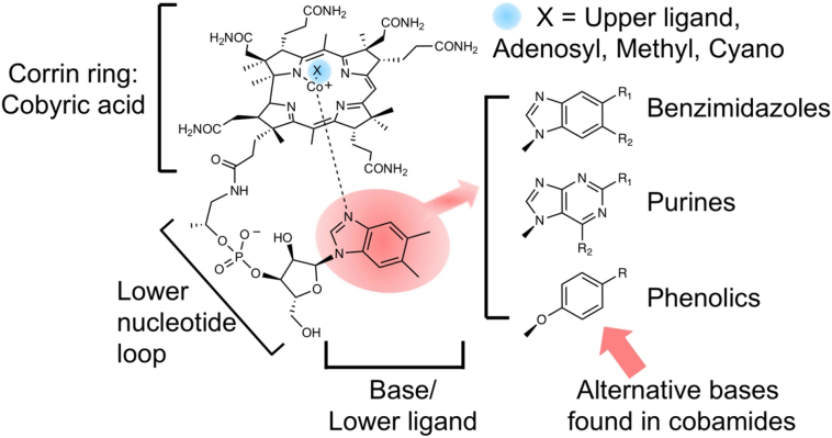
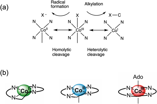
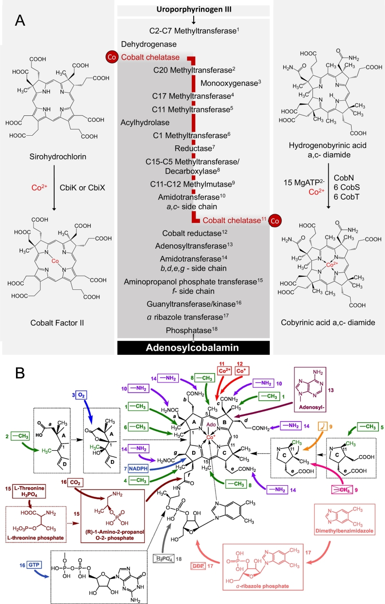
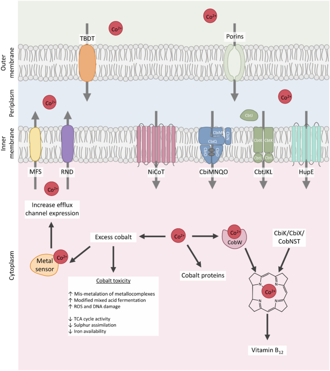
Vitamin B12, cobalamin, is a cobalt-containing ring-contracted modified tetrapyrrole that represents one of the most complex small molecules made by nature. In prokaryotes it is utilised as a cofactor, coenzyme, light sensor and gene regulator yet has a restricted role in assisting only two enzymes within specific eukaryotes including mammals. This deployment disparity is reflected in another unique attribute of vitamin B12 in that its biosynthesis is limited to only certain prokaryotes, with synthesisers pivotal in establishing mutualistic microbial communities. The core component of cobalamin is the corrin macrocycle that acts as the main ligand for the cobalt. Within this review we investigate why cobalt is paired specifically with the corrin ring, how cobalt is inserted during the biosynthetic process, how cobalt is made available within the cell and explore the cellular control of cobalt and cobalamin levels. The partitioning of cobalt for cobalamin biosynthesis exemplifies how cells assist metalation.

Keywords: Chelation; Cobalamin; Cobamide; Homeostasis; Metals; sensors.


Conflict of interest statement

The authors declare that they have no known competing financial interests or personal relationships that could have appeared to influence the work reported in this paper.

Figures

Fig. 1
Fig. 1
Structure of cobalamin (vitamin B12). The core component of cobalamin is the corrin ring, which houses a central cobalt ion. The corrin ring together with the cobalt is called cobyric acid. Attached to the propionic side chain of cobyric acid is a lower nucleotide loop, that contains an unusual base called dimethylbenzimidazole and which acts as a lower ligand to the cobalt ion. The upper ligand in vitamin B12, marked as a X in the diagram is a cyano group. In the biological forms of cobalamin, the upper ligand is usually a methyl or an adenosyl group. Some bacteria make variant forms of cobalamin where the dimethylbenzimidazole base is replaced with other bases such as other benzimidazoles (with variations around R1 and R2), purines (with variations around R1 and R2) and phenolics (with variations around R).
Fig. 2
Fig. 2
Cobalt coordination and chemistry associated with cobamides. (a) Co(III) corrinoids such as adenosylcobalamin (where X = deoxyadenosyl) can undergo homolytic cleavage to generate a Co(II) species and an adenosyl radical (X•), whereas methylcobalamin undergoes heterolytic cleavage to generate a Co(I) species and a methylated product. (b) The corrin ring naturally adopts a helical arrangement and this fits well with Co(I). More increasingly planar forms of the corrin ring are generated with Co(II) and Co(I) respectively. The ability to transition between planar and helical conformations means that the corrin ring acts as an entatic state module.
Fig. 3
Fig. 3
The biogenesis of cobalamin. (a) The biosynthesis of cobalamin from the common tetrapyrrole primogenitor uroporphyrinogen III involves in excess of twenty steps. Although there are two pathways for the synthesis of cobalamin, referred to as the aerobic (or cobalt-late) and anaerobic (cobalt early) routes, the series of methylation, amidation and rearrangement reactions are broadly similar and these steps are highlighted. The cobalt-insertion steps are also shown, with the cobalt-early insertion shown on the left hand side and the cobalt-late insertion stage shown on the right hand side. (b) The modifications that are associated with the cobalt-late pathway are highlighted and cross-referenced with (a). The Figure highlights the extensive modification that take place during the biogenesis of cobalamin.
Fig. 4
Fig. 4
Cobalt transporters. Cobalt is transported through the outer membrane by porins or by TonB-dependent transporters (TBDT). From the periplasm, cobalt is transported through the inner membrane by a range of primary and secondary transporters. When cobalt is in excess, cobalt is transported out of the cytoplasm by MFS (major facilitator) permeases and RND (resistance nodulation division pumps) whose expression is controlled by a variety of metal-sensing proteins.
Fig. 5
Fig. 5
Protein-based cobalt sensors. A, RcnR represses expression of rcnRAB (in E. coli and Salmonella, two RcnR tetramers bind the target site). RcnR can bind four Co(II) ions per tetramer, which weakens the affinity for DNA, de-repressing expression [83]. CoaR binds the coaT promoter in the absence of effector. Co(II) binding is predicted to induce a conformational change which distorts the operator-promoter, enabling recruitment of RNA polymerase and activation of expression. CoaR also harbours a tetrapyrrole (TP) binding domain including a hydrophobic patch which is capable of interacting with membranes [96]. Tetrapyrrole binding might tighten the affinity of CoaR for Co(II), and/or association with membrane-associated B12 biosynthetic machinery could confer a kinetic advantage for Co(II) and/or tetrapyrrole acquisition. CzrA represses expression of czrAB, and Co(II) binding weakens the DNA affinity of the CzrA dimer, alleviating repression. The structures of apo- and Zn(II)-CzrA are similar in the absence of DNA and an entropic mechanism for allosteric regulation has been demonstrated [103]. CzcS sensor histidine kinase detects periplasmic Co(II), and metal-binding induces autophosphorylation at a conserved histidine by Mg(II)-GTP. Transphosphorylation of the response regulator CzcR induces a conformational change promoting DNA-binding and activation of czcCBA [113]. CnrX is a transmembrane protein which forms a complex with CnrY and cytoplasmic CnrH. Co(II) binding to the periplasmic domain of CnrX, induces a conformational change which is transduced through the complex, releasing the CnrH extracytoplasmic function sigma factor. CnrH is recruited by RNA polymerase enabling expression of cnrCBA [112,113]. B–F, structural models of Co(II)-sensing proteins. B, Structural representation of RcnR tetramer (dimer of dimers) generated from PDB 5LYC. The ligands identified for co-ordination of Co(II) are shown. C, Dimeric representation of the deduced tetrapyrrole binding domain of CoaR bound to hydrogenobyrinic acid (HBA), modelled on the structure of CobH precorrin isomerase (PDB 1I1H) as described in [96] (main chain of Gly342 shown). The CoaR ligands which spatially overlay with the HBA-binding site of CobH are shown. D, The structures of Zn(II)- (PDB 2M30) and apo-CzrA (PDB 1R1U) reveal similar ‘open’ conformations which contrasts with the ‘closed’ conformation of the DNA-bound form (PDB 2KJB) [103]. The Zn(II) (and Co(II)) binding site at the dimer interface is shown. E, The sensor domain of P. aeruginosa Zn(II)-CzcS (PDB 5GPO) suggests Zn(II) binds to reciprocal residues at the dimer interface. His55, but not Asp60, is conserved in the Co(II)-responsive CzcS homolog from C. metallidurans [114]. F, The soluble periplasmic domain of CnrX (PDB 2Y3B) displays a similar structural fold to MerR-family sensors such as RcnR. The Co(II)-binding site is shown: a sulphur donor ligand is provided by a conserved methionine in contrast to the invariant cysteine in RcnR [83,120].
Fig. 6
Fig. 6
Riboswitches for B12 and Co(II). A, The binding site of AdoCbl and AqCbl in the AdoCbl riboswitch (Thermoanaerobacter tengcongensis; PDB 4GMA) and env8AqCbl riboswitch (marine metagenome; PDB 4FRN), respectively (adapted from [236]) . Although similar orientations of the J3/4 strand (red) are observed, the differential positioning of the central J6/3 strand (teal) enables accommodation of the deoxyadenosyl moiety (Ado) of AdoCbl which base pairs with A162 in the AdoCbl riboswitch, but is occluded by A20 and A68 in env8AqCbl [148]. B, The Co(II)-binding sites in the NiCo riboswitch from Erysipelotrichaceae bacterium (PDB 4RUM). The four Co(II) ions bind with octahedral co-ordination geometry and Co(II) ions I and II are co-ordinated by four conserved guanine nucleotides (G46, G47, G87, G88). Site III requires G45 which also forms the outer co-ordination sphere of site II implicating an allosteric linkage between sites I-III [148].
Fig. 7
Fig. 7
The chelatases associated with the cobalt-early pathway. (a) The type II chelatases are associated with the insertion of Co(II) into cobalamin, Fe(II) into siroheme and heme and Ni(II) into coenzyme F430. They all have a common fold despite often having limited sequence similarity. The CbiXS cobaltochelatase from Archaeoglobus fulgidus is a homo-dimer composed of two identical subunits (coloured orange and cyan). The active site is formed at the junction of the two subunits and is symmetrical. Because of the symmetry imposed by having two identical subunits, sirohydrochlorin is sandwiched by two catalytic histidines from each subunit. In other type II chelatases, such as CbiK from Salmonella, the two subunits have fused together to give a single polypeptide chain and only two active site histidines (His 145 and 207) are found here – in this case located below the sirohydrochlorin substrate. The pseudo two-fold symmetry of CbiK, reflecting the ancestral CbiXS heritage, is shown by the light and dark cyan colouring. (b) The evolution of the different type II chelatases is reflected in where the active site histidine residues are located. The cartoon highlights that the enzymes have the same basic architecture and are homologous, with those with a higher level of homology shown in green. For the small chelatases such as CbiXS and CfbA the enzyme has a symmetrical active site with a total of 4 histidines, with two from each subunit. In the larger single subunit enzymes the two histidines are located either in the N-terminal domain of the protein or the C-terminal domain. Thus CbiXL and CbiK (shown in pink) have evolved by retaining the active site histidine residues in different domains. Similarly, CbiXPP/RC and SirB, which are both ferrochelatases associated with siroheme biogenesis, have also evolved with differential retention of the active site histidines. HemH (shown in grey), which is a ferrochelatase associated with heme biogenesis has changed one of the histidine residues for a glutamate and retained these in its C-terminal domain.
Fig. 8
Fig. 8
Thermodynamic characterisation of a set of sensors. A, The set of cytosolic protein-based metal sensors from Salmonella with cognate metal ion(s) and allosteric mechanism of regulation shown, as described in [93]. B, Thermodynamic cycle depicting the allosteric coupling of metal (M) binding and DNA (D) binding by metal sensor proteins (P) [211]. C, Fractional occupancy of target DNA binding site(s) with respective metal sensor protein as a function of buffered metal concentration, following determination of K14, and number of sensor molecules and target DNA-sites per cell (B), sensor protein abundance and number of target DNA binding sites as described in [93]. D, the ranges of total (triangles) and the available (buffered; circles) metal concentrations in Salmonella. Total metal was determined in cells grown in minimal media with (closed triangles) and without (open triangles) metal supplementation [93]. Open triangles obscured for Mn(II) and Zn(II). Available metal reflects the mid-point of each sensor's dynamic range in (c). The concentration equating to one hydrated atom per cell is shown by a dashed line.
Fig. 9
Fig. 9
An associative cell biology of metals. A, The four allosteric end states of a metal sensor protein: apo-protein (P), metal-bound protein (PM), DNA-bound apo-protein (PD) and DNA-bound metal-protein ((PM)D). Metal (M) exchange occurs between the metal sensor protein and cellular buffer (B) via K5-K9 by an associative mechanism [57,93,211]. B, Proposed metal-acquisition by a metal-sensor protein (InrS, PDB 5FMN) from a cellular metal-buffer complex (Ni(II)His2, LHISNI01) via formation of a heterocomplex (modelled on PDB 4XKN). The Ni(II)-InrS model was generated using PDB 2HH7 plus free His (HIS_LFOH). InrS side chains are light blue, histidine from the cellular buffer is grey, nickel is green.
Fig. 10
Fig. 10
Metalation of CbiK. A, Metal availabilities (expressed as free-energy changes, ΔG) in the bacterial cytosol determined from the thermodynamic characterisation of a set of metal-sensor proteins and related riboswitch (Fig. 8) [93]. Bars show the ΔG range as each metal sensor shifts from 10 to 90% of its transcriptional response (i.e. 0.1–0.9 fractional DNA occupancy). In cases where there are two sensors for one metal, the combined range is also shown. An estimated Ni(II) availability from thermodynamic values for RcnR (RcnR*) is shown, using determined RcnR DNA-binding affinities and the Ni(II)-affinity of InrS, a related Ni(II)-sensor (as only a limit Ni(II) affinity is known for RcnR) [132,237]. The ΔG associated with metal binding to CbiK is also shown. The arrow for Mn(II) reflects a lower limiting value. B, Fractional occupancy of CbiK (dotted lines) with Co(II) (upper) or Fe(II) (lower) at the intracellular availabilities where RcnR and Fur (sold lines) shift from 1 to 99% of their responses, respectively.
Fig. 11
Fig. 11
Allosteric relationship between nucleotide and substrate binding in G3E GTPases. a–b Square-planar Ni(II) coordination sites at dimeric interfaces in (a) Klebsiella pneumoniae UreG (in complex with GMPPNP; PDB 5XKT [220]) and (b) Helicobacter pylori HypB (in complex with GDP; PDB 4LPS [222]); the two protomers of each homodimer are coloured green and magenta, respectively. c) Structure of Methylorubrum extorquens MeaB in complex with GMPPNP (green; PDB 4JYB [226]) or GDP (magenta; PDB 4LC1 [226]); bound nucleotides are shown in black and red, respectively. Arrow points to the ‘Switch III’ loop, which changes conformation upon nucleotide hydrolysis and is crucial for chaperone function [226]. d) The CXCC putative metal-binding motif in Escherichia coli YjiA (of the COG0523 family; PDB 1NIJ [227]), crystallised in the absence of nucleotides.

References

    1. Whipple G.H., Hooper C.W., Robscheit F.S. Blood regeneration following anemia, iv: in- fluence of meat, liver and various extrac- tives, alone or combined with standard diets. JAMA. 1920;53:236–262.
    1. Minot G.R., Murphy W.P. Treatment of perni- cious anemia by a special diet. JAMA. 1926;87:470–476.
    1. Shorb M.S. Activity of vitamin B12 for the growth of Lactobacillus lactis. Science. 1948;107(2781):397–398. - PubMed
    1. Rickes E.L., Brink N.G., Koniuszy F.R., Wood T.R., Folkers K. Crystalline vitamin B12. Science. 1948;107(2781):396–397. - PubMed
    1. Smith E.L. Purification of anti-pernicious anaemia factors from liver. Nature. 1948;161(4095):638. - PubMed

Publication types

LinkOut - more resources