. 2021 Jan:95:52-67.
doi: 10.1016/j.matbio.2020.10.004. Epub 2020 Oct 20.

Collagen XII mediated cellular and extracellular mechanisms regulate establishment of tendon structure and function

Affiliations

Collagen XII mediated cellular and extracellular mechanisms regulate establishment of tendon structure and function

Yayoi Izu et al. Matrix Biol. 2021 Jan.

Abstract

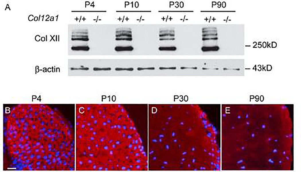
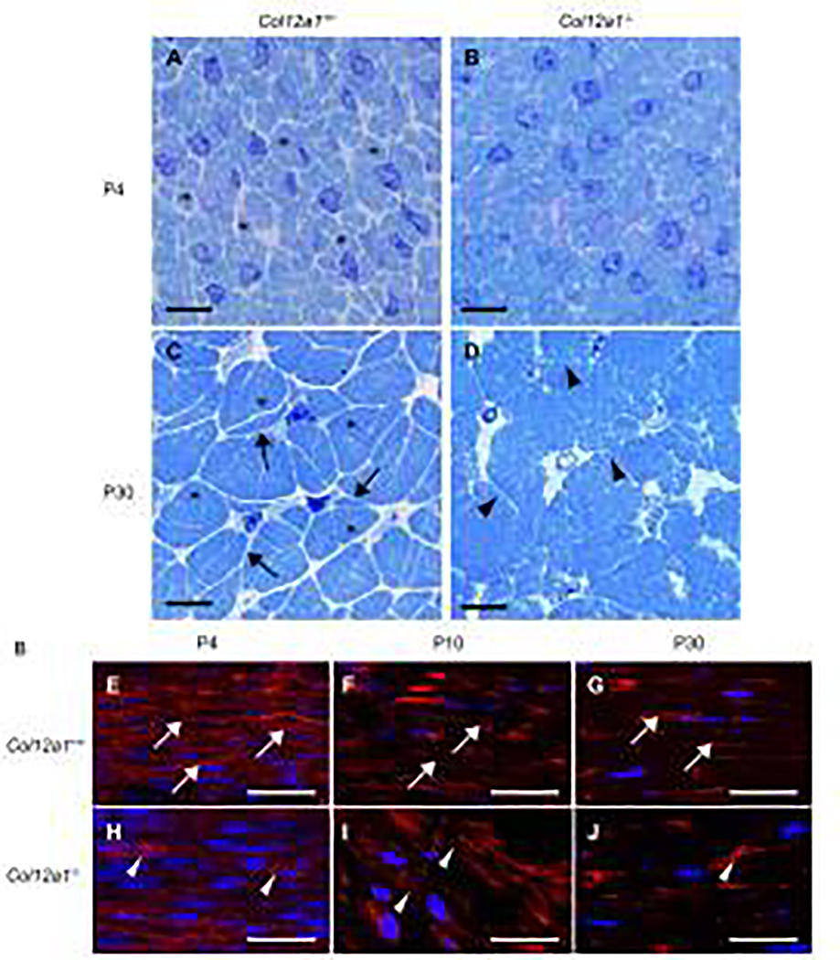
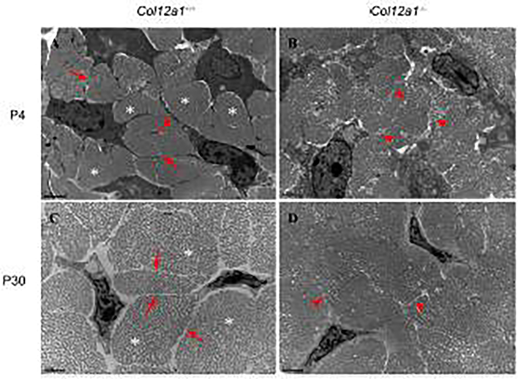
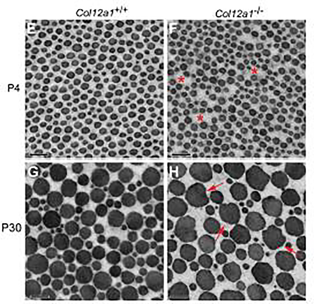
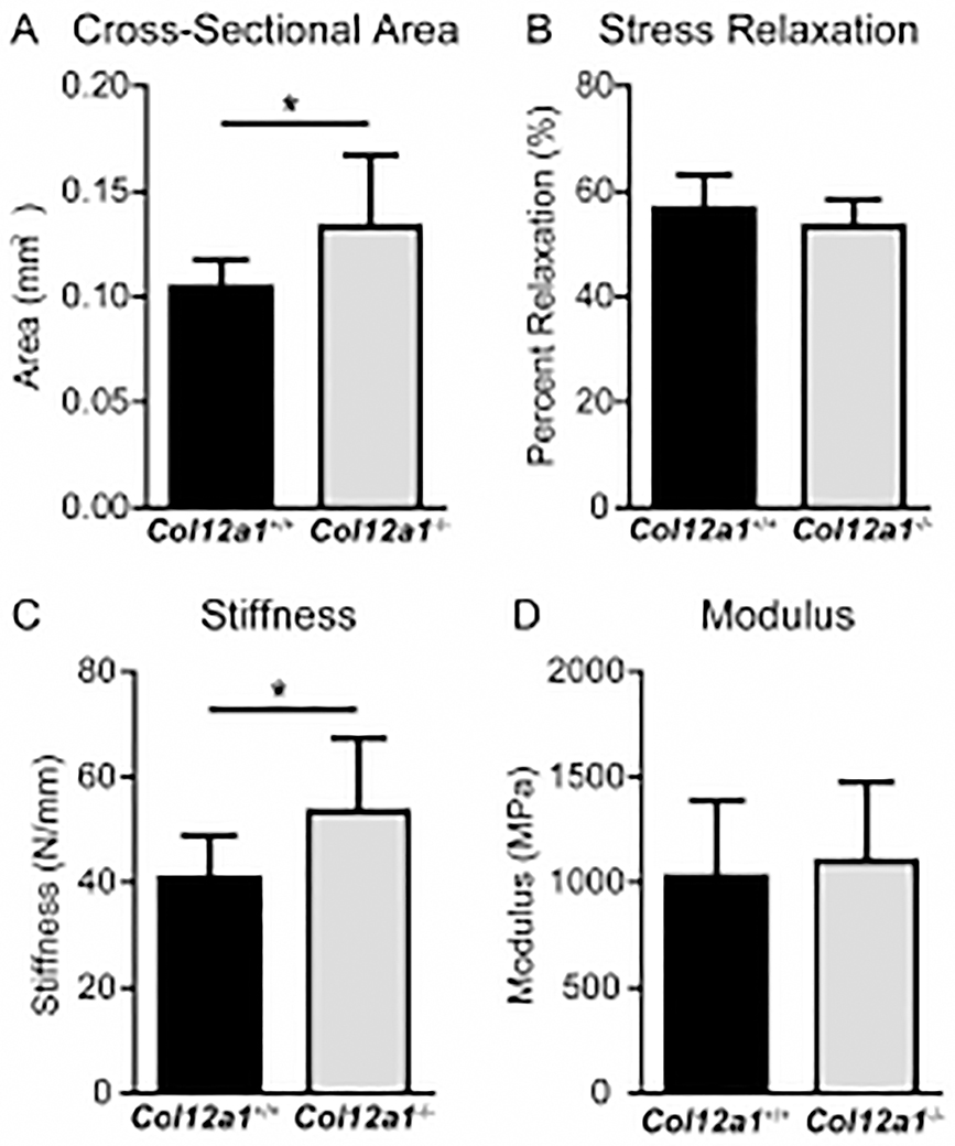
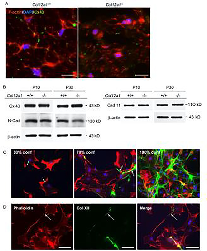
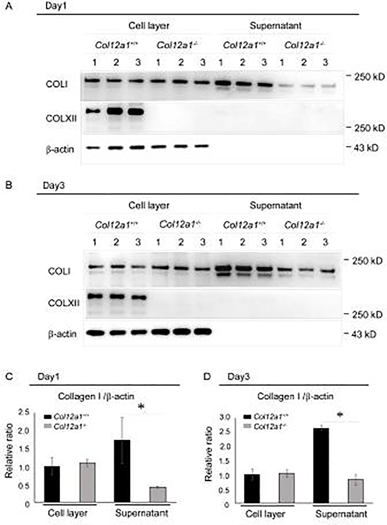
Tendons have a uniaxially aligned structure with a hierarchical organization of collagen fibrils crucial for tendon function. Collagen XII is expressed in tendons and has been implicated in the regulation of fibrillogenesis. It is a non-fibrillar collagen belonging to the Fibril-Associated Collagens with Interrupted Triple Helices (FACIT) family. Mutations in COL12A1 cause myopathic Ehlers Danlos Syndrome with a clinical phenotype involving both joints and tendons supporting critical role(s) for collagen XII in tendon development and function. Here we demonstrate the molecular function of collagen XII during tendon development using a Col12a1 null mouse model. Col12a1 deficiency altered tenocyte shape, formation of interacting cell processes, and organization resulting in impaired cell-cell communication and disruption of hierarchal structure as well as decreased tissue stiffness. Immuno-localization revealed that collagen XII accumulated on the tenocyte surface and connected adjacent tenocytes by building matrix bridges between the cells, suggesting that collagen XII regulates intercellular communication. In addition, there was a decrease in fibrillar collagen I in collagen XII deficient tenocyte cultures compared with controls suggesting collagen XII signaling specifically alters tenocyte biosynthesis. This suggests that collagen XII provides feedback to tenocytes regulating extracellular collagen I. Together, the data indicate dual roles for collagen XII in determination of tendon structure and function. Through association with fibrils it functions in fibril packing, fiber assembly and stability. In addition, collagen XII influences tenocyte organization required for assembly of higher order structure; intercellular communication necessary to coordinate long range order and feedback on tenocytes influencing collagen synthesis. Integration of both regulatory roles is required for the acquisition of hierarchal structure and mechanical properties.

Keywords: Cell–cell communication; Collagen XII; Collagen fibril assembly; Mechanical properties; Tendon; Tendon extracellular matrix assembly.


Conflict of interest statement

Declaration of Competing Interest The author(s) declare no competing interests.

Figures

Fig 1.
Fig 1.. Expression of collagen XII during mouse tendon development and maturation.
(A) Quantitative analysis of collagen XII was performed by Western blotting from P4, P10, P30 and P90 Col12a1+/+ and Col12a1−/− mouse FDL tendons. The collagen XII expression was stable during tendon development. (B-E) Collagen XII localization was analyzed by immunofluoresence in P4, P10, P30 and P90 mouse FDLs. Collagen XII reactivity was present throughout the tendon with no change in distribution with age. At P30 and P90 reactivity was less than in the younger tendons, but with same distribution. DAPI, blue for nuclei. Bar = 50 μm.
Fig 2.
Fig 2.. Tenocytes and fiber domain structure in cross and logitudinal sections.
(A-D) Cross sections of FDLs were stained with Toluidine blue and (E-J) longitudinal sections were stained with phalloidin and DAPI. At P4, Col12a1+/+ FDLs consisted of tenocytes and collagen fibers (asterisks). The domains, defined by the tenocytes containing fibers, are clearly defined (A), whereas no clear fiber domains are detected in Col12a1−/− FDLs (B). At P30, Col12a1+/+ tenocyte processes (arrows) interact with processes from neighboring cells and clearly define the fiber domains (asterisks) (C). In contrast, tenocyte processes (arrow heads) and fiber domains are disorganized and poorly defined in Col12a1−/− FDLs (D). Phalloidin staining of longitudinal sections demonstrates that tenocytes (arrows) are parallel and orientated along with the longitudinal axis at P4 (E). At P10 and P30, the tenocytes become attenuated along the tendon axis (F, G). In Col12a1−/− FDLs at P4, the actin cytoskeleton is less developed, and tenocytes are poorly organized (arrowheads) (H). Tenocyte structure (cytoskeleton) is disrupted, and the tenocytes are disorganized with the tendon axis hard to define at P10 (I) and P30 (J). Scale bars 50 μm (A-D) and 25 μm (E-J).
Fig 3.
Fig 3.. Altered tenocyte process formation and fibril spacing in the absence of collagen XII.
FDL cross sections from P4 and P30 Col12a1+/+ and Col12a1−/− mice were analyzed by transmission electron microscopy (TEM). At P4, Col12a1+/+ tenocytes extend their processes (arrows) and interact with adjacent cells. Fibers (asterisks) are surrounded by tenocyte processes (A). In contrast, tenocyte processes (arrowheads) are unclear and no obvious, well defined fibers are observed in Col12a1−/− FDLs (B). At P30, similar to P4, Col12a1+/+ tenocytes have fine processes (arrows) and clear fiber domains (asterisks) (C), whereas no clear tenocyte processes (arrowheads) and fiber domains are found in the Col12a1−/− FDL (D). Scale bars 2 μm.
Fig. 4.
Fig. 4.. Abnormal tendon collagen fibril packing in the absence of collagen XII.
Cross sections of fibrils from FDLs were analyzed using transmission electron microscopy. (A-B) At P4, fibril diameters are uniform with normal circular cross-sectional profiles in Col12a1+/+ FDLs. In Col12a1−/− FDLs, fibril spacing is increased (asterisks) and fibril packing is irregular. (C-D) At P30, Col12a1+/+ fibrils are well packed with circular crosssectional profiles (C), whereas in Col12a1−/− FDLs aberrant fibril packing is observed. In addition, abnormal fibrils are seen with altered fibril growth resulting in irregular cross-sectional profiles. Arrows indicate irregular fibril profiles (D.) Scale bars 200nm.
Fig 5.
Fig 5.. Biomechanics in Col12a1+/+ and Col12a1−/− FDLs.
Biomechanics were analyzed in P60 Col12a1+/+ and Col12a1−/− mouse FDL tendons. (A) Col12a1−/− FDLs are significantly larger in cross-sectional area than Col12a1+/+ FDLs. (B) There was no difference in percent relaxation, a measure of viscoelasticity, between the two groups. (C) Col12a1−/− FDLs exhibits increased stiffness compared to wild type controls. (D) No significant change is detected in modulus in Col12a1−/−, when compared to the wild type controls.
Fig 6.
Fig 6.. Collagen XII regulates establishment of a communicating tenocyte network.
(A) Connexin 43 immunolocalization was performed in primary tenocytes obtained from FDLs of Col12a1+/+ and Col12a1−/− neonatal mice. Tenocyte structure was visualized after staining of the cytoplasmic filamentous actin network with Alexa 594 Phalloidin (red) and nuclei were stained with DAPI (blue). Col12a1+/+ tenocytes are dendritic and connexin 43 (green) is localized where tenocyte processes interacted with other processes from adjacent tenocytes. In contrast, Col12a1− /− tenocytes have altered shapes with no clear processes, and no specific localization of connexin 43 is observed. Scale bars 25 μm. (B) Connexin 43 and cell adhesion molecules were analyzed by Western blotting in P10 and P30 FDLs from Col12a1+/+ and Col12a1−/− mice. Connexin 43 and cadherin 11 expression is equivalent between Col12a1+/+ and Col12a1−/− at both P10 and P30. N-cadherin expression is similar between genotypes at P4, but slightly decreased in P30 Col12a1−/− FDLs when compared to Col12a1+/+. b-actin was used as an internal control. (C,D) Collagen XII localization was analyzed in primary tenocytes obtained from Col12a1+/+ FDLs. (C) Immunostaining for collagen XII (green), phalloidin (red) and DAPI (blue) was analyzed in 30%, 70% and 100% confluent cell cultures. At 30% confluence, few intercellular connections are formed. Collagen XII is detected as punctate structures on the tenocyte surface (arrowheads). At 70% confluence, intercellular connections begin to form. Collagen XII is localized along the processes connecting neighboring cells (double arrows). At 100% confluence, collagen XII forms a network connecting neighboring cells. (D) Collagen XII forms bridges between neighboring cells before they establish physical connection. Scale bars 100 μm.
Fig 7.
Fig 7.. Collagen XII is associated with the pericellular matrix and influences collagen I secretion.
Collagen I secretion was analyzed in primary tenocytes obtained from Col12a1+/+ and Col12a1−/− FDLs. Cell layer and culture supernatant were harvested at 1 (A) and 3 (B) days after confluence and analyzed for collagens I and XII content by Western blotting. b-actin is used as an internal control. Collagen XII was found in cell layer at day1 and 3, but not in the supernatant (A, B). Collagen I was found in both the cell layer and culture supernatant as expected. In the cell layer, the collagen I content is comparable between genotypes on both culture days. However, collagen I in the supernatant is decreased in Col12a1−/− cultures at both time points when compared to wild type. (C,D) Quantitative analysis of collagen I at day 1 (C) and day 3 (D) was performed. In wild type controls, more collagen I is present in the supernatant than in the cell layer at both time points, whereas there is less in the Col12a1−/−cell layer. Compared to wild type controls, a significant reduction of collagen I in Col12a1−/− supernatants is found in both culture days.

References

    1. Birk DE, Trelstad RL, Extracellular compartments in tendon morphogenesis: collagen fibril, bundle, and macroaggregate formation., J Cell Biol. 103 (1986) 231–240. - PMC - PubMed
    1. Birk DE, Zycband E, Assembly of the tendon extracellular matrix during development., J. Anat. 184 (1994) 457–63. - PMC - PubMed
    1. Graham HK, Holmes DF, Watson RB, Kadler KE, Identification of collagen fibril fusion during vertebrate tendon morphogenesis. The process relies on unipolar fibrils and is regulated by collagen-proteoglycan interaction, J. Mol. Biol. 295 (2000) 891–902. 10.1006/jmbi.1999.3384. - DOI - PubMed
    1. Canty EG, Kadler KE, Procollagen trafficking, processing and fibrillogenesi, J Cell Sci. 118 (2005) 1341–1353. 10.1242/jcs.01731. - DOI - PubMed
    1. Zhang G, Young BB, Ezura Y, Favata M, Soslowsky LJ, Chakravarti S, Birk DE, Development of tendon structure and function: Regulation of collagen fibrillogenesis, J. Musculoskelet. Neuronal Interact. 5 (2005) 5–21. - PubMed

Publication types

LinkOut - more resources