. 2020 Dec:120:104926.
doi: 10.1016/j.archoralbio.2020.104926. Epub 2020 Oct 7.

Oral commensal bacteria differentially modulate epithelial cell death

Affiliations

Oral commensal bacteria differentially modulate epithelial cell death

Tyresia White et al. Arch Oral Biol. 2020 Dec.

Abstract

Objective: Epithelial cell death is an important innate mechanism at mucosal surfaces, which enables the elimination of pathogens and modulates immunoinflammatory responses. Based on the antimicrobial and anti-inflammatory properties of cell death, we hypothesized that oral epithelial cell (OECs) death is differentially modulated by oral bacteria.

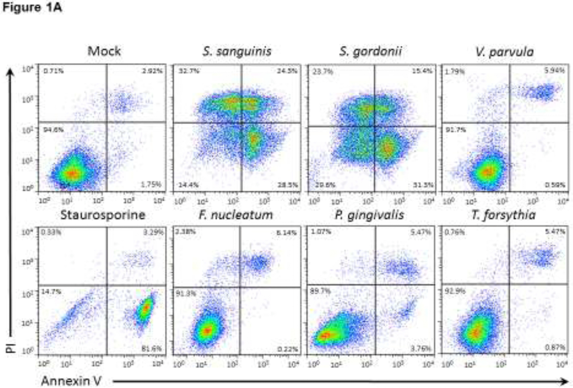
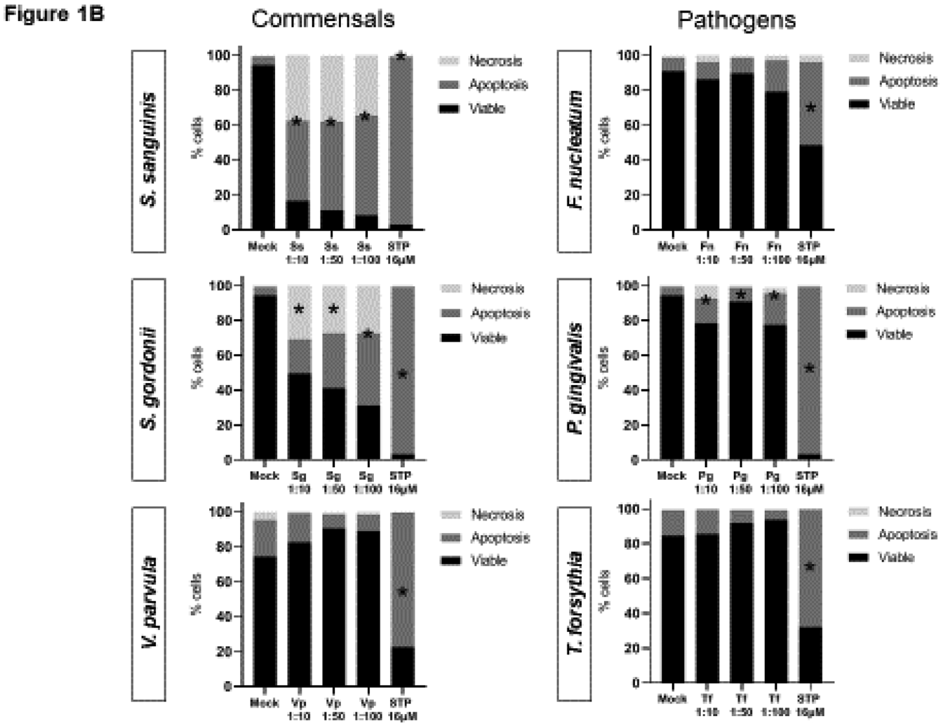
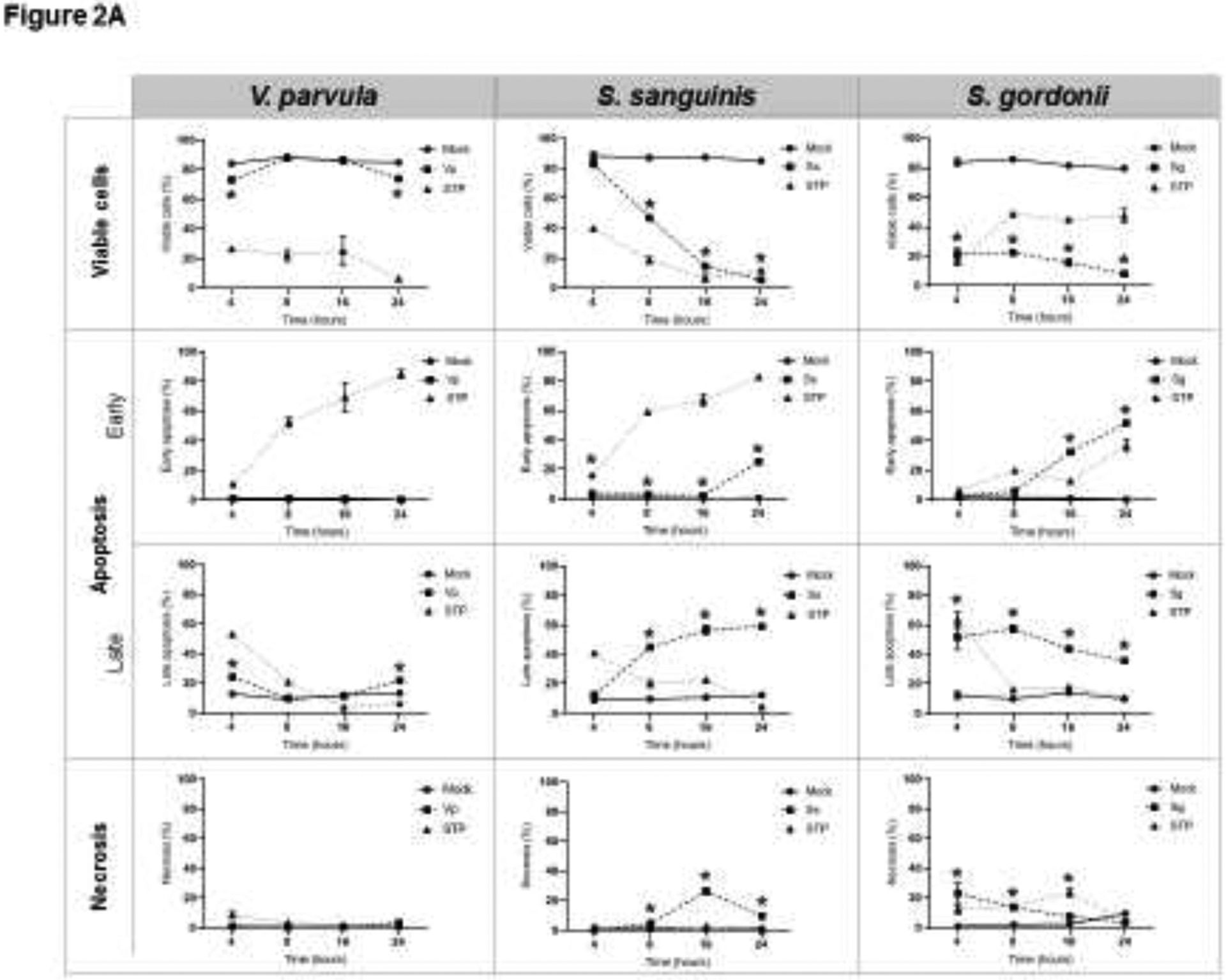
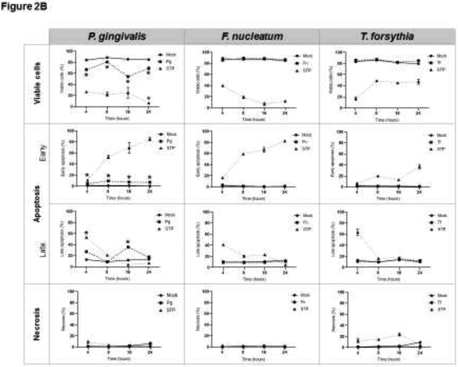
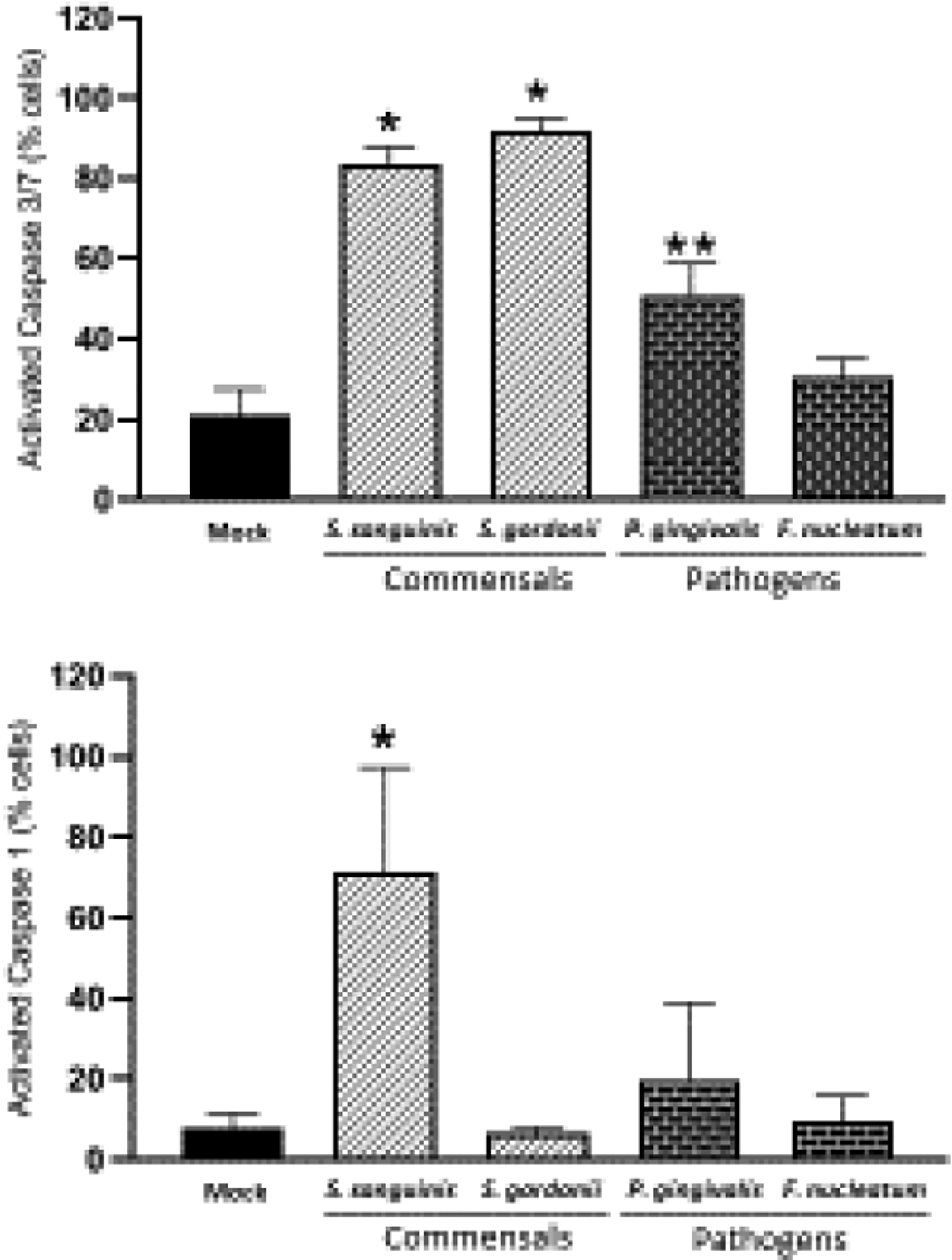
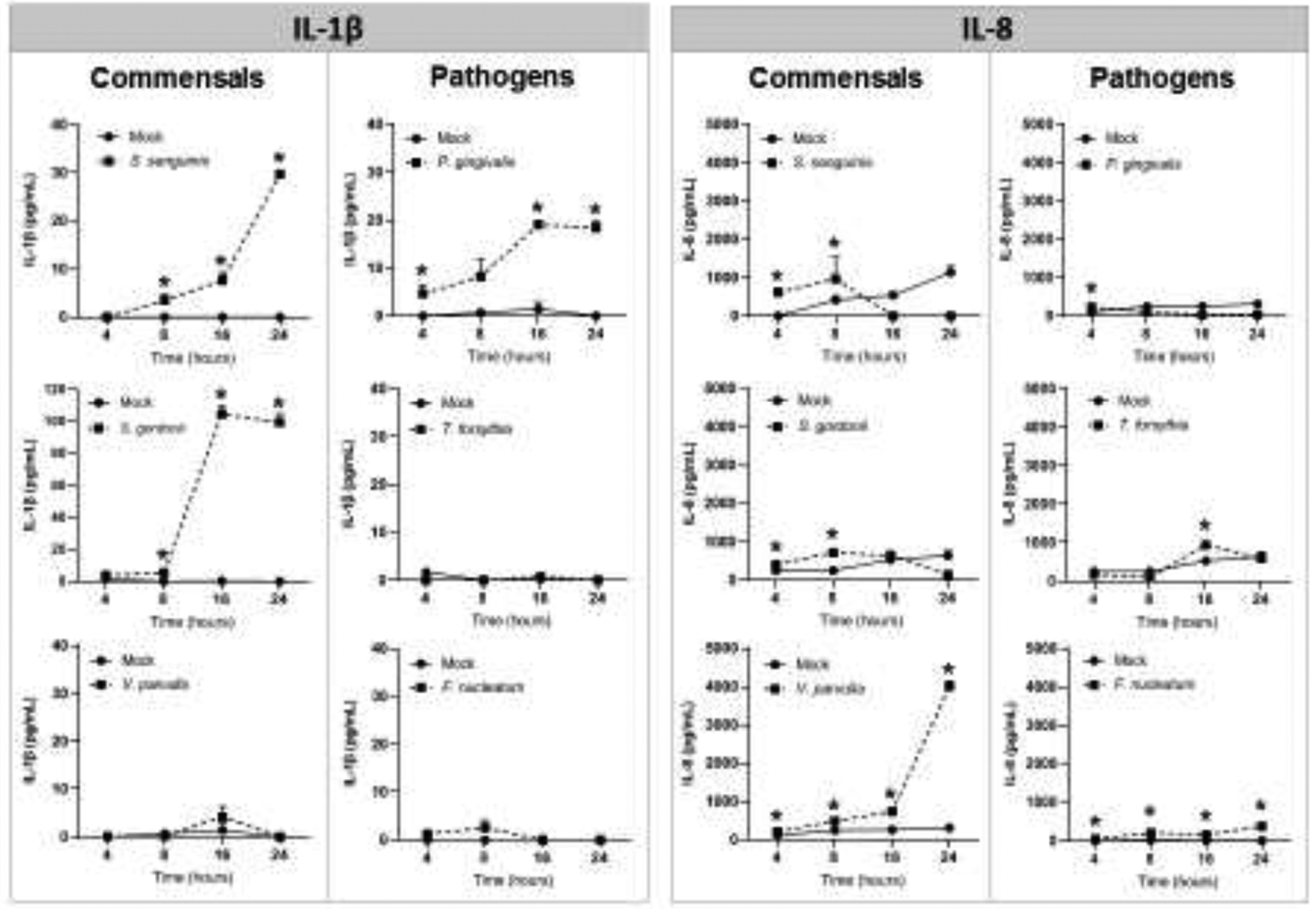
Material and methods: We evaluated the effect of oral commensals Streptococcus gordonii (Sg), Streptococcus sanguinis (Ss), and Veillonella parvula (Vp), and pathogens Porphyromonas gingivalis (Pg), Tannerella forsythia (Tf), and Fusobacterium nucleatum (Fn) on OEC death. Apoptosis and necrosis were evaluated by flow cytometry using FITC Annexin-V and Propidium Iodide staining. Caspase-3/7 and caspase-1 activities were determined as markers of apoptosis and pyroptosis, respectively. IL-1β and IL-8 protein levels were determined in supernatants by ELISA.

Results: Significant increases in apoptosis and necrosis were induced by Sg and Ss. Pg also induced apoptosis, although at a substantially lower level than the commensals. Vp, Tf, and Fn showed negligible effects on cell viability. These results were consistent with Sg, Ss, and Pg activating caspase-3/7. Only Ss significantly increased the levels of activated caspase-1, which correlated to IL-1β over-expression.

Conclusions: OEC death processes were differentially induced by oral commensal and pathogenic bacteria, with Sg and Ss being more pro-apoptotic and pro-pyroptotic than pathogenic bacteria. Oral commensal-induced cell death may be a physiological mechanism to manage the extent of bacterial colonization of the outer layers of mucosal epithelial surfaces. Dysbiosis-related reduction or elimination of pro-apoptotic oral bacterial species could contribute to the risk for persistent inflammation and tissue destruction.

Keywords: Apoptosis; Oral commensal bacteria; Oral epithelial cells; Oral pathogenic bacteria; Pyroptosis.


Conflict of interest statement

Declaration of Competing Interest

The authors declare no potential conflicts of interest with respect to authorship and/or publication of this manuscript.

Figures

Figure 1.
Figure 1.
Effect of oral commensal and pathogenic bacteria on epithelial cell death (Dose-response). (A) Representative dot plots and (B) mean of percentages for viable cells or undergoing cell death (apoptosis or necrosis) after 24h bacterial challenge of two independent experiments analyzing at least 10,000 events by FACS for each condition is shown. Viable cells (Annexin V−/PI−), early apoptosis (Annexin V+), late apoptosis (Annexin V+/PI+), and Necrosis (PI+). STP: 16 μM Staurosporine was used as a positive control. *Treated cells vs. Unstimulated (Mock) cells p<0.05 (ANOVA and post hoc Fisher LSD).
Figure 1.
Figure 1.
Effect of oral commensal and pathogenic bacteria on epithelial cell death (Dose-response). (A) Representative dot plots and (B) mean of percentages for viable cells or undergoing cell death (apoptosis or necrosis) after 24h bacterial challenge of two independent experiments analyzing at least 10,000 events by FACS for each condition is shown. Viable cells (Annexin V−/PI−), early apoptosis (Annexin V+), late apoptosis (Annexin V+/PI+), and Necrosis (PI+). STP: 16 μM Staurosporine was used as a positive control. *Treated cells vs. Unstimulated (Mock) cells p<0.05 (ANOVA and post hoc Fisher LSD).
Figure 2.
Figure 2.
Effect of oral (A) commensal, and (B) pathogenic bacteria on epithelial cell death (Time-response). MOI [1:50] was used for all bacteria. For each condition, at least 10,000 events were analyzed by FACS to identify viable cells (Annexin V−/PI−), or undergoing early apoptosis (Annexin V+), late apoptosis (Annexin V+/PI+), and Necrosis (PI+). STP: 16 μM Staurosporine was used as a positive control. The mean ± standard deviation from two independent experiments with each condition in duplicate is shown. *Treated cells vs. Unstimulated (Mock) cells p<0.05 (ANOVA and post hoc Fisher LSD).
Figure 2.
Figure 2.
Effect of oral (A) commensal, and (B) pathogenic bacteria on epithelial cell death (Time-response). MOI [1:50] was used for all bacteria. For each condition, at least 10,000 events were analyzed by FACS to identify viable cells (Annexin V−/PI−), or undergoing early apoptosis (Annexin V+), late apoptosis (Annexin V+/PI+), and Necrosis (PI+). STP: 16 μM Staurosporine was used as a positive control. The mean ± standard deviation from two independent experiments with each condition in duplicate is shown. *Treated cells vs. Unstimulated (Mock) cells p<0.05 (ANOVA and post hoc Fisher LSD).
Figure 3.
Figure 3.
Effect of oral commensal and pathogenic bacteria on Caspase 3/7 (apoptosis) and Caspase-1 (Pyroptosis) activation in epithelial cells after 24h. Mean and standard deviation bars of percentage of cells expressing activated Caspase-3/7 or activated Caspase-1 for each condition are shown. For each condition 10,000 events were analyzed by FACS. The mean ± standard deviation from two independent experiments with each condition in duplicate is shown. MOI=1:50 *Treated cells vs. Unstimulated (Mock) cells *p<0.01; **p<0.05 (ANOVA and post hoc Fisher LSD).
Figure 4.
Figure 4.
Effect of oral commensal and pathogenic bacteria [MOI=1:50] on IL-1β and IL-8 production in oral epithelial cells. Mean ± standard deviation bars of cytokine levels (pg/ml) detected by ELISA in cell culture supernatants from two independent experiments for each condition in duplicate is shown. *Treated cells vs. Unstimulated (Mock) cells p<0.05 (ANOVA and post hoc Fisher LSD).

References

    1. Abuhussein H, Bashutski JD, Dabiri D, Halubai S, Layher M, Klausner C, Makhoul H, & Kapila Y (2014). The role of factors associated with apoptosis in assessing periodontal disease status. J Periodontol, 85(8), 1086–1095. 10.1902/jop.2013.130095 - DOI - PubMed
    1. Al-Attar A, Alimova Y, Kirakodu S, Kozal A, Novak MJ, Stromberg AJ, Orraca L, Gonzalez-Martinez J, Martinez M, Ebersole JL, & Gonzalez OA (2018). Activation of Notch-1 in oral epithelial cells by P. gingivalis triggers the expression of the antimicrobial protein PLA2-IIA. Mucosal Immunol, 11(4), 1047–1059. 10.1038/s41385-018-0014-7 - DOI - PMC - PubMed
    1. Bergsbaken T, Fink SL, & Cookson BT (2009). Pyroptosis: host cell death and inflammation. Nat Rev Microbiol, 7(2), 99–109. 10.1038/nrmicro2070 - DOI - PMC - PubMed
    1. Bhattacharya R, Xu F, Dong G, Li S, Tian C, Ponugoti B, & Graves DT (2014). Effect of bacteria on the wound healing behavior of oral epithelial cells. PLoS One, 9(2), e89475 10.1371/journal.pone.0089475 - DOI - PMC - PubMed
    1. Bortoluci KR, & Medzhitov R (2010). Control of infection by pyroptosis and autophagy: role of TLR and NLR. Cell Mol Life Sci, 67(10), 1643–1651. 10.1007/s00018-010-0335-5 - DOI - PMC - PubMed

Supplementary concepts

LinkOut - more resources