

Regulatory Connections between Iron and Glucose Metabolism
- PMID: 33096618
- PMCID: PMC7589414
- DOI: 10.3390/ijms21207773
Regulatory Connections between Iron and Glucose Metabolism
Abstract
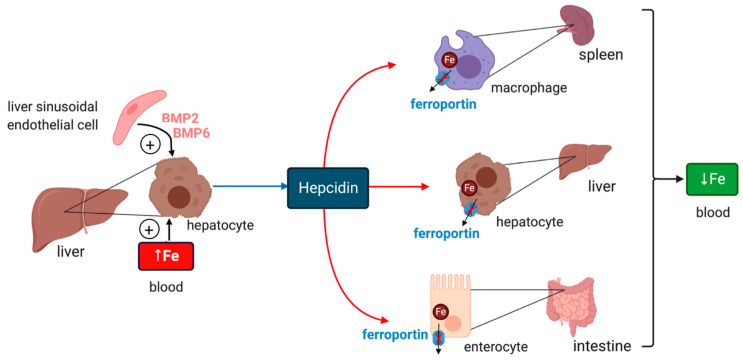
Iron is essential for energy metabolism, and states of iron deficiency or excess are detrimental for organisms and cells. Therefore, iron and carbohydrate metabolism are tightly regulated. Serum iron and glucose levels are subjected to hormonal regulation by hepcidin and insulin, respectively. Hepcidin is a liver-derived peptide hormone that inactivates the iron exporter ferroportin in target cells, thereby limiting iron efflux to the bloodstream. Insulin is a protein hormone secreted from pancreatic β-cells that stimulates glucose uptake and metabolism via insulin receptor signaling. There is increasing evidence that systemic, but also cellular iron and glucose metabolic pathways are interconnected. This review article presents relevant data derived primarily from mouse models and biochemical studies. In addition, it discusses iron and glucose metabolism in the context of human disease.
Keywords: IRP1; IRP2; adipokines; ferroportin; hepcidin; insulin.
Conflict of interest statement
The authors declare no conflict of interest.
Figures


Similar articles
-
Systemic iron homeostasis and erythropoiesis.IUBMB Life. 2017 Jun;69(6):399-413. doi: 10.1002/iub.1629. Epub 2017 Apr 6. IUBMB Life. 2017. PMID: 28387022 Review.
-
Excess capacity of the iron regulatory protein system.J Biol Chem. 2007 Aug 24;282(34):24650-9. doi: 10.1074/jbc.M703167200. Epub 2007 Jun 28. J Biol Chem. 2007. PMID: 17604281
-
A synergistic role of IRP1 and FBXL5 proteins in coordinating iron metabolism during cell proliferation.J Biol Chem. 2017 Sep 22;292(38):15976-15989. doi: 10.1074/jbc.M117.785741. Epub 2017 Aug 2. J Biol Chem. 2017. PMID: 28768766 Free PMC article.
-
Hepcidin and iron regulatory proteins coordinately regulate ferroportin 1 expression in the brain of mice.J Cell Physiol. 2019 May;234(5):7600-7607. doi: 10.1002/jcp.27522. Epub 2018 Oct 28. J Cell Physiol. 2019. PMID: 30370612
-
Role of nitric oxide in cellular iron metabolism.Biometals. 2003 Mar;16(1):125-35. doi: 10.1023/a:1020788603046. Biometals. 2003. PMID: 12572672 Review.
Cited by
-
Association between weight-adjusted-waist index and serum ferritin in patients with type 2 diabetes.Asia Pac J Clin Nutr. 2025 Jun;34(3):411-419. doi: 10.6133/apjcn.202506_34(3).0015. Asia Pac J Clin Nutr. 2025. PMID: 40419401 Free PMC article.
-
Iron Reshapes the Gut Microbiome and Host Metabolism.J Lipid Atheroscler. 2021 May;10(2):160-183. doi: 10.12997/jla.2021.10.2.160. Epub 2021 Mar 10. J Lipid Atheroscler. 2021. PMID: 34095010 Free PMC article. Review.
-
Myelodysplastic Syndromes and Metabolism.Int J Mol Sci. 2021 Oct 19;22(20):11250. doi: 10.3390/ijms222011250. Int J Mol Sci. 2021. PMID: 34681910 Free PMC article. Review.
-
Association Between Dietary Inflammatory Index and Triglyceride Glucose-Body Mass Index with Iron Deficiency in Reproductive Age Women: Evidence from NHANES 2005-2018.Int J Womens Health. 2025 Feb 10;17:355-367. doi: 10.2147/IJWH.S507765. eCollection 2025. Int J Womens Health. 2025. PMID: 39959754 Free PMC article.
-
A contemporary understanding of iron metabolism in active premenopausal females.Front Sports Act Living. 2022 Jul 28;4:903937. doi: 10.3389/fspor.2022.903937. eCollection 2022. Front Sports Act Living. 2022. PMID: 35966107 Free PMC article. Review.
References
-
- Frausto da Silva J.J.R., Williams R.J.P. The Biological Chemistry of the Elements. The Inorganic Chemistry of Life. Clarendon Press; Oxford, UK: 1991. pp. 319–369.
Publication types
MeSH terms
Substances
Grants and funding
LinkOut - more resources
Full Text Sources
Medical
