Review
. 2020 Oct 21;8(4):621.
doi: 10.3390/vaccines8040621.

Next-Generation Pertussis Vaccines Based on the Induction of Protective T Cells in the Respiratory Tract

Affiliations
Review

Next-Generation Pertussis Vaccines Based on the Induction of Protective T Cells in the Respiratory Tract

Caitlín Ní Chasaide et al. Vaccines (Basel). .

Abstract

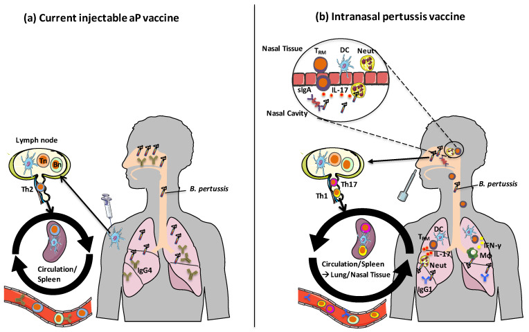
Immunization with current acellular pertussis (aP) vaccines protects against severe pertussis, but immunity wanes rapidly after vaccination and these vaccines do not prevent nasal colonization with Bordetella pertussis. Studies in mouse and baboon models have demonstrated that Th1 and Th17 responses are integral to protective immunity induced by previous infection with B. pertussis and immunization with whole cell pertussis (wP) vaccines. Mucosal Th17 cells, IL-17 and secretory IgA (sIgA) are particularly important in generating sustained sterilizing immunity in the nasal cavity. Current aP vaccines induce potent IgG and Th2-skewed T cell responses but are less effective at generating Th1 and Th17 responses and fail to prime respiratory tissue-resident memory T (TRM) cells, that maintain long-term immunity at mucosal sites. In contrast, a live attenuated pertussis vaccine, pertussis outer membrane vesicle (OMV) vaccines or aP vaccines formulated with novel adjuvants do induce cellular immune responses in the respiratory tract, especially when delivered by the intranasal route. An increased understanding of the mechanisms of sustained protective immunity, especially the role of respiratory TRM cells, will facilitate the development of next generation pertussis vaccines that not only protect against pertussis disease, but prevent nasal colonization and transmission of B. pertussis.

Keywords: Bordetella pertussis; T cells; Th1 cells; Th17 cells; memory T cells; pertussis vaccine.


Conflict of interest statement

Kingston Mills is inventor on a patent application on B. pertussis-derived TLR2 agonists as novel adjuvants and has collaborative research funding from and acts as consultant to Vaccine manufacturers. The funders had no role in the writing of the manuscript or in the decision to publish this review.

Figures

Figure 1
Figure 1
Mechanisms of action of current and future pertussis vaccines. (a) Following immunization with current injectable aP vaccines formulated with alum, DCs at the site of injection take up antigens and migrate to draining lymph nodes where they activate differentiation of Th2 cells from naive T cells. Memory Th2 cells circulate, but do not home to respiratory tissues. IgG4 (IgG1 in mice) is produced by antigen-specific B cells, which protects against toxin-mediated pertussis disease but does not protect against nasal colonization with B. pertussis. (b) Following intranasal immunization with aP vaccines and novel adjuvants, pertussis OMV vaccines or live attenuated pertussis vaccines, local DCs are activated in the respiratory tract. These DCs migrate to draining lymph nodes and activate differentiation of naïve T cells into Th1 and Th17 cells, which have homing properties that facilitate migration to respiratory tissues. Th1 and Th17-type respiratory TRM cells produce IFN-γ and IL-17, which promote recruitment of macrophages and neutrophil to the lungs and nasal mucosae. Intranasal immunization with these vaccines also leads to B cell activation and the production of IgG1 (IgG2a/c in mice) antibodies as well as sIgA. Images from Servier Medical Art (www.smart.servier.com). Tn: naïve T cell, Bn: naïve B cell, TRM: tissue-resident memory T cell, DC: Dendritic cell, sIgA: secretory immunoglobulin-A, MΦ: macrophage.
Figure 2
Figure 2
Mechanisms of action of novel adjuvants under investigation in experimental aP vaccines. Activation of signaling pathways in APC by various adjuvants can be harnessed to generate Th1/Th17 polarizing conditions following immunization. These include the TLR2 agonist LP1569 and the TLR4 agonists MPL, LpxL1 and LpxL2 which are synthetic analogues of LPS. SMIP7.10 and CpG ligate the endosomal TLRs, TLR7 and TLR 9 respectively. TLR 2, 4, 7 and 9 signal through the adaptor molecule MyD88, with TLR2 and TLR4 requiring the bridging adaptor Mal to activate MyD88-mediated signaling. MyD88 signaling results in downstream activation of NFκB and MAPKs. MAPKs activate transcription factors such as AP-1. NFκB and AP-1 translocate to the nucleus leading to the transcription of pro-inflammatory and T cell-polarizing cytokines, chemokines, MHC class II and costimulatory molecules. The CLR, Mincle, is ligated by the TDB component (shown in red) of liposomes, which signals via SYK, and subsequently activates CARD9-Bcl10-Malt signalosome and NFκB. c-di-GMP activates the intracellular DNA sensor, STING, located at the ER, and complexed with TBK1 translocates to perinuclear regions, activating NFκB and IRF3. LP-GMP, which combines LP1569 and c-di-GMP adjuvants, signals through TLR2 and STING respectively, resulting in synergistic induction of Th1/Th17 responses. Block arrows denote activation and dashed arrows denote translocation of transcription factors to the nucleus. Images from Servier Medical Art (www.smart.servier.com). APC: antigen presenting cell, TLR: Toll-like receptor, Th: T helper cell, MPL: Monophosphoryl lipid A, LPS: Lipopolysaccharide, MyD88: Myeloid differentiation primary response gene 88, Mal: MyD88 adaptor-like, NF-κB: Nuclear Factor kappa B, MAPK: Mitogen-activated tyrosine kinase, TDB: α,α trehalose 6,6’-dibehenate, SYK: Spleen tyrosine kinase, CARD9: Caspase recruitment domain family member 9, Bcl10: B-cell lymphoma 10, Malt1: Mucosa-associated lymphoid tissue 1, AP1: Activator protein 1, IRF3: Interferon response factor 3, STING: Stimulator of interferon genes, TBK1: TANK-binding kinase 1, MHC Class II: Major histocompatibility complex class II, Costim: costimulatory molecules.

References

    1. Mattoo S., Cherry J.D. Molecular pathogenesis, epidemiology, and clinical manifestations of respiratory infections due to Bordetella pertussis and other Bordetella subspecies. Clin. Microbiol. Rev. 2005;18:326–382. doi: 10.1128/CMR.18.2.326-382.2005. - DOI - PMC - PubMed
    1. Hewlett E.L., Burns D.L., Cotter P.A., Harvill E.T., Merkel T.J., Quinn C.P., Stibitz E.S. Pertussis Pathogenesis—What We Know and What We Don’t Know. J. Infect. Dis. 2014;209:982–985. doi: 10.1093/infdis/jit639. - DOI - PMC - PubMed
    1. Janda W.M., Santos E., Stevens J., Celig D., Terrile L., Schreckenberger P.C. Unexpected isolation of Bordetella pertussis from a blood culture. J. Clin. Microbiol. 1994;32:2851–2853. doi: 10.1128/JCM.32.11.2851-2853.1994. - DOI - PMC - PubMed
    1. Cody C.L., Baraff L.J., Cherry J.D., Marcy S.M., Manclark C.R. Nature and Rates of Adverse Reactions Associated with DTP and DT Immunizations in Infants and Children. Pediatrics. 1981;68:650–660. - PubMed
    1. Decker M.D., Edwards K.M., Steinhoff M.C., Rennels M.B., Pichichero M.E., Englund J.A., Anderson E.L., Deloria M.A., Reed G.F. Comparison of 13 acellular pertussis vaccines: Adverse reactions. Pediatrics. 1995;96:557–566. - PubMed

LinkOut - more resources