Review
. 2020 Oct 21;8(10):441.
doi: 10.3390/biomedicines8100441.

Application of 3D Bioprinting Technologies to the Management and Treatment of Diabetic Foot Ulcers

Affiliations
Review

Application of 3D Bioprinting Technologies to the Management and Treatment of Diabetic Foot Ulcers

Chew Teng Tan et al. Biomedicines. .

Abstract

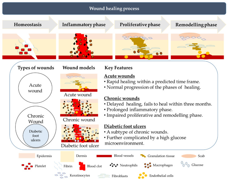
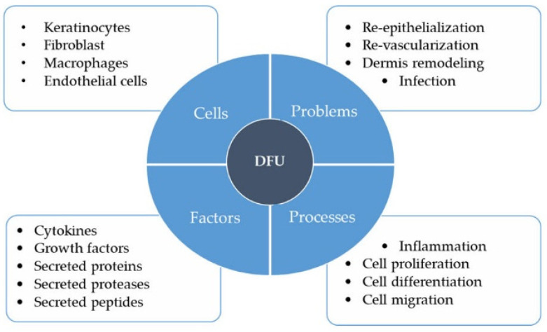
Diabetes mellitus (DM) is a chronic metabolic disease with increasing prevalence worldwide. Diabetic foot ulcers (DFUs) are a serious complication of DM. It is estimated that 15-25% of DM patients develop DFU at least once in their lifetime. The lack of effective wound dressings and targeted therapy for DFUs often results in prolonged hospitalization and amputations. As the incidence of DM is projected to rise, the demand for specialized DFU wound management will continue to increase. Hence, it is of great interest to improve and develop effective DFU-specific wound dressings and therapies. In the last decade, 3D bioprinting technology has made a great contribution to the healthcare sector, with the development of personalized prosthetics, implants, and bioengineered tissues. In this review, we discuss the challenges faced in DFU wound management and how 3D bioprinting technology can be applied to advance current treatment methods, such as biomanufacturing of composite 3D human skin substitutes for skin grafting and the development of DFU-appropriate wound dressings. Future co-development of 3D bioprinting technologies with novel treatment approaches to mitigate DFU-specific pathophysiological challenges will be key to limiting the healthcare burden associated with the increasing prevalence of DM.

Keywords: 3D bioprinting; diabetic foot ulcers; wound healing.


Conflict of interest statement

The authors declare no conflict of interest. The funders had no role in the design of the study; in the collection, analyses, or interpretation of data; in the writing of the manuscript, or in the decision to publish the results.

Figures

Figure 1
Figure 1
An illustration of the different phases of cutaneous wound healing and the key differences between acute wounds, chronic and diabetic foot ulcers. In normal wounds, healing is initiated by hemostasis to minimize further blood loss, followed by the inflammatory phase to prevent microbial infection. In the proliferative phase, granulation tissue is formed accompanied by the proliferation of epidermal keratinocytes and dermal fibroblasts, facilitating the final phase of tissue remodeling to restore the structure and function of the skin. Wounds can be classified as acute and chronic. Acute wounds heal rapidly within a short time frame with normal progression of the phases of healing, while chronic wounds take more than three months to heal due to a prolonged inflammatory phase and impaired proliferative and remodeling phases. Diabetic foot ulcers exhibit similar characteristics as other chronic wounds, but the healing of diabetic foot ulcers (DFUs) is further complicated by a sustained high glucose microenvironment.
Figure 2
Figure 2
Major factors that contribute to the pathophysiological status of diabetic foot ulcers (DFUs). Keratinocytes, fibroblasts, macrophages, and endothelial cells play important roles in wound healing such as re-epithelialization, revascularization, dermal remodeling, and infection prevention. These functions may be disrupted in the high glucose microenvironment in the skin of diabetic patients, leading to impaired cell proliferation, migration, and differentiation, and prolonged or exaggerated inflammation. Altered cytokine, growth factor, protein, protease, and peptide secretion profiles of different cell types in the skin in response to high glucose microenvironment further define the pathophysiological status of DFUs.

References

    1. Raghav A., Khan Z.A., Labala R.K., Ahmad J., Noor S., Mishra B.K. Financial Burden of Diabetic Foot Ulcers to World: A Progressive Topic to Discuss Always. Ther. Adv. Endocrinol. Metab. 2018;9:29–31. doi: 10.1177/2042018817744513. - DOI - PMC - PubMed
    1. Zhang P., Lu J., Jing Y., Tang S., Zhu D., Bi Y. Global Epidemiology of Diabetic Foot Ulceration: A Systematic Review and Meta-Analysis. Ann. Med. 2017;49:106–116. doi: 10.1080/07853890.2016.1231932. - DOI - PubMed
    1. Hicks C.W., Selvarajah S., Mathioudakis N., Sherman R.E., Hines K.F., Black J.H., Abularrage C.J. Burden of Infected Diabetic Foot Ulcers on Hospital Admissions and Costs. Ann. Vasc. Surg. 2016;33:149–158. doi: 10.1016/j.avsg.2015.11.025. - DOI - PMC - PubMed
    1. Jiang Y., Ran X., Jia L., Yang C., Wang P., Ma J., Chen B., Yu Y., Feng B., Chen L., et al. Epidemiology of Type 2 Diabetic Foot Problems and Predictive Factors for Amputation in China. Int. J. Low. Extrem. Wounds. 2015;14:19–27. doi: 10.1177/1534734614564867. - DOI - PubMed
    1. Falanga V. Wound Healing and Its Impairment in the Diabetic Foot. Lancet. 2005;366:1736–1743. doi: 10.1016/S0140-6736(05)67700-8. - DOI - PubMed