. 2020 Oct 21;8(10):442.
doi: 10.3390/biomedicines8100442.

Treatment with Luteolin Improves Lipopolysaccharide-Induced Periodontal Diseases in Rats

Affiliations

Treatment with Luteolin Improves Lipopolysaccharide-Induced Periodontal Diseases in Rats

Giovanna Casili et al. Biomedicines. .

Abstract

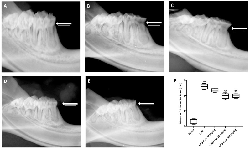
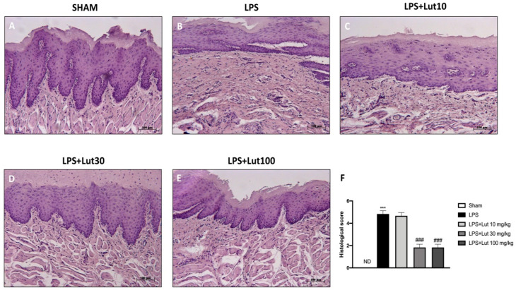
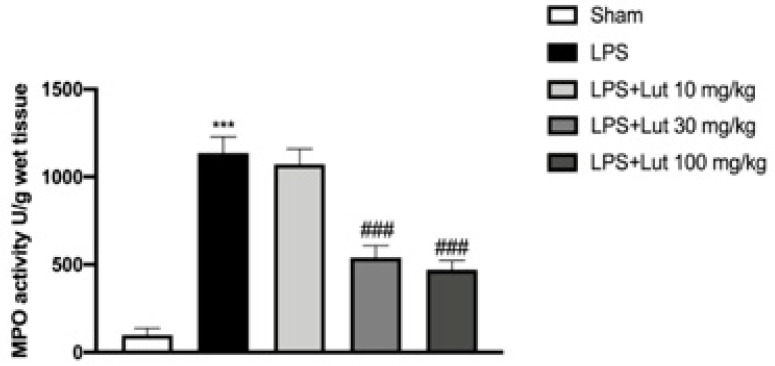
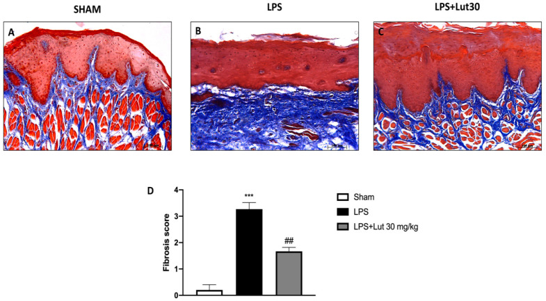
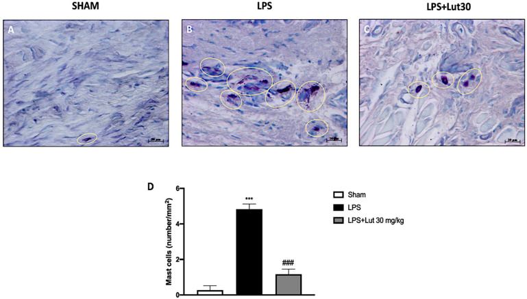
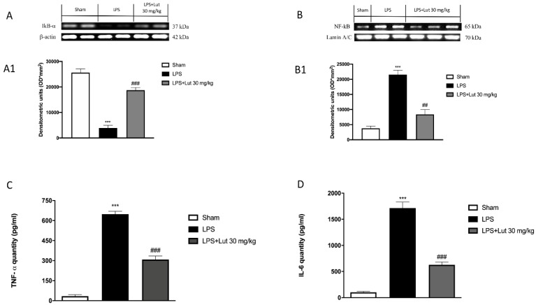
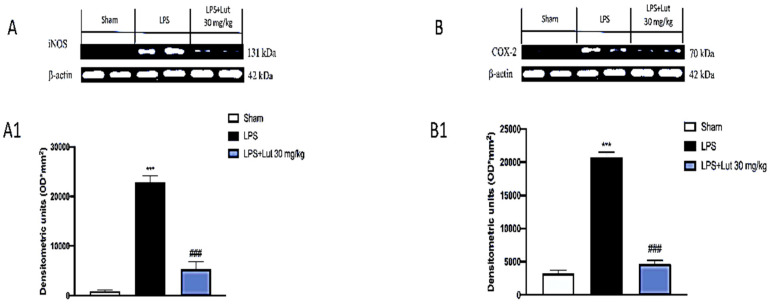
Periodontitis is a dental disease that produces the progressive destruction of the bone surrounding the tooth. Especially, lipopolysaccharide (LPS) is involved in the deterioration of the alveolar bone, inducing the release of pro-inflammatory mediators, which cause periodontal tissue inflammation. Luteolin (Lut), a molecule of natural origin present in a large variety of fruits and vegetables, possess beneficial properties for human health. On this basis, we investigated the anti-inflammatory properties of Lut in a model of periodontitis induced by LPS in rats. Animal model predicted a single intragingival injection of LPS (10 μg/μL) derived from Salmonella typhimurium. Lut administration, was performed daily at different doses (10, 30, and 100 mg/kg, orally), starting from 1 h after the injection of LPS. After 14 days, the animals were sacrificed, and their gums were processed for biochemical analysis and histological examinations. Results showed that Lut (30 and 100 mg/kg) was equally able to reduce alveolar bone loss, tissue damage, and neutrophilic infiltration. Moreover, Lut treatment reduced the concentration of collagen fibers, mast cells degranulation, and NF-κB activation, as well as the presence of pro-inflammatory enzymes and cytokines. Therefore, Lut implementation could represent valid support in the pharmacological strategy for periodontitis, thus improving the well-being of the oral cavity.

Keywords: anti-inflammatory; dental diseases; flavonoids; lipopolysaccharide; luteolin; periodontitis.


Conflict of interest statement

The authors declare no conflict of interest.

Figures

Figure 1
Figure 1
Luteolin (Lut) administration decreased the alveolar bone distance. Fourteen days after the lipopolysaccharide (LPS) injection, the X-rays of the rats LPS-induce periodontitis showed a greater distance from the cement–enamel junction (CEJ) to the bone (B,F), compared to the sham group rats (A,F). Lut 30 mg/kg (D,F) and 100 mg/kg (E,F) were effective in reducing this distance, as opposed to treatment with Lut 10 mg/kg which proved ineffective (C,F). Values reported in the box plot are expressed as mean ± SEM of 10 rats for each group. *** p < 0.001 vs. sham; ## p < 0.01 vs. LPS group.
Figure 2
Figure 2
Lut administration reduced histological damage LPS-induced periodontitis. No histological damage was found in the gingivomucosal tissues from sham-group rats (A), see histological score (F). Extensive damage, accompanied by edema, tissue injury, and inflammatory cells infiltration, was assessed in LPS rats (B), see histological score (F). The administration of Lut 30 mg/kg (D), see histological score (F) and 100 mg/kg (E), see histological score (F), reduced LPS tissue damage as opposed to treatment with Lut 10 mg/kg which proved ineffective (C), see histological score (F). Data are representative of at least three independent experiments; One-Way ANOVA test. *** p < 0.001 vs. sham; ### p < 0.001 vs. LPS group. ND = not detectable.
Figure 3
Figure 3
Lut treatment moderated neutrophilic infiltration. An increase in MPO levels was found in LPS-induced periodontitis rats, compared to the sham group. Only the 30 and 100 mg/kg dosages proved to be equally effective in reducing MPO levels. One-Way ANOVA test.*** p < 0.001 vs. sham; ### p < 0.001 vs. LPS group.
Figure 4
Figure 4
Lut treatment reduced collagen formation. Masson’s trichrome stain presented an increase in the concentration of collagen fibers in gingivomucosal tissues in the LPS group (B,D), compared to the control group (A,D). Lut 30 mg/kg significantly attenuated collagen formation (C,D). One-Way ANOVA test.*** p < 0.001 vs. sham; ## p < 0.01 vs. LPS group.
Figure 5
Figure 5
Effects of Lut treatment on mast cell degranulation. Toluidine blue staining allowed mast cell count. In gingivomucosal tissues of rats belonging to the LPS group, an increased number of mast cells was identified (B,D), as compared to control group (A,D). Lut 30 mg/kg considerably reduced mast cell infiltration (C,D). Yellow circles indicate the mast cells degranulated appeared in the tissue. One-Way ANOVA test.*** p < 0.001 vs. sham; ### p < 0.001 vs. LPS group.
Figure 6
Figure 6
Effects of Lut treatment on NF-κB pathway and pro-inflammatory cytokines. Western blot analysis demonstrated an increase in the degradation of IκB-α in the LPS group (A) and densitometric analysis (A1) compared to the sham group (A) and densitometric analysis (A1). Lut 30 mg/kg has proven to be truly effective in restoring these levels (A) and densitometric analysis (A1). NF-κB was significantly increased in the LPS group (B) and densitometric analysis (B1), as compared to the sham group (B) and densitometric analysis (B1); Lut 30 mg/kg effectively decreased the levels of NF-κB (B) and densitometric analysis (B1). The levels of TNF-α (C) and IL-6 (D) were significantly increased in rats injected with LPS. The increases in levels of TNF-α and IL-6 were significantly attenuated in rats administrated with Lut 30 mg/kg. Data are representative of at least three independent experiments. One-Way ANOVA test.*** p < 0.001 vs. sham; ### p < 0.001 vs. LPS group. ## p < 0.01 vs. LPS group.
Figure 7
Figure 7
Effects of Lut treatment on pro-inflammatory enzymes. Western blot analysis of iNOS (A) and densitometric analysis (A1) and COX-2 (B) and densitometric analysis (B1) revealed minimal levels in the sham group that conversely were increased in the LPS group. Treatment with Lut 30 mg/kg proved effective to reduce COX-2 and iNOS expressions. Data are representative of at least three independent experiments. One-Way ANOVA test. *** p < 0.001 vs. sham; ### p < 0.001 vs. LPS group.

Similar articles

Cited by

References

    1. Gasner N.S., Schure R.S. Periodontal Disease. StatPearls; Treasure Island, FL, USA: 2020.
    1. Van Dyke T.E., Sima C. Understanding resolution of inflammation in periodontal diseases: Is chronic inflammatory periodontitis a failure to resolve? Periodontol. 2000. 2020;82:205–213. doi: 10.1111/prd.12317. - DOI - PubMed
    1. Nazir M.A. Prevalence of periodontal disease, its association with systemic diseases and prevention. Int. J. Health Sci. (Qassim) 2017;11:72–80. - PMC - PubMed
    1. Riley M. Incorrect nutrition as a risk factor for periodontal disease. Alpha Omegan. 2007;100:85–88. doi: 10.1016/j.aodf.2007.04.008. - DOI - PubMed
    1. Leite F.R.M., Nascimento G.G., Scheutz F., Lopez R. Effect of Smoking on Periodontitis: A Systematic Review and Meta-regression. Am. J. Prev. Med. 2018;54:831–841. doi: 10.1016/j.amepre.2018.02.014. - DOI - PubMed

LinkOut - more resources