. 2020 Oct 21;12(10):1197.
doi: 10.3390/v12101197.

Maturation of Pseudo-Nucleus Compartment in P. aeruginosa, Infected with Giant phiKZ Phage

Affiliations

Maturation of Pseudo-Nucleus Compartment in P. aeruginosa, Infected with Giant phiKZ Phage

Yana A Danilova et al. Viruses. .

Abstract

The giant phiKZ phage infection induces the appearance of a pseudo-nucleus inside the bacterial cytoplasm. Here, we used RT-PCR, fluorescent in situ hybridization (FISH), electron tomography, and analytical electron microscopy to study the morphology of this unique nucleus-like shell and to demonstrate the distribution of phiKZ and bacterial DNA in infected Pseudomonas aeruginosa cells. The maturation of the pseudo-nucleus was traced in short intervals for 40 min after infection and revealed the continuous spatial separation of the phage and host DNA. Immediately after ejection, phage DNA was located inside the newly-identified round compartments; at a later infection stage, it was replicated inside the pseudo-nucleus; in the mature pseudo-nucleus, a saturated internal network of filaments was observed. This network consisted of DNA bundles in complex with DNA-binding proteins. On the other hand, the bacterial nucleoid underwent significant rearrangements during phage infection, yet the host DNA did not completely degrade until at least 40 min after phage application. Energy dispersive x-ray spectroscopy (EDX) analysis revealed that, during the infection, the sulfur content in the bacterial cytoplasm increased, which suggests an increase of methionine-rich DNA-binding protein synthesis, whose role is to protect the bacterial DNA from stress caused by infection.

Keywords: Pseudomonas aeruginosa; analytical electron microscopy; electron tomography; fluorescent in situ hybridization; giant phage; nucleoid; phiKZ; pseudo-nucleus; stress response.


Conflict of interest statement

The authors declare no conflict of interest.

Figures
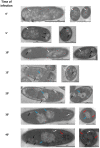
Figure 1
Figure 1
Time-course of the phiKZ infection and pseudo-nucleus maturation in P. aeruginosa cells. Left column—minutes after infection. White arrows—bacterial nucleoid; black arrows—round compartments (RC); blue arrows—pseudo-nuclei at different stages of maturation; red arrows—new phage capsids.
Figure 2
Figure 2
Phage and bacterial DNA distribution in control and infected P. aeruginosa cells. TEM image (a,d,g,j); HAADF image (b,e,h,k), and electron energy loss spectroscopy (EELS) Phosphorus distribution maps through the pseudo-nucleus (c,f,i,l). Phosphorous (P) signal was mapped after background subtraction and multiple scattering correction with Fourier-ratio deconvolution. Arrows are pointing to the DNA-containing structures; arrowheads—to the areas, which do not contain the P signal; yellow outlines in (g,h,i) roughly mark the pseudo-nucleus border.
Figure 3
Figure 3
EDX spectra of P. aeruginosa control cells, 15 and 30 min after phiKZ infection. (a) Colored outlines mark the areas of bacteria subjected for EDX analysis. Bars – 0.5 um.; (b) Superimposed EDX spectra from the selected areas, marked in (a), normalized to the C peak. Peaks are labeled as follows: P—phosphorus; S—sulfur; Cl—chloride; Ca—calcium.
Figure 4
Figure 4
Analysis of the total DNA content from phiKZ-infected cells. (a) agarose gel-electrophoresis. Above the gel, the time points are mentioned, where ‘0′ is a non-infected cell, and the last line is the total DNA from phiKZ virions. PAO1/SmaI—bacterial DNA cleaved by SmaI REase. (b) real-time PCR results. Blue diamonds-bacterial DNA, orange diamonds-phage DNA.
Figure 5
Figure 5
Analysis of bacterial and phage DNA at different times before and after infection of P. aeruginosa cells by the phiKZ phage. Results of fluorescent in situ hybridization (FISH) (a) with bacterial Cy5-probes, (b) with phage Cy5-probes. DAPI signal—cyan color, Cy5 signal is colored magenta. Bar—1 um. Time points are indicated on the left.
Figure 6
Figure 6
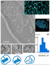
Electron tomography of a phiKZ infected P. aeruginosa cell. (a) A central slice through the tomogram of an infected cell (30 min after infection). Bar—200 nm. P-N, pseudo-nucleus. Insert—3D representation of the P-N network. White arrows are pointing to DNA strands, arrowheads—to phage DNA-binding proteins. The phage capsids and corresponding gallery images are labeled with rectangles one and two. Bar size—100 nm. Below each gallery image is the matching schematic of the capsid with the inner body (IB) marked with a black arrow; rectangle three and the corresponding gallery image represent a RC. The electron-dense material is clearly visible inside (black arrows). (b) The subtomogram representation of the pseudo-nucleus network. Each blue strand represents one DNA helix. Bar—50 nm. (c) The distribution (in %) of the average DNA strand’s thickness in the pseudo-nucleus network.

References

    1. Hertveldt K., Lavigne R., Pleteneva E., Sernova N., Kurochkina L., Korchevskii R., Robben J., Mesyanzhinov V., Krylov V.N., Volckaert G. Genome comparison of pseudomonas aeruginosa large phages. J. Mol. Biol. 2005;354:536–545. doi: 10.1016/j.jmb.2005.08.075. - DOI - PubMed
    1. Mesyanzhinov V.V., Robben J., Grymonprez B., Kostyuchenko V.A., Bourkaltseva M.V., Sykilinda N.N., Krylov V.N., Volckaert G. The genome of bacteriophage φKZ of Pseudomonas aeruginosa. J. Mol. Biol. 2002;317:1–19. doi: 10.1006/jmbi.2001.5396. - DOI - PubMed
    1. Ceyssens P.-J., Minakhin L., Van den Bossche A., Yakunina M., Klimuk E., Blasdel B., De Smet J., Noben J.-P., Blasi U., Severinov K., et al. Development of giant bacteriophage ϕKZ is independent of the host transcription apparatus. J. Virol. 2014;88:10501–10510. doi: 10.1128/JVI.01347-14. - DOI - PMC - PubMed
    1. Yakunina M., Artamonova T., Borukhov S., Makarova K.S., Severinov K., Minakhin L. A non-canonical multisubunit RNA polymerase encoded by a giant bacteriophage. Nucleic Acids Res. 2015:gkv1095. doi: 10.1093/nar/gkv1095. - DOI - PMC - PubMed
    1. Lecoutere E., Ceyssens P.-J., Miroshnikov K.A., Mesyanzhinov V.V., Krylov V.N., Noben J.-P., Robben J., Hertveldt K., Volckaert G., Lavigne R. Identification and comparative analysis of the structural proteomes of ϕKZ and EL, two giant Pseudomonas aeruginosa bacteriophages. Proteomics. 2009;9:3215–3219. doi: 10.1002/pmic.200800727. - DOI - PubMed

Publication types

MeSH terms

LinkOut - more resources