. 2020 Oct 21;12(10):1202.
doi: 10.3390/v12101202.

How HIV-1 Integrase Associates with Human Mitochondrial Lysyl-tRNA Synthetase

Affiliations

How HIV-1 Integrase Associates with Human Mitochondrial Lysyl-tRNA Synthetase

Xaysongkhame Phongsavanh et al. Viruses. .

Abstract

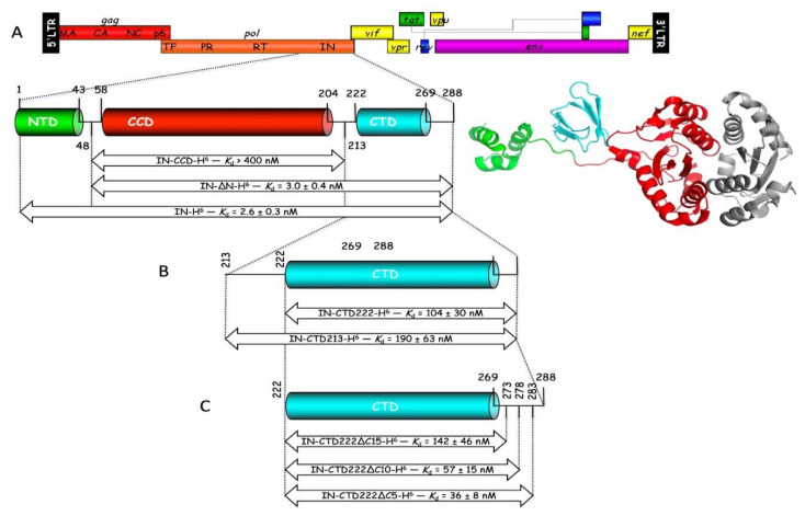
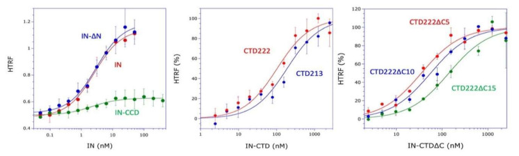
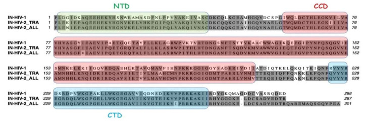
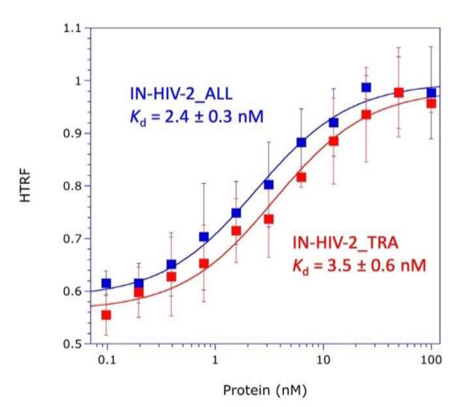
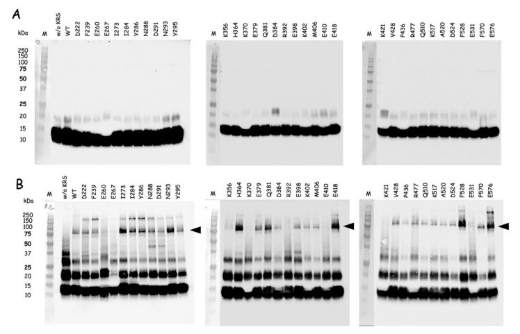
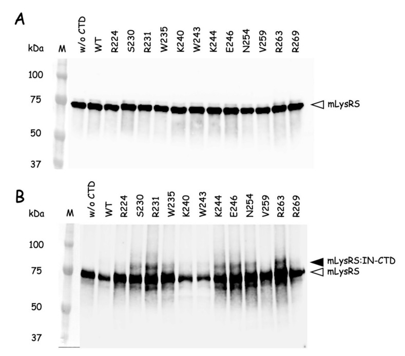
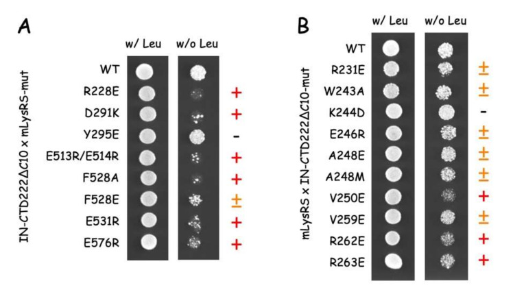
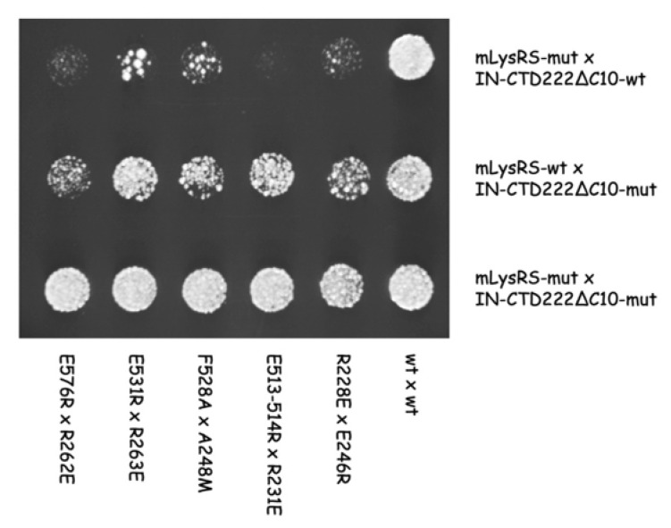
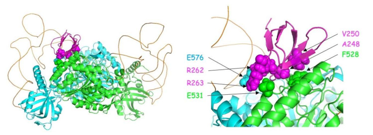
Replication of human immunodeficiency virus type 1 (HIV-1) requires the packaging of tRNALys,3 from the host cell into the new viral particles. The GagPol viral polyprotein precursor associates with mitochondrial lysyl-tRNA synthetase (mLysRS) in a complex with tRNALys, an essential step to initiate reverse transcription in the virions. The C-terminal integrase moiety of GagPol is essential for its association with mLysRS. We show that integrases from HIV-1 and HIV-2 bind mLysRS with the same efficiency. In this work, we have undertaken to probe the three-dimensional (3D) architecture of the complex of integrase with mLysRS. We first established that the C-terminal domain (CTD) of integrase is the major interacting domain with mLysRS. Using the pBpa-photo crosslinking approach, inter-protein cross-links were observed involving amino acid residues located at the surface of the catalytic domain of mLysRS and of the CTD of integrase. In parallel, using molecular docking simulation, a single structural model of complex was found to outscore other alternative conformations. Consistent with crosslinking experiments, this structural model was further probed experimentally. Five compensatory mutations in the two partners were successfully designed which supports the validity of the model. The complex highlights that binding of integrase could stabilize the tRNALys:mLysRS interaction.

Keywords: 3D model; HIV-1; integrase; mitochondrial lysyl-tRNA synthetase; tRNA packaging complex.


Conflict of interest statement

The authors declare no conflict of interest. The funders had no role in the design of the study; in the collection, analyses, or interpretation of data; in the writing of the manuscript, or in the decision to publish the results.

Figures

Figure 1
Figure 1
The three-domain structure of integrase from HIV-1, and the constructs of IN used in this study. (A): The complete genome of HIV-1 is shown. Integrase (IN) is encoded in the very C-terminal region of the Pol domain from the GagPol polyprotein precursor. The schematic view and three-dimensional (3D) representation of IN are shown. The domain structure of IN is formed by the N-terminal domain (NTD, in green) and the C-terminal domain (CTD, in cyan) connected to the catalytic core domain (CCD, in red). The second CCD of the IN dimer visible in the cryo-EM structure is shown in grey [22]. Full-length integrase with a C-terminal His-tag (IN-H6), with a deletion of the NTD (IN-∆N), or with a deletion of the NTD and the CTD (IN-CCD) were constructed. (B) Constructs of the CTD on IN starting at residues 213 (IN-CTD213) or 222 (IN-CTD222) are shown. (C) Constructs of IN-CTD222 with a deletion of 5, 10, or 15 C-terminal residues. These derivatives of IN were tested for their association with mLysRS (Kd determined as reported in Figure 2 are indicated; values are the means ± SEM of three independent experiments).
Figure 2
Figure 2
Association of the derivatives of IN with mLysRS. The binding affinities of mLysRS to IN constructs were determined in an HTRF assay using 1.5 nM of HA-tagged mLysRS and increasing concentrations of His-tagged IN derivatives, expressed as dimer (IN, IN-∆N, IN-CCD) or monomer (CTD213, CTD222, CTD222∆C5, CTD222∆C10, CTD222∆C15) concentrations. Experimental values (symbols) were fit (curves) to a binding equation assuming that one molecule of IN binds one molecule of mLysRS. The binding constants and the associated standard deviations (n = 3) are indicated in Figure 1.
Figure 3
Figure 3
Sequence alignment of integrases from HIV-1 and HIV-2. Integrase from HIV-1 of pNL4-3 (IN_HIV-1) and two integrase species from two HIV-2 isolates (IN-HIV-2_TRA and IN-HIV-2_ALL) are aligned. Residues conserved in at least two sequences are outlined in dark grey, similar residues are in light grey. The three structural domains (NTD, CCD, and CTD) are indicated according to Figure 1A.
Figure 4
Figure 4
Association of IN from HIV-2 with mLysRS. The binding affinities of mLysRS to IN-HIV-2_TRA and IN-HIV-2_ALL were determined in an HTRF assay using 1.5 nM of HA-tagged mLysRS and increasing concentrations of His-tagged IN-HIV-2 species, expressed as dimer concentrations. Experimental values (symbols) were fit (curves) to a binding equation assuming that one molecule of IN binds one molecule of mLysRS. The binding constants and the associated standard deviations (n = 3) are indicated.
Figure 5
Figure 5
Cross-linking of the pBpa-containing mLysRS variants with IN-CTD222. Wild-type mLysRS (WT) or mLysRS containing pBpa inserted at different positions (D222 to E576) were incubated at a dimer concentration of 85 nM with IN-CTD222 (monomer concentration of 1.12 µM) and exposed to UV light at 365 nm for 70 min. Samples recovered before (A) or after (B) exposure to UV were analyzed by SDS-PAGE and western blotting using anti-IN-CTD antibodies. The lanes containing the stained size-marker are added on the left (in kDa). The migration level corresponding to an expected cross-linked polypeptide (79 kDa) containing one monomer of IN-CTD222 and one monomer of mLysRS is indicated by a black arrowhead. This experiment is the representative of two independent experiments.
Figure 6
Figure 6
Cross-linking of the pBpa-containing IN-CTD222 variants with mLysRS. Wild-type IN-CTD222 (WT) or IN-CTD222 containing pBpa inserted at different positions (R224 to R269) were incubated at a monomer concentration of 1 µM with mLysRS (dimer concentration of 250 nM) and exposed to UV light at 365 nm for 90 min. Samples recovered before (A) or after (B) exposure to UV were analyzed by SDS-PAGE and western blotting using anti-LysRS antibodies. The lanes containing the stained size-marker are added on the left (in kDa). The migration level of mLysRS (68 kDa) and of the polypeptide corresponding to an expected cross-linked species containing one monomer of IN-CTD222 and one monomer of mLysRS (79 kDa) are indicated by a white and black arrowhead, respectively. This experiment is the representative of two independent experiments.
Figure 7
Figure 7
3D model of the mLysRS:IN-CTD complex obtained by molecular docking. The two subunits of LysRS (3BJU) are in cyan and green, the two tRNA molecules in orange, the monomer of IN-CTD (5U1C) in magenta. Only one IN-CTD is shown. The two tRNA molecules were anchored according to the crystal structure of the complex of yeast aspartyl-tRNA synthetase with two tRNA molecules (1ASY). The coordinates of the 3D model can be accessed at https://modelarchive.org/doi/10.5452/ma-bxirn.
Figure 8
Figure 8
Two-hybrid analysis of mLysRS:IN-CTD interaction. The CTD of HIV-1 integrase (IN-CTD222∆C10) was expressed fused to LexA in pEG202, and mLysRS was expressed fused to the B42 transcription activator under the control of a galactose-inducible promoter in pJG4-5. (A) Wild-type IN-CTD222∆C10 was co-expressed with wild-type or mutants of mLysRS. (B) Wild-type mLysRS was co-expressed with wild-type or mutants of IN-CTD222∆C10. Yeast cells were grown in a galactose-containing medium in the presence (w/ Leu) or in the absence of leucine (w/o Leu). When the two proteins interact, yeast cells are able to grow in a medium lacking leucine. Mutations have either no effect (-), or have a moderate (±) or strong effect (+) on yeast growth.
Figure 9
Figure 9
Two-hybrid analysis of compensatory mutations in mLysRS and IN-CTD. Mutants of the CTD of HIV-1 integrase (IN-CTD222∆C10) were co-expressed with mutants of mLysRS in the two-hybrid system, as described in the legend of Figure 8. Cells co-expressing wild-type or mutants of mLysRS with wild-type IN-CTD222∆C10 (first row), wild-type mLysRS with wild-type or mutants of IN-CTD222∆C10 (second row), or a mutant of mLysRS with a mutant of IN-CTD222∆C10 (third row) were grown in a galactose-containing medium in the absence of leucine. Yeast cells expressing compensatory mutations were growing much faster than cells expressing a single of these mutations and were able to grow similarly to cells expressing the wild-type proteins.
Figure 10
Figure 10
3D model of the platform for association of IN-CTD with mLysRS. Left: The structural model shows a dimer of human LysRS in association with a monomer of the CTD of integrase from HIV-1. The two monomers of LysRS (3BJU) are in cyan and green, the two tRNA molecules in orange, the monomer of IN-CTD (5U1C) in magenta. The residues that build the platform for the assembly of the complex are represented by spheres. Docking of the two tRNA molecules was performed using the crystal structure of yeast aspartyl-tRNA synthetase complexed with two tRNA molecules (1ASY). Right: Zoom of the IN-CTD:LysRS interaction platform. Residues from the platform are labeled and colored according to the main chains.

References

    1. Jiang M., Mak J., Ladha A., Cohen E., Klein M., Rovinski B., Kleiman L. Identification of tRNAs incorporated into wild-type and mutant human immunodeficiency virus type-1. J. Virol. 1993;67:3246–3253. doi: 10.1128/JVI.67.6.3246-3253.1993. - DOI - PMC - PubMed
    1. Pavon-Eternod M., Wei M., Pan T., Kleiman L. Profiling non-lysyl tRNAs in HIV-1. RNA. 2010;16:267–273. doi: 10.1261/rna.1928110. - DOI - PMC - PubMed
    1. Abbink T.E., Berkhout B. HIV-1 reverse transcription: Close encounters between the viral genome and a cellular tRNA. Adv. Pharmacol. 2007;55:99–135. - PubMed
    1. Seif E., Niu M., Kleiman L. In virio SHAPE analysis of tRNA(Lys3) annealing to HIV-1 genomic RNA in wild type and protease-deficient virus. Retrovirology. 2015;12:40. doi: 10.1186/s12977-015-0171-7. - DOI - PMC - PubMed
    1. Gabor J., Cen S., Javanbakht H., Niu M.J., Kleiman L. Effect of altering the tRNA3Lys concentration in human immunodeficiency virus type 1 upon its annealing to viral RNA, GagPol incorporation, and viral infectivity. J. Virol. 2002;76:9096–9102. doi: 10.1128/JVI.76.18.9096-9102.2002. - DOI - PMC - PubMed

Publication types

MeSH terms

Substances

LinkOut - more resources