

Case report: anti-N-Methyl-D-Aspartate receptor encephalitis and bilateral temporal calcifications
- PMID: 33097034
- PMCID: PMC7583296
- DOI: 10.1186/s12883-020-01962-3
Case report: anti-N-Methyl-D-Aspartate receptor encephalitis and bilateral temporal calcifications
Abstract
Background: In this study, we report a case of a young female who was hospitalized for seizures and diagnosed with anti-N-methyl-D-aspartate receptor (NMDAR) encephalitis.
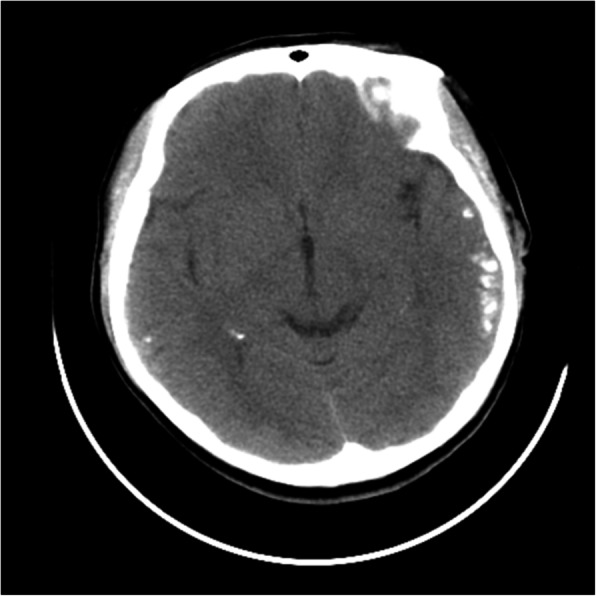
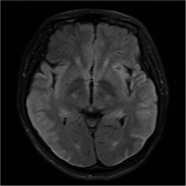
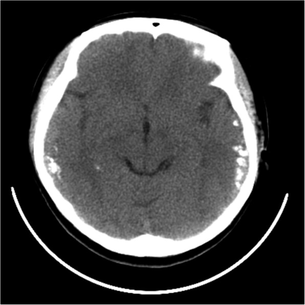
Case presentation: The main feature of this patient was bilateral temporal calcifications detected by routine head computed tomography (CT). The co-existence of anti-NMDAR encephalitis and cerebral calcifications has not been reported. We supposed that the patient had an incomplete form of celiac disease (CD), epilepsy and cerebral calcifications syndrome (CEC). The patient's symptoms were alleviated by a series of treatments, and she remained stable during the follow-ups.
Conclusions: Our findings confirm the rarity co-existing anti-NMDAR encephalitis and cerebral calcifications. In future clinical work, we need to elucidate the relationship between anti-NMDAR encephalitis and cerebral calcifications, and the association between anti-NMDAR encephalitis and other co-existing autoimmune disorders.
Keywords: Anti-NMDAR encephalitis; Bilateral temporal calcifications; CEC; Epilepsy.
Conflict of interest statement
None of the authors declared any conflict of interest.
Figures
References
-
- Ferlazzo E, Polidoro S, Gobbi G, Gasparini S, Sueri C, Cianci V, Sofia V, Giuliano L, Giallonardo AT, Di Bonaventura C, et al. Epilepsy, cerebral calcifications, and gluten-related disorders: Are anti-transglutaminase 6 antibodies the missing link? Seizure. 2019;73:17–20. - PubMed
-
- Gobbi G, Ambrosetto P, Zaniboni MG, Lambertini A, Ambrosioni G, Tassinari CA. Celiac disease, posterior cerebral calcifications and epilepsy. Brain Develop. 1992;14(1):23–29. - PubMed
Publication types
MeSH terms
LinkOut - more resources
Full Text Sources
Medical
