Case Reports
. 2020 Oct 23;18(1):271.
doi: 10.1186/s12957-020-02054-9.

Lung metastases from intraductal papillary neoplasm of the bile duct: a case report

Affiliations
Case Reports

Lung metastases from intraductal papillary neoplasm of the bile duct: a case report

Rika Fujino et al. World J Surg Oncol. .

Abstract

Background: Intraductal papillary neoplasm of the bile duct (IPNB) is considered a pre-cancerous biliary lesion and/or an early cancer lesion, although its classification remains unclear. The 2019 revised edition of the World Health Organization Classification of Tumors of the Digestive System proposed type 1 and type 2 as new classification categories, and meta-analyses and/or multi-center cohort studies are beginning to be reported. However, treatment for IPNB recurrence and metastasis remains unclear.

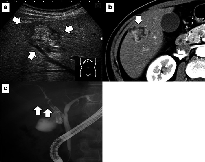
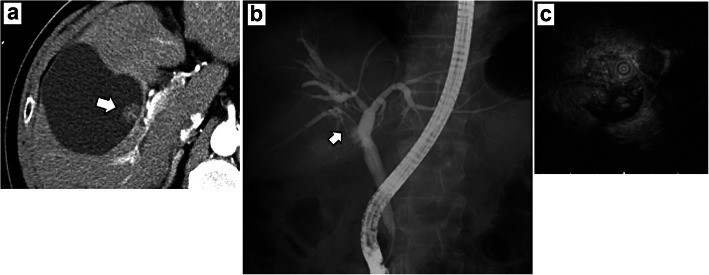
Case presentation: A 60-year-old man who was referred to our hospital after a suspected liver tumor was diagnosed using abdominal ultrasonography. Imaging findings revealed an irregularly shaped tumor in segment 5 (S5) of the liver (size 20 mm). The S5 lesion was suspected as IPNB, and segmentectomy was performed. The pathological findings revealed invasive carcinoma derived from IPNB, and immunohistochemistry revealed positive expression of MUC1, MUC5AC, and MUC6, but negative expression of CDX2 and MUC2. At 9 months after the surgery, computed tomography revealed a tumor in the right bile duct, which was diagnosed as liver recurrence of IPNB, and right hepatectomy was performed. The histopathological findings were the same as for the first resected specimen (i.e., IPNB). At 45 months after the second surgery, computed tomography revealed nodules in both lungs, which were diagnosed as lung metastases from IPNB and resected in two separate procedures. The pathological findings were metastatic carcinoma from IPNB for both lung lesions. The patient is currently alive and undergoing adjuvant chemotherapy (S-1), which was initiated 64 months after the first resection and 12 months after resection of the lung metastases.

Conclusion: We encountered a rare case of lung metastases from IPNB, which were diagnosed immunohistologically. Because IPNB is generally a slow-growing tumor, resection may be feasible for IPNB recurrence and/or metastasis, which may be detected during long-term follow-up. Thus, even if resection is performed for primary IPNB, additional surgical treatment may be feasible in this setting.

Keywords: IPNB; Intraductal papillary neoplasm of the bile duct; Lung metastasis; Re-resection; Recurrence; Surgery.


Conflict of interest statement

The authors declare that they have no competing interests.

Figures

Fig. 1
Fig. 1
Preoperative imaging findings. a Abdominal ultrasonography revealed a hyperechoic tumor (core size of 20 mm) in the intrahepatic area and peripheral to the hypoechoic area (white arrow). The hyperechoic area is even more intense at the posterior aspect of the tumor. b Dynamic computed tomography revealed an unevenly shaped and enhanced tumor (white arrow) in S5 of the liver during the arterial phase. c Endoscopic retrograde cholangiography revealed a filling defect (white arrow) from the origin of the right area to the periphery and severe dilation of the S5 branch of the bile duct
Fig. 2
Fig. 2
Macroscopic and pathological findings from the first resection. a The papillary and nodular tumor in the dilatation of intrahepatic bile duct (white arrow). b Complex papillary or tubular proliferation of atypical columnar or cuboidal cells with focal fine vascular stroma was observed in the dilated intrahepatic bile ducts. c Most of the tumor was composed of high-grade atypical cells that had enlarged nuclei, whereas low-grade dysplasia components were focally observed. d Tumor focally invaded into the stroma of portal area with mucinous trabecular or tubular fashion, although there was no invasion to the liver parenchyma. Immunohistochemistry findings are shown for e MUC1, f MUC2, g MUC5AC, and h MUC6
Fig. 3
Fig. 3
Imaging findings from the liver recurrence. a Dynamic computed tomography revealed an enhanced tumor within a cystic substance during the arterial phase (white arrow). b Endoscopic retrograde cholangiography revealed a filling defect (white arrow). c Intraductal ultrasonography showed a papillary tumor in the dilatation of right hepatic duct
Fig. 4
Fig. 4
Macroscopic and pathological findings from the recurrence. a Macroscopic findings from the recurrence revealed a papillary tumor in the cystic tumor (white arrow). b Hematoxylin and eosin staining revealed papillary growth at the epithelium and abundant mucus production. Immunohistochemistry findings are shown for c MUC1, d MUC2, e MUC5AC, and f MUC6
Fig. 5
Fig. 5
Imaging findings for both of the lung metastases. Computed tomography showed a 5-mm-sized frosted glass shadow in the right lung apex (a), and a 5-mm-sized node from the left superior mediastinum to the ventral pleura in the left lung (b) 42 months after first resection. Forty-six months after first resection, the right lesion changed to a nodule and expanded to 10 mm (c), the left lesion disappeared (d). Both the right (e) and left lesions (f) indicated growth again 50 months after first resection. The size of both tumors, the right (g: white arrow) and left lesion (h: white arrow) had increased to 20 mm right before resection 54 months after first resection
Fig. 6
Fig. 6
Pathological findings for both of the lung metastases. a Whole microscopic view showing nodular and cystic tumors in the resected right lung. b Tumor showing papillary, tubular, or lepidic proliferation of atypical columnar cells. Immunohistochemistry findings for the right lung specimen for c MUC1, d MUC2, e MUC5AC, f MUC6, and g TTF-1. h Whole microscopic view showing nodular and cystic tumors in the resected left lung. A small daughter lesion near the main tumor was also noted in the left lung (white arrow). i Tumor showing papillary, tubular, or lepidic proliferation of atypical columnar cells. Immunohistochemistry findings for the left lung specimen for j MUC1, k MUC2, l MUC5AC, m MUC6, and n TTF-1

Similar articles

Cited by

References

    1. Chen TC, Nakanuma Y, Zen Y, Chen MF, Jan YY, Yeh TS, et al. Intraductal papillary neoplasia of the liver associated with hepatolithiasis. Hepatology. 2001;34:651–658. doi: 10.1053/jhep.2001.28199. - DOI - PubMed
    1. Gordon-Weeks AN, Jones K, Harriss E, Smith A, Silva M. Systematic review and meta-analysis of current experience in treating IPNB: clinical and pathological correlates. Ann Surg. 2016;263:656–663. doi: 10.1097/SLA.0000000000001426. - DOI - PubMed
    1. Nakamura Y, Bastruk O, Esposito I, Klimstra DS, Komuta M, Zen Y. Intraductal papillary neoplasm of the bile duct. In: WHO Classification of Tumors Editorial Board. Digestive System Tumors WHO Classification of Tumors. 5th ed: World Health Organization; 2019. p. 279–82.
    1. Nakanuma Y, Jang KT, Fukushima N, Furukawa T, Hong SM, Kim H, et al. A statement by the Japan-Korea expert pathologists for future clinicopathological and molecular analyses toward consensus building of intraductal papillary neoplasm of the bile duct through several opinions at the present stage. J Hepatobiliary Pancreat Sci. 2018;25:181–187. doi: 10.1002/jhbp.532. - DOI - PubMed
    1. You Y, Choi SH, Choi DW, Heo JS, Han IW, Jang KT, et al. Recurrence after resection for intraductal papillary neoplasm of bile duct (IPNB) according to tumor location. J Gastrointest Surg. 2019;24:804–812. doi: 10.1007/s11605-019-04235-8. - DOI - PubMed

Publication types