. 2020 Oct 23;11(1):5379.
doi: 10.1038/s41467-020-19165-2.

Polymerization and editing modes of a high-fidelity DNA polymerase are linked by a well-defined path

Affiliations

Polymerization and editing modes of a high-fidelity DNA polymerase are linked by a well-defined path

Thomas Dodd et al. Nat Commun. .

Abstract

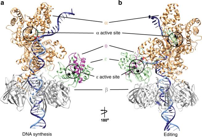
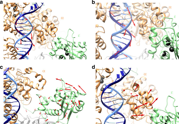
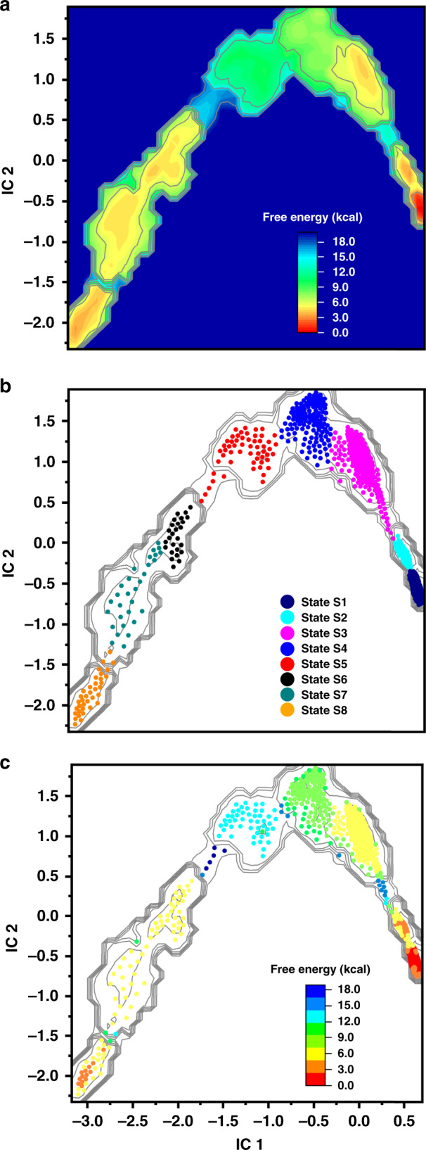
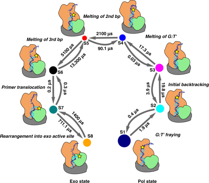
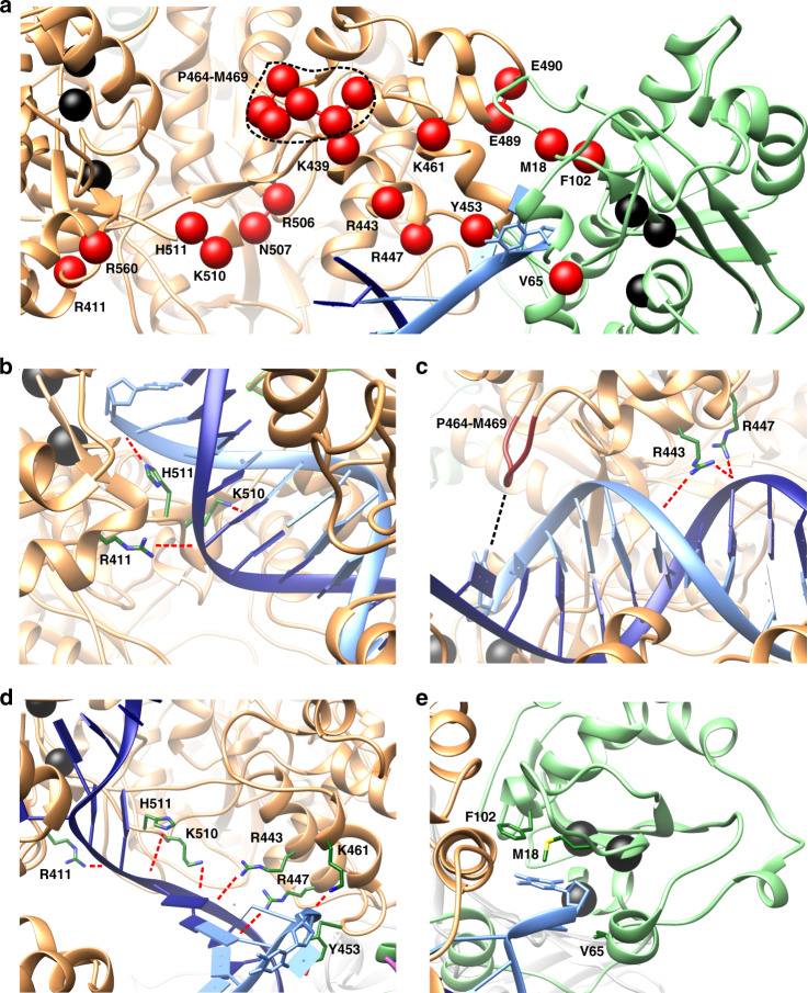
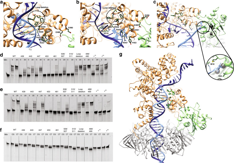
Proofreading by replicative DNA polymerases is a fundamental mechanism ensuring DNA replication fidelity. In proofreading, mis-incorporated nucleotides are excised through the 3'-5' exonuclease activity of the DNA polymerase holoenzyme. The exonuclease site is distal from the polymerization site, imposing stringent structural and kinetic requirements for efficient primer strand transfer. Yet, the molecular mechanism of this transfer is not known. Here we employ molecular simulations using recent cryo-EM structures and biochemical analyses to delineate an optimal free energy path connecting the polymerization and exonuclease states of E. coli replicative DNA polymerase Pol III. We identify structures for all intermediates, in which the transitioning primer strand is stabilized by conserved Pol III residues along the fingers, thumb and exonuclease domains. We demonstrate switching kinetics on a tens of milliseconds timescale and unveil a complete pol-to-exo switching mechanism, validated by targeted mutational experiments.


Conflict of interest statement

The authors declare no competing interests.

Figures

Fig. 1
Fig. 1. Structural models of DNA polymerase III in polymerization and editing mode.
The Pol III core (α, ε, and θ) bound to the DNA sliding clamp β in (a) polymerization mode and (b) editing mode. Pol III subunits are colored and labeled as follows: α in orange, ε in green, θ in magenta, and β in gray. The primer and template DNA strands are shown in light and dark gray respectively. The polymerase active site (in the α subunit) and exonuclease active site (in the ε subunit) are highlighted with circles.
Fig. 2
Fig. 2. Concerted motions of Pol III holoenzyme guide the primer along the path toward the exonuclease state.
a Initial backtracking motion of the DNA duplex away from polymerase active site observed in the MEP. b Subsequent rotational motion of the DNA and additional backtracking facilitates sequential unpairing at the primer/template junction. c Tilting motion of the ε subunit toward the α subunit shortens the distance between the polymerase and exonuclease active sites. d Outward shift of the thumb domain with respect to the PHP domain creates an opening to accommodate repositioning of the ε subunit. Red arrows indicate direction of motions observed in the minimum free energy path (MEP). Shifts in atomic positions for consecutive replicas of the MEP during different stages of the pol-to-exo transition were computed as vectors and mapped onto the structural elements of the Pol III holoenzyme. The α subunit is shown in orange; the ε subunit is shown in light green; the primer and template DNA strands are shown in light and dark blue, respectively; residues in the pol and exo active sites are shown as black spheres. The θ subunit has been omitted for clarity.
Fig. 3
Fig. 3. Analysis of the Pol III conformational ensemble reveals distinct kinetic intermediates in the pol-to-exo transition.
a Effective free energy profile projected onto the first two independent components (ICs) from TICA analysis. Inset denotes ΔG scale in kcal/mol and is set relative to the polymerization state. b Multi-ensemble Markov model (MEMM) constructed by combining the biased and unbiased simulation ensembles. Microstates (dots) are colored by the macrostate (intermediate) they belong to. Macrostate identities were computed with the PCCA+ algorithm. Color scheme for the macrostates is shown in the inset. c Microstates (dots) colored by their computed free energies from the MEMM analysis. Inset denotes ΔG scale in kcal/mol and is set relative to polymerization state.
Fig. 4
Fig. 4. Complete kinetic model for the pol-to-exo mode transition connecting all on-path intermediates identified by the MEMM analysis.
Macrostates S1–S8 are denoted by circles. Larger circles correspond to more populated macrostates. Transition between states are indicated with arrows and computed timescales for transitioning in and out of each macrostate are shown above the arrows. Each microstate is also represented by a cartoon, indicating the position and the extent of unpairing of the DNA primer end. The position of the mismatch nucleotide on the primer strand is indicated by a yellow star.
Fig. 5
Fig. 5. Specific interactions along the optimal path accommodate the transitioning primer end to ensure facile pol-to-exo switching.
a Key residues (critical nodes) for pol-to-exo mode switching determined from dynamic network, conservation, and persistent contacts analyses and mapped onto the Pol III structure. Critical nodes are shown as spheres, labeled and colored in red. Polymerase and exonuclease active site residues are shown as spheres and colored in black. bd Palm and thumb domain residues of the α subunit forming contacts important for polymerization (b, c) and for transitioning the primer end (d). Residue sidechains are shown in stick representation and labeled and colored by atom type (C is green, N is blue, and S is yellow). Salt-bridge and polar interactions to the DNA are shown as dashed red lines. Hydrophobic interactions are shown as a dashed black line. e Stabilization of the incoming mismatched nucleotide by the hydrophobic cluster of the ε subunit. Residues from the ε subunit hydrophobic patch are shown as sticks, labeled and colored in green.
Fig. 6
Fig. 6. Residues experimentally determined to be critical for transfer of DNA primer strand from polymerase to exonuclease active site.
ac Close up of the transition path between polymerase and exonuclease active site in (a) polymerase mode, (b) intermediate mode, and (c) exonuclease mode. Polymerase is colored in orange, exonuclease in green, template DNA strand in dark blue, and primer strand in light blue. Mutated residues are shown in dark green sticks. d Denaturing gel analysis of polymerase activity of wild type and mutant proteins on matched DNA. Mutants showing W-T activity are highlighted in green, mutations that are moderately affected in orange, and mutations that render the protein inactive in red. e Similar analysis using a DNA substrate with a terminal C–T mismatch. f Exonuclease activity on matched (C–G) and mismatched (C–T) DNA measured in the same DNA substrates as in panels d and e in the absence of nucleotides. g Overview of Pol III core complex in polymerase mode. The mutated residues are highlighted in dark green and the β-clamp in gray. The experiments in panels d, e have been reproduced more than three times, the experiment in panel f have been reproduced twice.

References

    1. Benkovic SJ, Valentine AM, Salinas F. Replisome-mediated DNA replication. Annu Rev. Biochem. 2001;70:181–208. doi: 10.1146/annurev.biochem.70.1.181. - DOI - PubMed
    1. Joyce CM, Benkovic SJ. DNA polymerase fidelity: kinetics, structure, and checkpoints. Biochemistry. 2004;43:14317–14324. doi: 10.1021/bi048422z. - DOI - PubMed
    1. Johnson KA. The kinetic and chemical mechanism of high-fidelity DNA polymerases. Biochim. Biophys. Acta. 2010;1804:1041–1048. doi: 10.1016/j.bbapap.2010.01.006. - DOI - PMC - PubMed
    1. Steitz TA. A mechanism for all polymerases. Nature. 1998;391:231–232. doi: 10.1038/34542. - DOI - PubMed
    1. Steitz TA. DNA polymerases: structural diversity and common mechanisms. J. Biol. Chem. 1999;274:17395–17398. doi: 10.1074/jbc.274.25.17395. - DOI - PubMed

Publication types