. 2021 Jun;73(6):2251-2265.
doi: 10.1002/hep.31604. Epub 2021 May 14.

Hepatocyte Nuclear Factor 4α Prevents the Steatosis-to-NASH Progression by Regulating p53 and Bile Acid Signaling (in mice)

Affiliations

Hepatocyte Nuclear Factor 4α Prevents the Steatosis-to-NASH Progression by Regulating p53 and Bile Acid Signaling (in mice)

Yanyong Xu et al. Hepatology. 2021 Jun.

Abstract

Background and aims: Hepatocyte nuclear factor 4α (HNF4α) is highly enriched in the liver, but its role in the progression of nonalcoholic liver steatosis (NAFL) to NASH has not been elucidated. In this study, we investigated the effect of gain or loss of HNF4α function on the development and progression of NAFLD in mice.

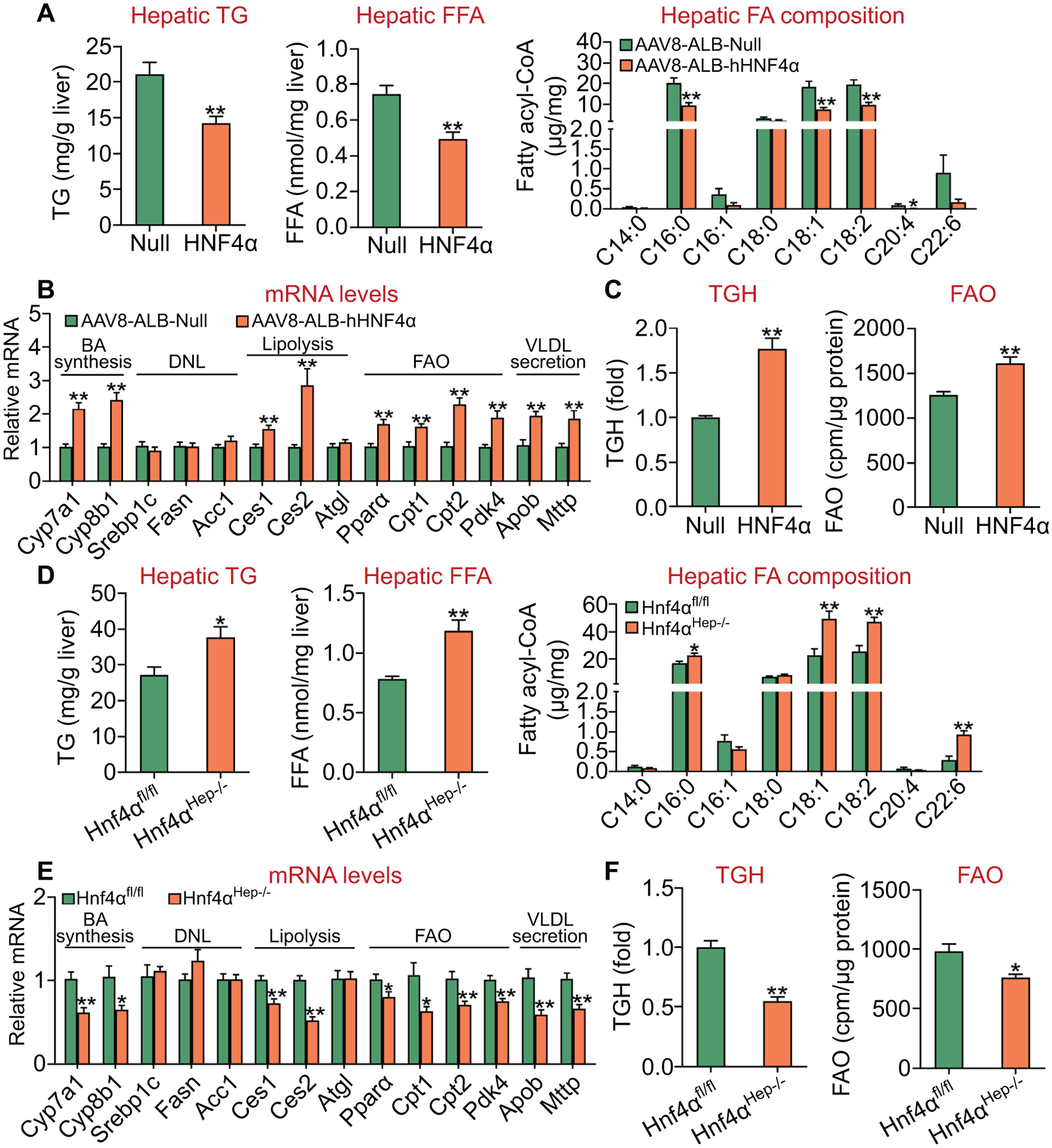
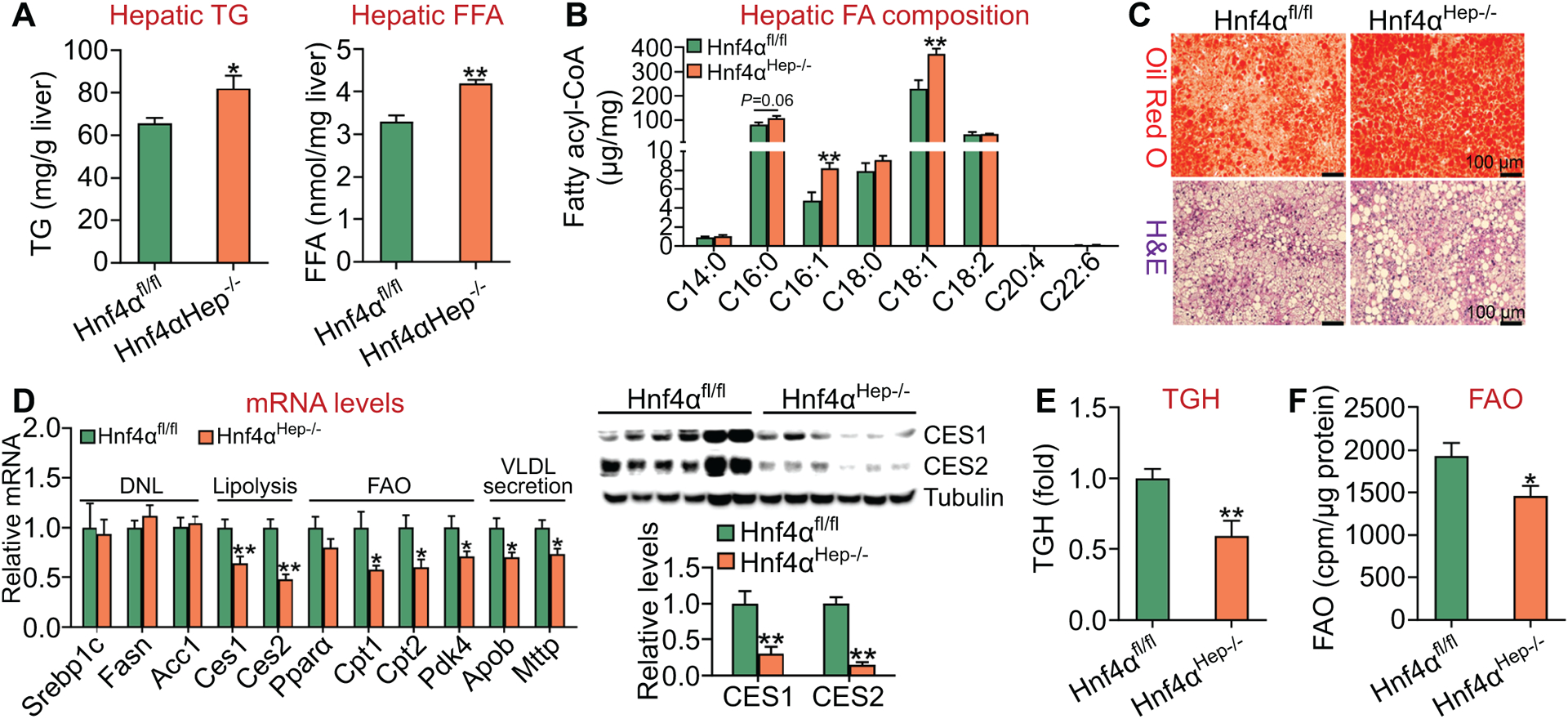
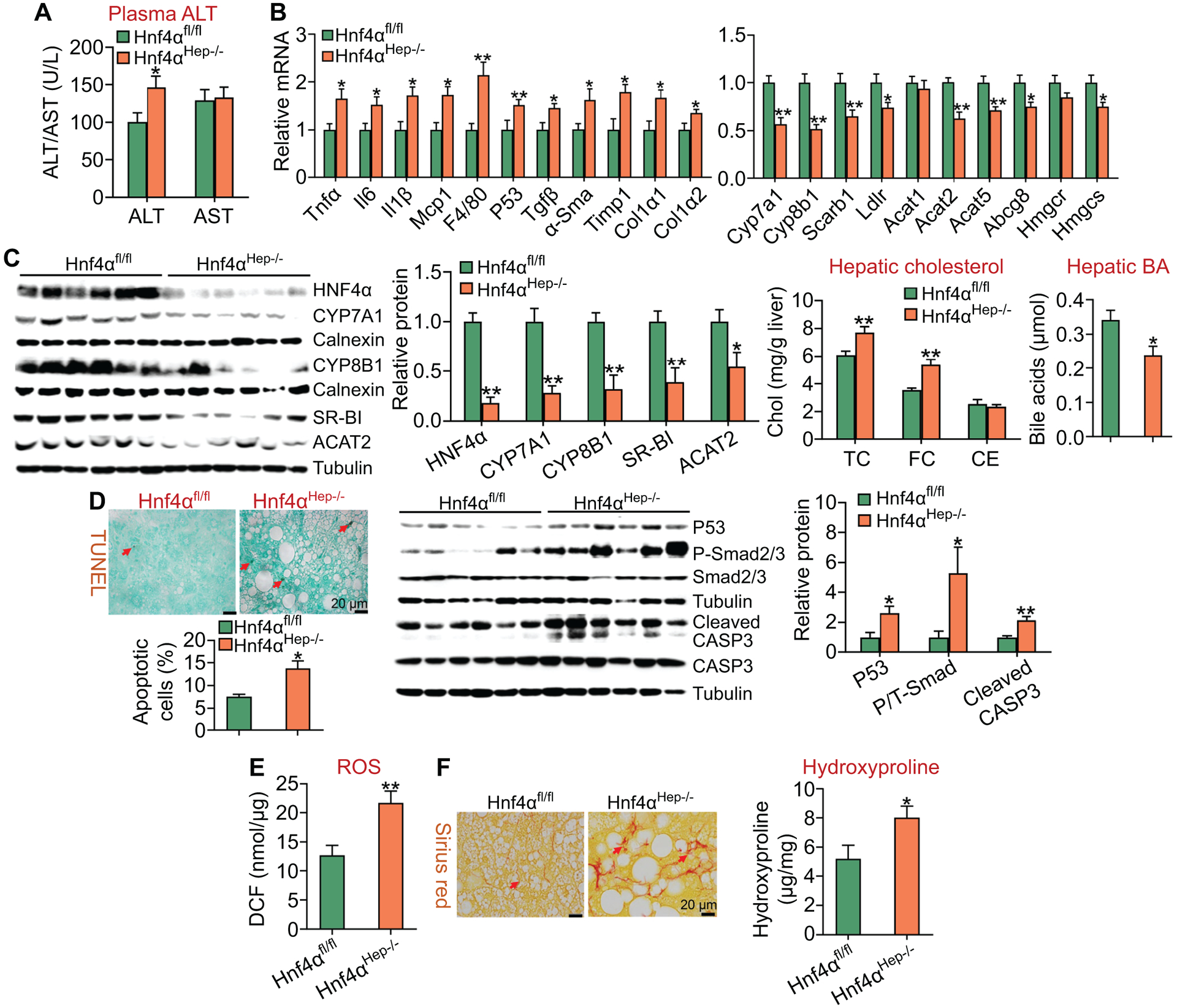
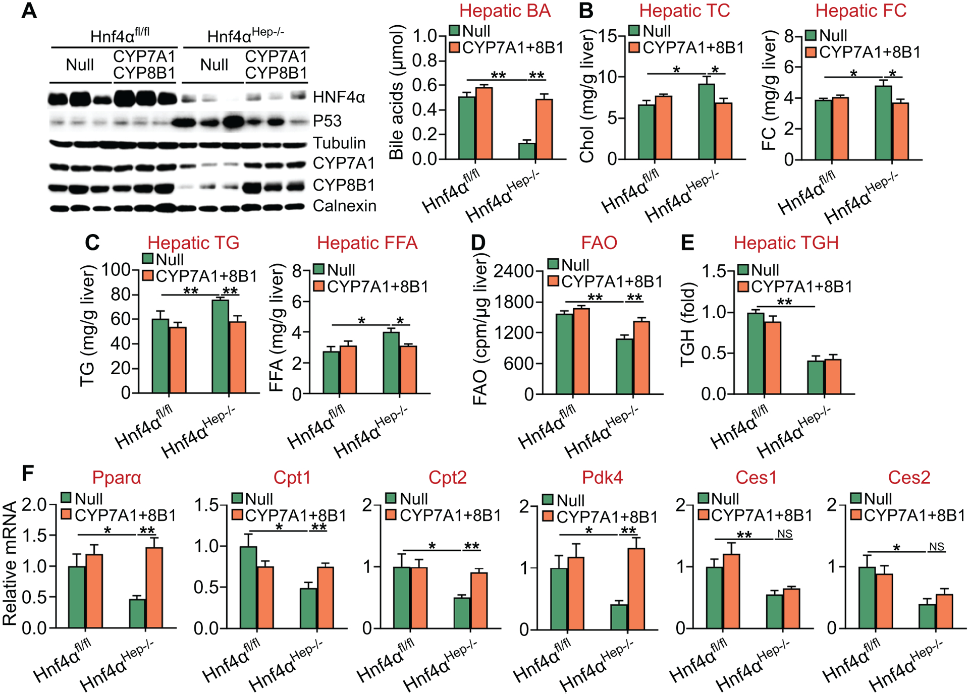
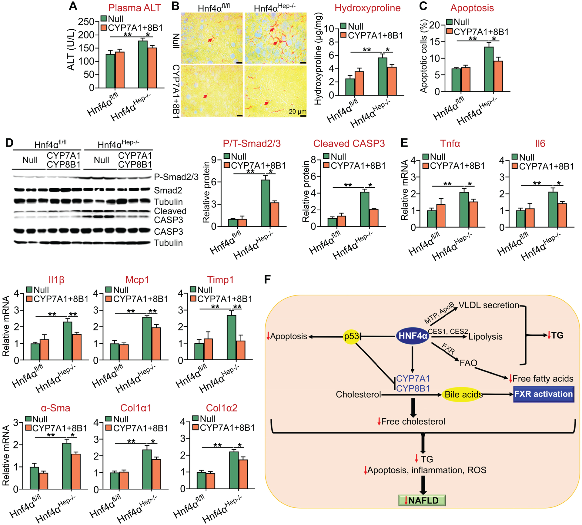
Approach and results: Overexpression of human HNF4α protected against high-fat/cholesterol/fructose (HFCF) diet-induced steatohepatitis, whereas loss of Hnf4α had opposite effects. HNF4α prevented hepatic triglyceride accumulation by promoting hepatic triglyceride lipolysis, fatty acid oxidation, and VLDL secretion. Furthermore, HNF4α suppressed the progression of NAFL to NASH. Overexpression of human HNF4α inhibited HFCF diet-induced steatohepatitis in control mice but not in hepatocyte-specific p53-/- mice. In HFCF diet-fed mice lacking hepatic Hnf4α, recapitulation of hepatic expression of HNF4α targets cholesterol 7α-hydroxylase and sterol 12α-hydroxylase and normalized hepatic triglyceride levels and attenuated steatohepatitis.

Conclusions: The current study indicates that HNF4α protects against diet-induced development and progression of NAFLD by coordinating the regulation of lipolytic, p53, and bile acid signaling pathways. Targeting hepatic HNF4α may be useful for treatment of NASH.


Figures

Figure 1.
Figure 1.. HNF4α is an essential regulator of hepatic TGH activity and fatty acid oxidation.
(A-C) C57BL/6J mice were injected i.v. with AAV8-ALB-Null or AAV8-ALB-hHNF4α and then fed a chow diet for 8 weeks (n=8 per group). Hepatic levels of TG and FFA as well as fatty acid composition were determined (A). Hepatic mRNA levels were quantified (B). Hepatic TGH activity (left panel) and FAO in hepatocytes (right panel) were determined (C). (D-F) Hnf4αfl/fl mice and Hnf4αHep−/− mice were fed a chow diet (n=8 per group). Hepatic levels of TG and FFA as well as fatty acid composition were determined (D). Hepatic mRNA levels were quantified (E). Hepatic TGH activity (left panel) and FAO in hepatocytes (right panel) were determined (F). *P<0.05, **P<0.01
Figure 2.
Figure 2.. Hepatocyte-specific expression of human HNF4α is sufficient to prevent HFCF diet-induced hepatosteatosis.
C57BL/6J mice were injected i.v. with AAV8-ALB-Null or AAV8-ALB-hHNF4α and then fed an HFCF diet for 20 weeks (n=8 per group). (A) Hepatic TG (left panel) and FFA (right panel) levels. (B) Hepatic fatty acid composition was analyzed by GC-MS. (C) Liver sections were stained by Oil Red O (top panel) or H&E (lower panel). (D) Hepatic mRNA levels (left panel) or protein levels (right panel). (E) Hepatic TGH activity. (F) FAO was analyzed using isolated hepatocytes. *P<0.05, **P<0.01
Figure 3.
Figure 3.. Hepatocyte-specific expression of human HNF4α ameliorates HFCF diet-induced steatohepatitis.
C57BL/6J mice were injected i.v. with AAV8-ALB-Null or AAV8-ALB-hHNF4α and then fed an HFCF diet for 20 weeks (n=8 per group). (A) Plasma ALT and AST levels. (B) Hepatic mRNA levels (left and right panels). (C) Hepatic proteins were analyzed by Western blot assays and then quantified (left panels). Hepatic cholesterol and BA levels were also analyzed (right panels). (D) Hepatic apoptosis was analyzed by TUNEL assays and then quantified (left panel). Hepatic protein levels were analyzed by Western blot assays and then quantified (middle and right panels). P/T-Smad, ratio of phosphorylated Smad2/3 to total Smad2/3. (E) Hepatic reactive oxygen species levels. (F) Hepatic sections were stained by Sirius Red (left panel), and hepatic hydroxyproline levels were quantified (right panel). *P<0.05, **P<0.01
Figure 4.
Figure 4.. Hepatocyte HNF4α is required for protection against HFCF diet-induced hepatosteatosis.
Hnf4αfl/fl mice and Hnf4αHep−/− mice were fed an HFCF diet for 20 weeks (n=8 per group). (A) Hepatic TG (left panel) and FFA (right panel) levels. (B) Hepatic fatty acid composition was analyzed by GC-MS. (C) Liver sections were stained by Oil Red O (top panel) or H&E (lower panel). (D) Hepatic mRNA levels (left panel) or protein levels (right panel). (E) Hepatic TGH activity. (F) FAO was analyzed in hepatocytes. *P<0.05, **P<0.01
Figure 5.
Figure 5.. Loss of hepatocyte HNF4α aggravates HFCF diet-induced steatohepatitis.
Hnf4αfl/fl mice and Hnf4αHep−/− mice were fed an HFCF diet for 20 weeks (n=8 per group). (A) Plasma ALT and AST levels. (B) Hepatic mRNA levels (left and right panels). (C) Hepatic proteins were analyzed by Western blot assays and then quantified (left panels). Hepatic cholesterol and BA levels were also analyzed (right panels). (D) Hepatic apoptosis was analyzed by TUNEL assays and then quantified (left panel). Hepatic protein levels were analyzed by Western blot assays and then quantified (middle and right panels). (E) Hepatic reactive oxygen species levels. (F) Hepatic sections were stained by Sirius Red (left panel), and hepatic hydroxyproline levels were quantified (right panel). *P<0.05, **P<0.01
Figure 6.
Figure 6.. Hepatocyte-specific over-expression of HNF4α attenuates HFCF diet-induced steatohepatitis via inhibition of p53.
p53fl/fl mice and p53Hep−/− mice were injected i.v. with AAV8-ALB-Null or AAV8-ALB-hHNF4α, and then fed an HFCF diet for 20 weeks (n=8 per group). (A) Plasma ALT levels. (B and C) Hepatic mRNA levels. (D) Hepatic levels of hydroxyproline (left panel), TG, FFA and cholesterol (middle and right panels). (E) Hepatic apoptosis was analyzed by TUNEL assays. (F) Hepatic proteins were analyzed by Western blot assays (left panel) and then quantified (right panel). P/T-Smad, ratio of phosphorylated Smad2/3 to total Smad2/3. *P<0.05, **P<0.01
Figure 7.
Figure 7.. Recapitulation of hepatic CYP7A1 and CYP8B1 expression normalizes hepatic TG levels and FAO in Hnf4αHep−/− mice.
Hnf4αfl/fl mice and Hnf4αHep−/− mice were injected i.v. with 0.5×10¹¹ GC of AAV8-ALB-Null (Null) or AAV8-ALB-hCYP7A1 plus AAV8-ALB-hCYP8B1 (CYP7A1+8B1), and then fed an HFCF diet for 20 weeks (n=8 per group). (A) Hepatic protein levels were analyzed by Western blot assays (left panel) and hepatic BA levels were quantified (right panel). (B) Hepatic TC (left panel) and FC (right panel) levels. (C) Hepatic TG (left panel) and FFA (right panel) levels. (D) FAO was analyzed using isolated hepatocytes. (E) Hepatic TGH activity was analyzed. (F) Hepatic mRNA levels. NS, not significant. *P<0.05, **P<0.01
Figure 8.
Figure 8.. Recapitulation of hepatic CYP7A1 and CYP8B1 expression attenuates diet-induced steatohepatitis in Hnf4αHep−/− mice.
Hnf4αfl/fl mice and Hnf4αHep−/− mice were injected i.v. with 0.5×10¹¹ GC of AAV8-ALB-Null or AAV8-ALB-hCYP7A1 plus AAV8-ALB-hCYP8B1, and then fed an HFCF diet for 20 weeks (n=8 per group). (A) Plasma ALT levels. (B) Liver sections were stained with Sirius Red (left panel) and hepatic hydroxyproline levels were quantified (right panel). (C) Hepatic apoptosis was analyzed by TUNEL assays. (D) Hepatic proteins were analyzed by Western blot assays (left panel) and then quantified (middle and right panels). (E) Hepatic mRNA levels. (F) A model for hepatic HNF4α to regulate the development and progression of NAFLD. HNF4α regulates VLDL secretion, lipolysis, fatty acid oxidation (FAO), bile acid synthesis and p53 expression. As a result, FXR is activated, hepatic free fatty acid, free cholesterol and triglyceride (TG) levels are decreased, and hepatic apoptosis, inflammation, and ROS production are inhibited. These multifacted effects of HNF4α lead to inhibition of development and progression of NAFLD. *P<0.05, **P<0.01

References

    1. Edmison J, McCullough AJ. Pathogenesis of non-alcoholic steatohepatitis: human data. Clin Liver Dis 2007;11:75–104, ix. - PubMed
    1. Day CP, James OF. Steatohepatitis: a tale of two “hits”? Gastroenterology 1998;114:842–845. - PubMed
    1. Jou J, Choi SS, Diehl AM. Mechanisms of disease progression in nonalcoholic fatty liver disease. Semin Liver Dis 2008;28:370–379. - PubMed
    1. Lewis JR, Mohanty SR. Nonalcoholic fatty liver disease: a review and update. Dig Dis Sci 2010;55:560–578. - PubMed
    1. Adams LA, Lymp JF, St Sauver J, Sanderson SO, Lindor KD, Feldstein A, Angulo P. The natural history of nonalcoholic fatty liver disease: a population-based cohort study. Gastroenterology 2005;129:113–121. - PubMed

Publication types

MeSH terms