. 2021 Jan;89(1):165-176.
doi: 10.1002/ana.25944. Epub 2020 Nov 9.

Retinal Thickness Predicts the Risk of Cognitive Decline in Parkinson Disease

Affiliations

Retinal Thickness Predicts the Risk of Cognitive Decline in Parkinson Disease

Ane Murueta-Goyena et al. Ann Neurol. 2021 Jan.

Abstract

Objective: This study was undertaken to analyze longitudinal changes of retinal thickness and their predictive value as biomarkers of disease progression in idiopathic Parkinson's disease (iPD).

Methods: Patients with Lewy body diseases were enrolled and prospectively evaluated at 3 years, including patients with iPD (n = 42), dementia with Lewy bodies (n = 4), E46K-SNCA mutation carriers (n = 4), and controls (n = 17). All participants underwent Spectralis retinal optical coherence tomography and Montreal Cognitive Assessment, and Unified Parkinson's Disease Rating Scale score was obtained in patients. Macular ganglion cell-inner plexiform layer complex (GCIPL) and peripapillary retinal nerve fiber layer (pRNFL) thickness reduction rates were estimated with linear mixed models. Risk ratios were calculated to evaluate the association between baseline GCIPL and pRNFL thicknesses and the risk of subsequent cognitive and motor worsening, using clinically meaningful cutoffs.

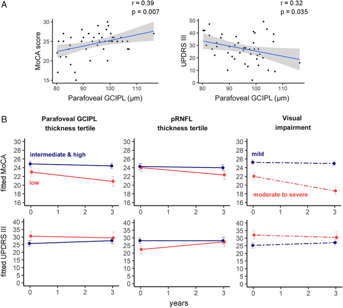
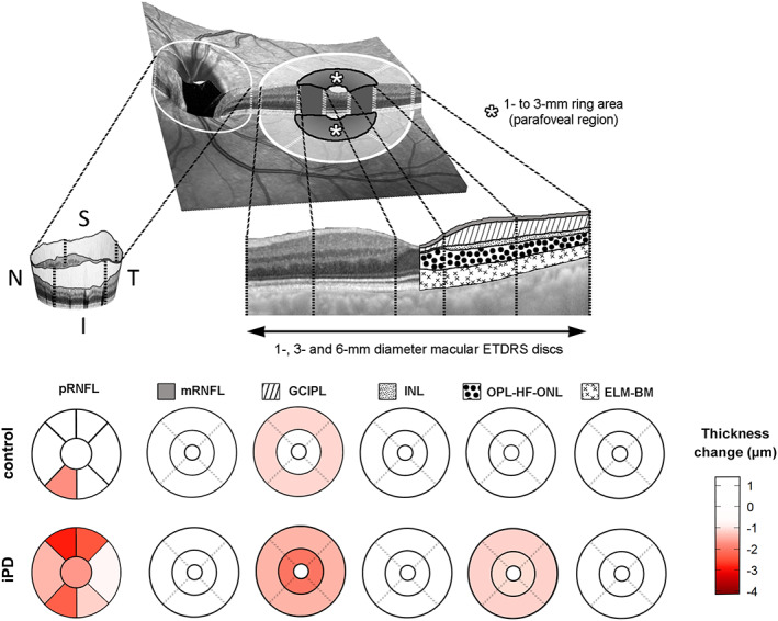
Results: GCIPL thickness in the parafoveal region (1- to 3-mm ring) presented the largest reduction rate. The annualized atrophy rate was 0.63μm in iPD patients and 0.23μm in controls (p < 0.0001). iPD patients with lower parafoveal GCIPL and pRNFL thickness at baseline presented an increased risk of cognitive decline at 3 years (relative risk [RR] = 3.49, 95% confidence interval [CI] = 1.10-11.1, p = 0.03 and RR = 3.28, 95% CI = 1.03-10.45, p = 0.045, respectively). We did not identify significant associations between retinal thickness and motor deterioration.

Interpretation: Our results provide evidence of the potential use of optical coherence tomography-measured parafoveal GCIPL thickness to monitor neurodegeneration and to predict the risk of cognitive worsening over time in iPD. ANN NEUROL 2021;89:165-176.


Conflict of interest statement

Nothing to report.

Figures

FIGURE 1
FIGURE 1
Visual outcomes. (A) Differences in baseline visual outcomes between controls and idiopathic Parkinson disease (iPD) patients. Significant differences are indicated with asterisks (Wilcoxon rank sum test, *p < 0.05, **p < 0.01, ***p < 0.001). (B) Longitudinal changes in visual outcomes. Relative changes (percentage) of visual outcomes were calculated as ([follow‐up visual score – baseline visual score] / baseline visual score) × 100. #p < 0.05 for the interaction term between group and time in linear mixed models. Note that the results of n = 50 iPD patients and n = 29 controls are represented in A and the results of n = 42 iPD patients and n = 17 controls in B. BLOJ = Benton Line Orientation Judgment; CA = cube analysis subitem; CDT = Clock Drawing Test, reproduction, corrected with Rouleau method; CS = contrast sensitivity; HCVA = high‐contrast visual acuity (number of correct letters); LCVA = low‐contrast visual acuity (number of correct letters); NL = number location subitem; SDMT = Symbol Digit Modality Test; SPCT = Salthouse Perceptual Comparison Test; TMT, part‐A (s) = time to complete Trail Making Test, part A, in seconds; VOSP = Visual Object and Space Perception battery; WAIS = figure completion part of Wechsler Adult Intelligence Scale IV. [Color figure can be viewed at www.annalsofneurology.org]
FIGURE 2
FIGURE 2
(A) Scatterplots represent the relationship between ganglion cell–inner plexiform layer complex (GCIPL) thickness in the parafoveal area (1‐ to 3mm) and Montreal Cognitive Assessment (MoCA) and Unified Parkinson Disease Rating Scale Part III (motor examination; UPDRS III) scores at baseline. (B) Progression of cognitive and motor manifestations. Parameter estimates from linear mixed‐effect models were converted to and plotted as condition means and standard error. Idiopathic Parkinson disease (iPD) patients were divided into subgroups according to baseline thickness in the parafoveal area (1‐ to 3mm ring) of GCIPL in the macula, baseline peripapillary retinal nerve fiber layer (pRNFL) thickness, and baseline visual outcomes. Retinal thicknesses of iPD patients were divided into tertiles according to the reference population, and visual impairment was determined using K‐means clustering (see Subjects and Methods). Eleven iPD patients from the lowest retinal thickness tertile and 31 iPD patients from the intermediate and highest tertiles completed a follow‐up visit. Thirty‐one patients who were classified as mild visual dysfunction and 11 classified as moderate to severe visual dysfunction completed the 3‐year visit. r = Pearson correlation coefficient. [Color figure can be viewed at www.annalsofneurology.org]
FIGURE 3
FIGURE 3
Progression of retinal atrophy in peripapillary peripapillary retinal nerve fiber layer (pRNFL), and in 5 layer complexes of the macula divided in 1mm, 1‐ to 3mm, and 3‐ to 6mm areas. The 1‐ to 3mm area corresponds to the parafoveal area of the macula. Estimated changes of macular and peripapillary thickness between baseline and year 3 follow‐up, adjusted for age and sex, are displayed, and negative values represent retinal thinning over time. ELM‐BM = retinal complex including external limiting membrane, ellipsoid band, retinal pigment epithelium, and Bruch membrane; ETDRS = Early Treatment Diabetic Retinopathy Study; GCIPL = ganglion cell–inner plexiform layer complex; I = inferior; INL = inner nuclear layer of the retina; iPD = idiopathic Parkinson disease; mRNFL, macular retinal nerve fiber layer; N = nasal; OPL‐HF‐ONL = outer plexiform–Henle fiber–outer nuclear layer complex; S = superior; T = temporal. [Color figure can be viewed at www.annalsofneurology.org]

References

    1. Lanskey JH, McColgan P, Schrag AE, et al. Can neuroimaging predict dementia in Parkinson's disease? Brain 2018;141:2545–2560. - PMC - PubMed
    1. Espay AJ, Kalia LV, Gan‐Or Z, et al. Disease modification and biomarker development in Parkinson disease: revision or reconstruction? Neurology 2020;94:481–494. - PMC - PubMed
    1. Zarranz JJ, Alegre J, Gomez‐Esteban JC, et al. The new mutation, E46K, of alpha‐synuclein causes Parkinson and Lewy body dementia. Ann Neurol 2004;55:164–173. - PubMed
    1. Hamilton JM, Salmon DP, Galasko D, et al. Visuospatial deficits predict rate of cognitive decline in autopsy‐verified dementia with Lewy bodies. Neuropsychology 2008;22:729–737. - PMC - PubMed
    1. Somme JH, Gomez‐Esteban JC, Molano A, et al. Initial neuropsychological impairments in patients with the E46K mutation of the alpha‐synuclein gene (PARK 1). J Neurol Sci 2011;310:86–89. - PubMed

Publication types