Review
. 2021 Jan;141(1):1-24.
doi: 10.1007/s00401-020-02235-6. Epub 2020 Oct 24.

Brain arteriolosclerosis

Affiliations
Review

Brain arteriolosclerosis

Brittney L Blevins et al. Acta Neuropathol. 2021 Jan.

Abstract

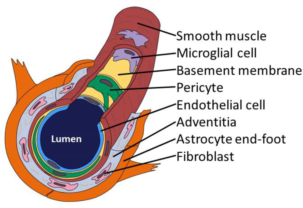
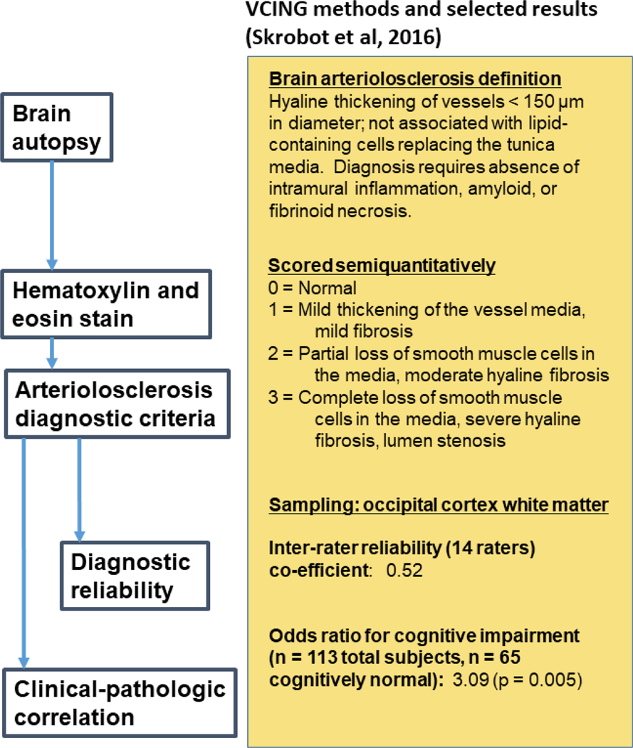
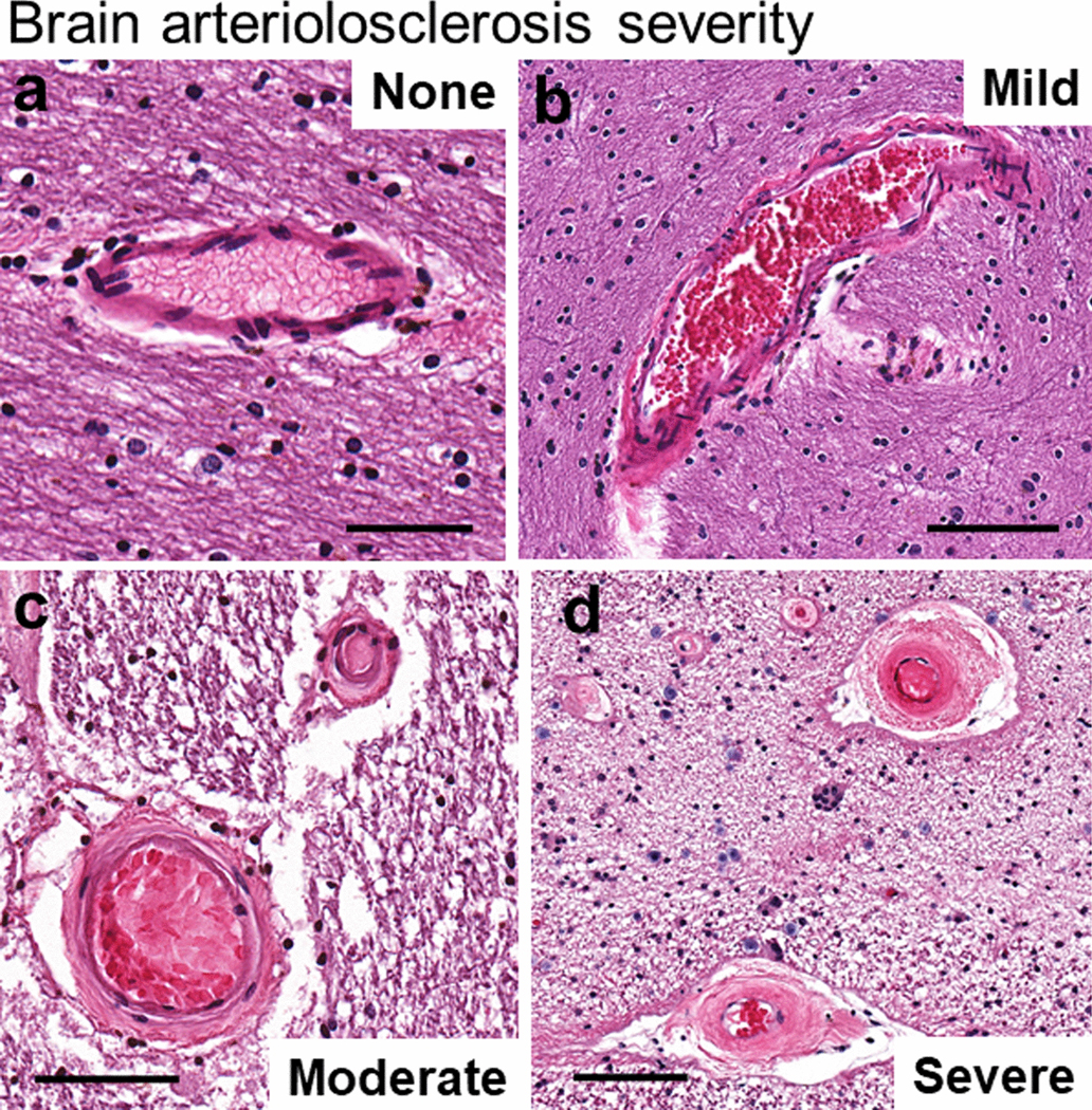
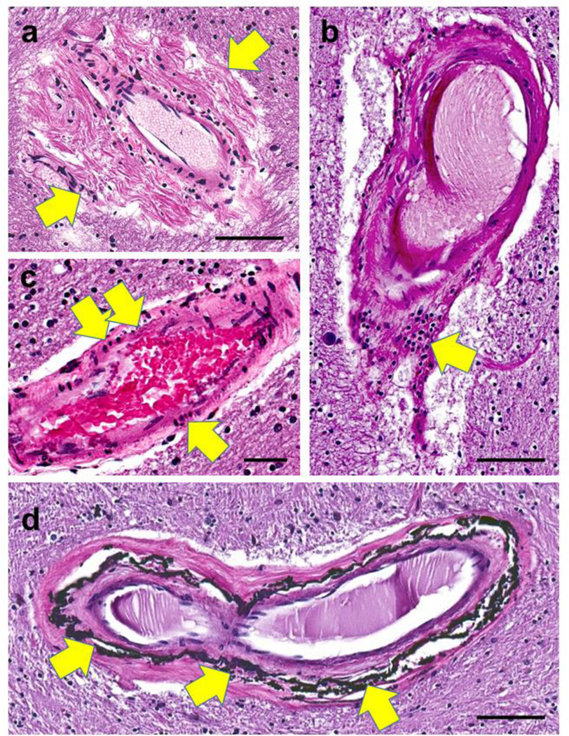
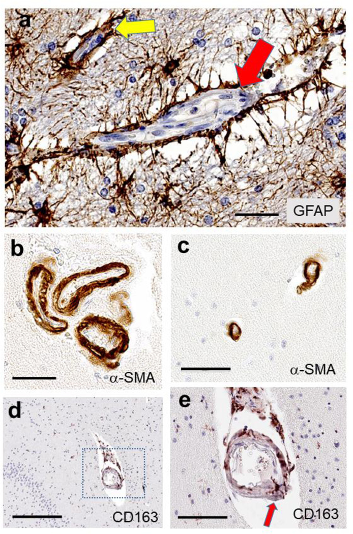
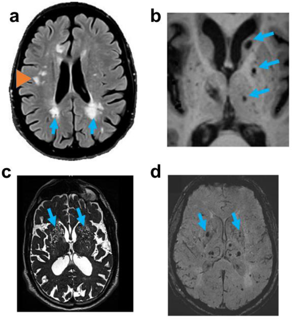
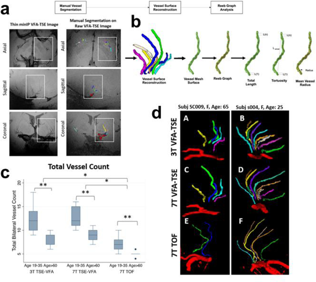
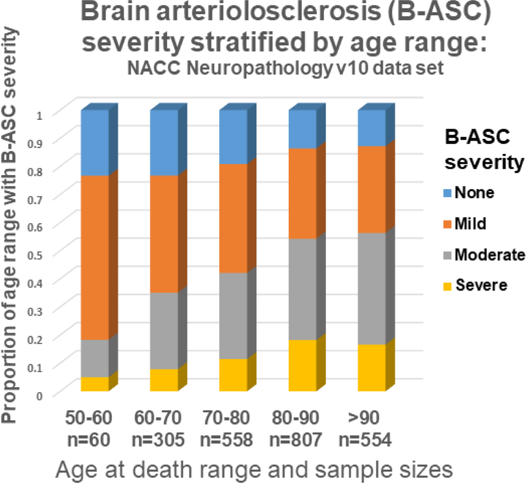
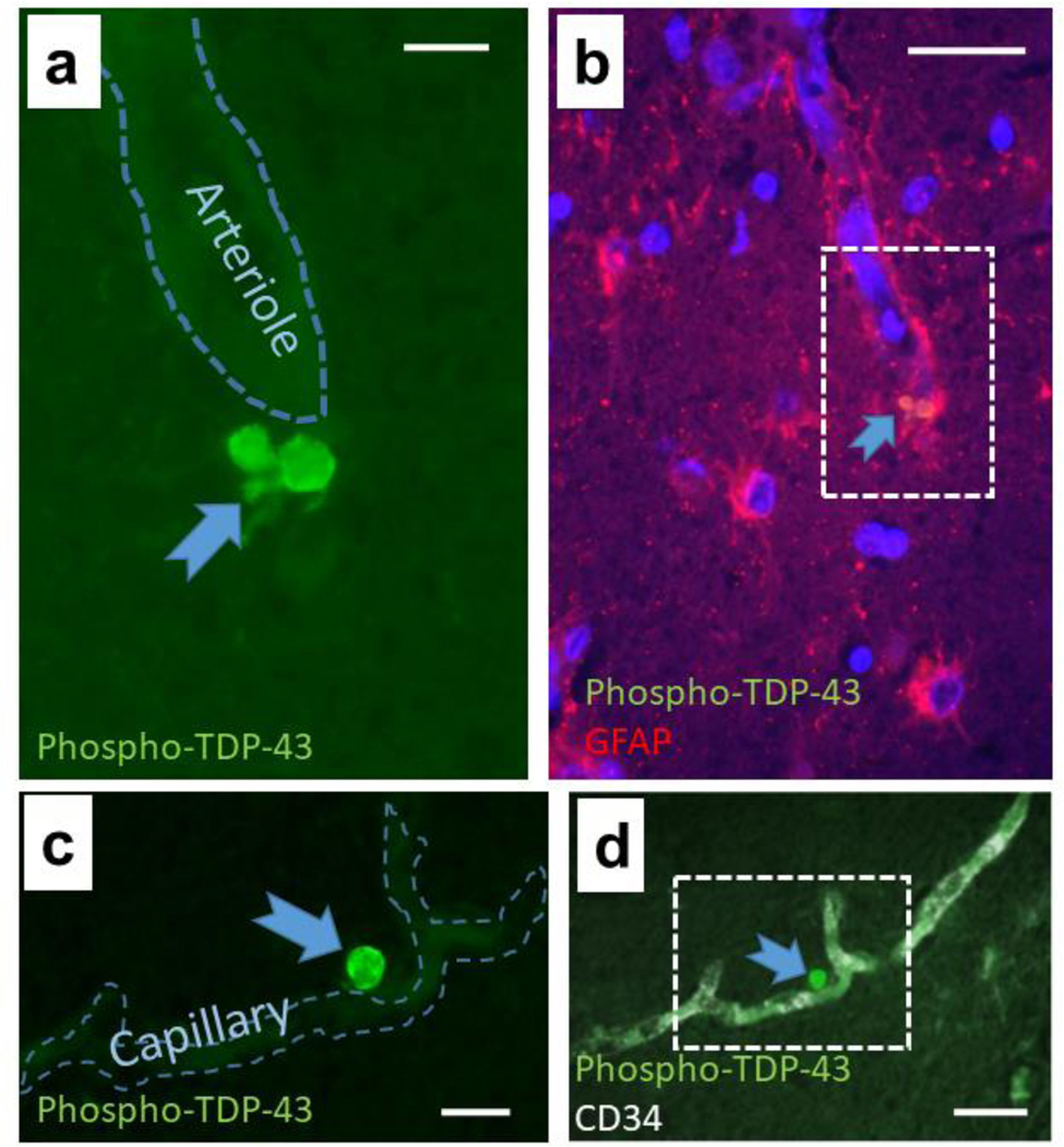
Brain arteriolosclerosis (B-ASC), characterized by pathologic arteriolar wall thickening, is a common finding at autopsy in aged persons and is associated with cognitive impairment. Hypertension and diabetes are widely recognized as risk factors for B-ASC. Recent research indicates other and more complex risk factors and pathogenetic mechanisms. Here, we describe aspects of the unique architecture of brain arterioles, histomorphologic features of B-ASC, relevant neuroimaging findings, epidemiology and association with aging, established genetic risk factors, and the co-occurrence of B-ASC with other neuropathologic conditions such as Alzheimer's disease and limbic-predominant age-related TDP-43 encephalopathy (LATE). There may also be complex physiologic interactions between metabolic syndrome (e.g., hypertension and inflammation) and brain arteriolar pathology. Although there is no universally applied diagnostic methodology, several classification schemes and neuroimaging techniques are used to diagnose and categorize cerebral small vessel disease pathologies that include B-ASC, microinfarcts, microbleeds, lacunar infarcts, and cerebral amyloid angiopathy (CAA). In clinical-pathologic studies that factored in comorbid diseases, B-ASC was independently associated with impairments of global cognition, episodic memory, working memory, and perceptual speed, and has been linked to autonomic dysfunction and motor symptoms including parkinsonism. We conclude by discussing critical knowledge gaps related to B-ASC and suggest that there are probably subcategories of B-ASC that differ in pathogenesis. Observed in over 80% of autopsied individuals beyond 80 years of age, B-ASC is a complex and under-studied contributor to neurologic disability.

Keywords: Arteriosclerosis; Neuroimaging; Neuropathology; SVD; Senescence; cAVU.


Figures

Fig. 1.
Fig. 1.. A brain arteriole and constituents of the cerebral arteriolar vascular unit (cAVU).
Brain arterioles are not all alike, but this schematic depiction may orient readers to some cAVU components. Shown here is a relatively thin-walled arteriole--no internal elastic lamina or adventitial nerve fibers are depicted. Inflammatory cells including macrophages and microglia are commonly found in and around the arteriolar wall. Acellular strands of elastin and collagen are interwoven with fibroblast-like cells in the adventitia.
Fig. 2.
Fig. 2.. Autopsy diagnosis of B-ASC involves staining with hematoxylin and eosin (H&E).
The VCING collaborative group provided a consensus among experts describing criteria for defining B-ASC and for generating parameters that reflect different severity of the pathology. These diagnostic criteria are presented on the right side of the figure. In this study [183], which included evaluation of n=113 brains and sampling from many different brain areas, stained slides of the occipital cortex white matter produced moderately strong inter-rater reliability among different neuropathologists, and a robust association with cognitive impairment, i.e., B-ASC pathology was more often present in cases with dementia than those with normal cognitive status.
Fig. 3.
Fig. 3.. One approach to grading arteriolosclerosis (B-ASC) pathologic severity is on a semiquantitative scale of “none”, “mild”, “moderate”, or “severe” B-ASC.
A score of 0 corresponds to no/very minimal changes (a); a score of 1 indicates mild changes (b); a score of 2 is moderately severe B-ASC (c); and a score of 3 indicates severe B-ASC (d). Note that in (d), the arterioles are partly occluded by severe hyperplastic (“onion-skin”-type) arteriolosclerotic changes. Scale bars = (a): 80 μm; (b): 200 μm; (c): 100 μm; and, (d): 100 μm. All of these 4 panels show vascular profiles from occipital cortex white matter of human subjects.
Fig. 4.
Fig. 4.. Arteriolar walls can show different histomorphologies with aging.
In panel (a), periarteriolar (adventitial) fibrosis is extensive and non-concentric (arrows). Panel (b) shows a collection of lymphocytes (arrow) in portions of the vessel wall. In panel (c), vessel wall changes include pyknotic-appearing smooth muscle cells (arrows). Siderocalcinosis, distinct from B-ASC, has been associated with dementia [194] and is usually seen preferentially in the globus pallidus (arrows in d). Synonyms for this include medial vascular calcification, calcific medial arteriosclerosis, and Monckeberg’s medial sclerosis. Scale bars = (a, b, and d): 100 μm; (c): 60 μm.
Fig. 5.
Fig. 5.. Photomicrographs to highlight features of the cAVU with immunohistochemistry.
Astrocytes and astrocytic end feet processes (a) are shown immunostained for GFAP, surrounding a capillary (yellow arrow) and a larger arteriole (red arrow). Smooth muscle cells, immunolabeled for α-SMA (panels b, c) in arterioles cut in cross-section, may highlight that multiple arterioles can exist in a single Virchow-Robin space (b), and the vascular profiles may be rather narrow, <20 μm lumen diameter (c). Some cellular constituents have only recently been appreciated, such as the “scavenger microglia” that express CD163 antigen and are arranged in and around small blood vessels, such as this hippocampal arteriole (d, e). In panel (e), a magnified view of the panel (d) inset, a CD163 immunoreactive cell spans the full thickness of the arteriolar wall (red arrow). Scale bars = (a): 60 μm; (b): 50 μm; (c): 60 μm; (d): 300 μm; and, (e): 90 μm.
Fig. 6.
Fig. 6.. Magnetic resonance imaging (MRI) modalities are commonly used in the study of cerebral small vessel disease.
FLAIR imaging (a) showing regions of periventricular white matter hyperintensities (WMHs; arrows) and deep WMHs (orange arrowhead); T1-weighted imaging (b) showing lacunes (arrows); T2-weighted imaging (c) showing expanded perivascular spaces (arrows); and, susceptibility-weighted imaging (d) showing presumed microbleeds (arrows).
Fig. 7.
Fig. 7.. High resolution MRI for the characterization of basal ganglia lenticulostriate arteries using 3T and 7T magnets.
This relatively high-resolution in vivo imaging [122] enables visualization of slender arteries/arterioles. In aging, there is increased tortuosity and decreased number of blood vessels resolved. In the raw images (a), blood is rendered black. Workflow is illustrated in panel (b), beginning with manual vessel segmentation on the raw turbo spin echo variable refocusing flip angles (TSE-VFA) image. The vessel volumes are reconstructed, and a mesh surface is created in preparation for shape analysis. Quantitative measures of vessel length, tortuosity and radius can be calculated. The box plot (c) shows that more vessels were detected in young participants than in older participants and more vessels were detected using 7T VFA-TSE compared to the more conventional 7T time-of-flight magnetic resonance angiography (7T TOF-MRA) [122]. An example comparing an older and younger adult (d) illustrates the smaller number of vessels detected in an older participant (A,C,E) than a younger participant (B, D, F).
Fig. 8.
Fig. 8.. Brain arteriolosclerosis (B-ASC) pathology by age at death, from the NACC Neuropathology version 10 data set (n=2,284).
B-ASC pathology increases with advanced age. Note that <20% of subjects had moderate or severe B-ASC pathology before age 60, whereas in subjects over 80, >50% had moderate or severe B-ASC pathology, and >80% had some degree of B-ASC.
Fig. 9.
Fig. 9.. Phospho-TDP-43 immunoreactive proteinopathy can be seen immediately adjacent to small blood vessels in brains with limbic-predominant age-related TDP-43 encephalopathy (LATE).
The micrograph shows perivascular TDP-43 proteinopathy near an arteriole (a, b) and a capillary (c, d) in hippocampi of 2 subjects with autopsy-proven LATE. In panel (a), the green filter shows the TDP-43 proteinopathy adjacent to an arteriole. Panel (b) shows lower magnification and the red filter shows GFAP immunoreactivity, revealing the colocalization with GFAP and TDP-43 (arrow). Panel (c) shows a phospho-TDP-43 deposit adjacent to a capillary as indicated by CD34 immunofluorescence at lower magnification (d). The presence of TDP-43 proteinopathy in astrocyte end-feet in FTLD-TDP was shown previously by Lin et al. [116]. LATE neuropathologic change is associated with relatively severe B-ASC [101, 154]. Thus, there may be parallel or synergistic disease-driving mechanisms related to the pathologies that usually are categorized separately as cerebrovascular or neurodegenerative. Scale bars = (a): 15 μm; (b): 40 μm; (c): 20 μm; and, (d): 40 μm.

References

    1. Abbott NJ, Pizzo ME, Preston JE, Janigro D, Thorne RG (2018) The role of brain barriers in fluid movement in the CNS: is there a ‘glymphatic’ system? Acta Neuropathol 135: 387–407. 10.1007/s00401-018-1812-4 - DOI - PubMed
    1. Abbott NJ, Revest PA, Romero IA (1992) Astrocyte-endothelial interaction: physiology and pathology. Neuropathol Appl Neurobiol 18: 424–433. 10.1111/j.13652990.1992.tb00808.x - DOI - PubMed
    1. Abner EL, Nelson PT, Kryscio RJ, et al. (2016) Diabetes is associated with cerebrovascular but not Alzheimer neuropathology. Alzheimers Dement ePub Jan 23, Available on PubMed. 10.1016/j.jalz.2015.12.006 - DOI - PMC - PubMed
    1. Abrahamson EE, Ikonomovic MD (2020) Brain injury-induced dysfunction of the blood brain barrier as a risk for dementia. Exp Neurol 328: 113257. 10.1016/j.expneurol.2020.113257 - DOI - PubMed
    1. Adib-Samii P, Devan W, Traylor M, et al. (2015) Genetic architecture of white matter hyperintensities differs in hypertensive and nonhypertensive ischemic stroke. Stroke 46: 348–353. 10.1161/STROKEAHA.114.006849 - DOI - PMC - PubMed

Publication types

Grants and funding