Observational Study
. 2021 Apr;51(3):663-674.
doi: 10.1007/s11239-020-02318-x. Epub 2020 Oct 24.

COVID-19 associated coagulopathy in critically ill patients: A hypercoagulable state demonstrated by parameters of haemostasis and clot waveform analysis

Affiliations
Observational Study

COVID-19 associated coagulopathy in critically ill patients: A hypercoagulable state demonstrated by parameters of haemostasis and clot waveform analysis

Bingwen Eugene Fan et al. J Thromb Thrombolysis. 2021 Apr.

Abstract

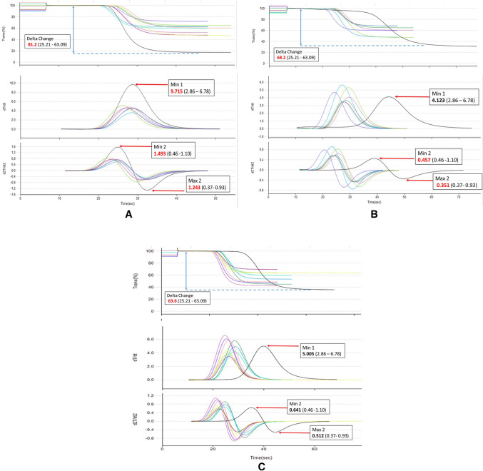
Patients with COVID-19 are known to be at risk of developing both venous, arterial and microvascular thrombosis, due to an excessive immuno-thrombogenic response to the SARS-CoV-2 infection. Overlapping syndromes of COVID-19 associated coagulopathy with consumptive coagulopathy and microangiopathy can be seen in critically ill patients as well. Blood was collected from 12 Intensive Care Unit (ICU) patients with severe COVID-19 who were on either mechanical ventilation or on high flow oxygen with a PaO2/FiO2 ratio of <300 mmHg. Laboratory tests were performed for parameters of haemostasis, clot waveform analysis and anti-phospholipid antibodies. CWA parameters were raised with elevated aPTT median Min1 (clot velocity) 9.3%/s (IQR 7.1-9.9%/s), elevated PT median Min1 10.3%/s (IQR 7.1-11.1%/s), elevated aPTT median Min2 (clot acceleration) 1.5%/s2 (IQR 1.0-1.6%/s2), elevated PT median Min2 5.2%/s2 (3.6-5.7%/s2), elevated aPTT median Max2 (clot deceleration) 1.3%/s2 (IQR 0.8-1.4%/s2) elevated PT median Max2 3.8%/s2 (IQR 2.6-4.2%/s2), increased aPTT median Delta change (decreased light transmission due to increased clot formation) 87.8% (IQR 70.2-91.8%) and PT median Delta change 33.0%. This together with raised median Factor VIII levels of 262.5%, hyperfibrinogenemia (median fibrinogen levels 7.5 g/L), increased median von Willebrand factor antigen levels 320% and elevated median D-dimer levels 1.7 μg/dl support the diagnosis of COVID-19 associated coagulopathy. A lupus anticoagulant was present in 50% of patients. Our laboratory findings further support the view that severe SARS-CoV-2 infection is associated with a state of hypercoagulability.

Keywords: Coronavirus; Hypercoagulability; Sepsis; Thrombophilia; Thrombosis.


Conflict of interest statement

The authors declare that they have no conflict of interest. No funding for this study was obtained.

Figures

Fig. 1
Fig. 1
a Hypercoagulable aPTT Clot Waveform tracing and parameters of a 35-year-old male with severe COVID-19 infection complicated by acute right ischaemic limb secondary to extensive thromboembolic disease in the abdominal aorta, right external iliac and right popliteal arteries. *Patient’s clot waveform tracing in black, healthy controls in multicolour (for reference). Patient’s haemostatic and clot waveform parameters: PT 14.2 s (11.7–14.0 s), aPTT 38.7 s (27.0–37.0 s), fibrinogen 5.0 g/L (1.8–4.5 g/L), D-Dimer 2.26 μg/ml (<0.50 μg/ml), Platelets 337 × 109/L (150–360 x109/L). Factor II 122% (70–120%), Factor VIII 196% (60–150%), Factor × 106% (70–120%), vWF 200% (56–160%), anti-thrombin III 74% (80–130%), Protein C 61% (70–150%), Protein S 79% (55–130%). Absent lupus anticoagulant and antiphospholipid antibodies. CWA: Min1 9.715%/s (2.86–6.78%/s), Min2 1.493%/s2 (0.46–1.10%/s2), Max2 1.243%/s2 (0.37–0.93%/s2), Delta change 81.2% (25.21–63.09%). b aPTT Clot Waveform tracing of a 40-year-old male on mechanical ventilation for severe COVID-19 pneumonia, Day 18 of illness (Day 2 ECMO), with probable pulmonary intravascular coagulopathy with concurrent bacterial pneumonia and sepsis induced coagulopathy, not initiated on anticoagulation yet. Clots were seen in the ECMO oxygenator as well as the dialysis circuit. *Patient’s clot waveform tracing in black, healthy controls in multicolour (for reference). Patient’s haemostatic and clot waveform parameters. PT 18.2 s (11.7–14.0 s), aPTT 53.2 s (27.0–37.0 s), fibrinogen 3.8 g/L (1.8–4.5 g/L), D-Dimer >20 μg /ml (<0.5 μg /ml), Platelets 115 × 109/L (150–360 × 109). Factor II 46% (70–120%), Factor V 68% (70–120%), Factor VIII 196% (60–150%), Factor IX 115% (60–150%) Factor × 68% (70–120%), vWF 387% (56–160%), anti-thrombin III 54% (80–130%), Protein C 28% (70–130%), Protein S 19% (55–130%). Lupus anticoagulant moderately present and anti cardiolipin IgG < 20 (0–20 GPL units), anti cardiolipin IgM 23 (0–20 MPL units), anti B2 glycoprotein-1 < 2 RU/ml (0–20 RU/ml). CWA: Min1 4.123%/s (2.86–6.78%/s), Min2 0.457%/s2 (0.46–1.10%/s2), Max2 0.351%/s2 (0.37–0.93), Delta change 68.2% (25.21–63.09%). c aPTT Clot Waveform tracing of a 73-year-old male on Day 13 of illness with severe COVID-19 pneumonia with probable pulmonary intravascular coagulopathy and concurrent sepsis induced coagulopathy from stenotrophomonas maltophilia bacteraemia and ventilator associated pneumonia. *Patient’s clot waveform tracing in black, healthy controls in multicolour (for reference). Patient’s haemostatic and clot waveform parameters. PT 15.3 s (11.7–14.0 s), aPTT 41.3 s (27.0–37.0 s), fibrinogen 4.0 g/L (1.8–4.5 g/L), D-Dimer >20 μg/ml (<0.5 μg/ml), Platelets 120 × 109/L. Factor II 56% (70–120%), Factor V 93% (70–120%), Factor VIII 195% (60–150%), Factor IX 105% (60–150%), Factor × 68% (70–120%), Factor XI 47% (60–150%), vWF 200% (56–160%), anti-thrombin III 74% (80–130%), Protein C 61% (70–150%), Protein S 79% (55–130%). Lupus anticoagulant and antiphospholipid antibodies absent. CWA: Min1 5.005%/s (2.86–6.78%/s), Min2 0.641%/s2 (0.46–1.10%/s2), Max2 0.512%/s2 (0.37–0.93%/s2), Delta change 63.6% (25.21–63.09%)

References

    1. Iba T, Levy JH, Levi M, Connors JM, Thachil J (2020) Coagulopathy of coronavirus disease 2019 [published online ahead of print, 2020 May 27]. Crit Care Med. 10.1097/CCM.0000000000004458 - PMC - PubMed
    1. Becker RC. COVID-19 update: COVID-19-associated coagulopathy. J Thromb Thrombolysis. 2020;50(1):54–67. doi: 10.1007/s11239-020-02134-3. - DOI - PMC - PubMed
    1. Thachil J, Tang N, Gando S, et al. ISTH interim guidance on recognition and management of coagulopathy in COVID-19. J Thromb Haemost. 2020;18(5):1023–1026. doi: 10.1111/jth.14810. - DOI - PMC - PubMed
    1. Hunt B, Retter A, McClintock C (2020) Practical guidance for the prevention of thrombosis and management of coagulopathy and disseminated intravascular coagulation of patients infected with COVID-19. https://b-s-h.org.uk/media/18171/th-and-covid-25-march-2020-final.pdf
    1. Kreuziger LB, Lee A, Garcia D, et al (2020) COVID-19 and VTE/anticoagulation: frequently asked questions (Version 4.0; last reviewed July 20, 2020) https://www.hematology.org/covid-19/covid-19-and-vte-anticoagulation

Publication types

Substances