. 2020 Nov 19;80(4):736-743.e4.
doi: 10.1016/j.molcel.2020.10.003. Epub 2020 Oct 23.

Insights into Lysosomal PI(3,5)P2 Homeostasis from a Structural-Biochemical Analysis of the PIKfyve Lipid Kinase Complex

Affiliations

Insights into Lysosomal PI(3,5)P2 Homeostasis from a Structural-Biochemical Analysis of the PIKfyve Lipid Kinase Complex

Joshua A Lees et al. Mol Cell. .

Abstract

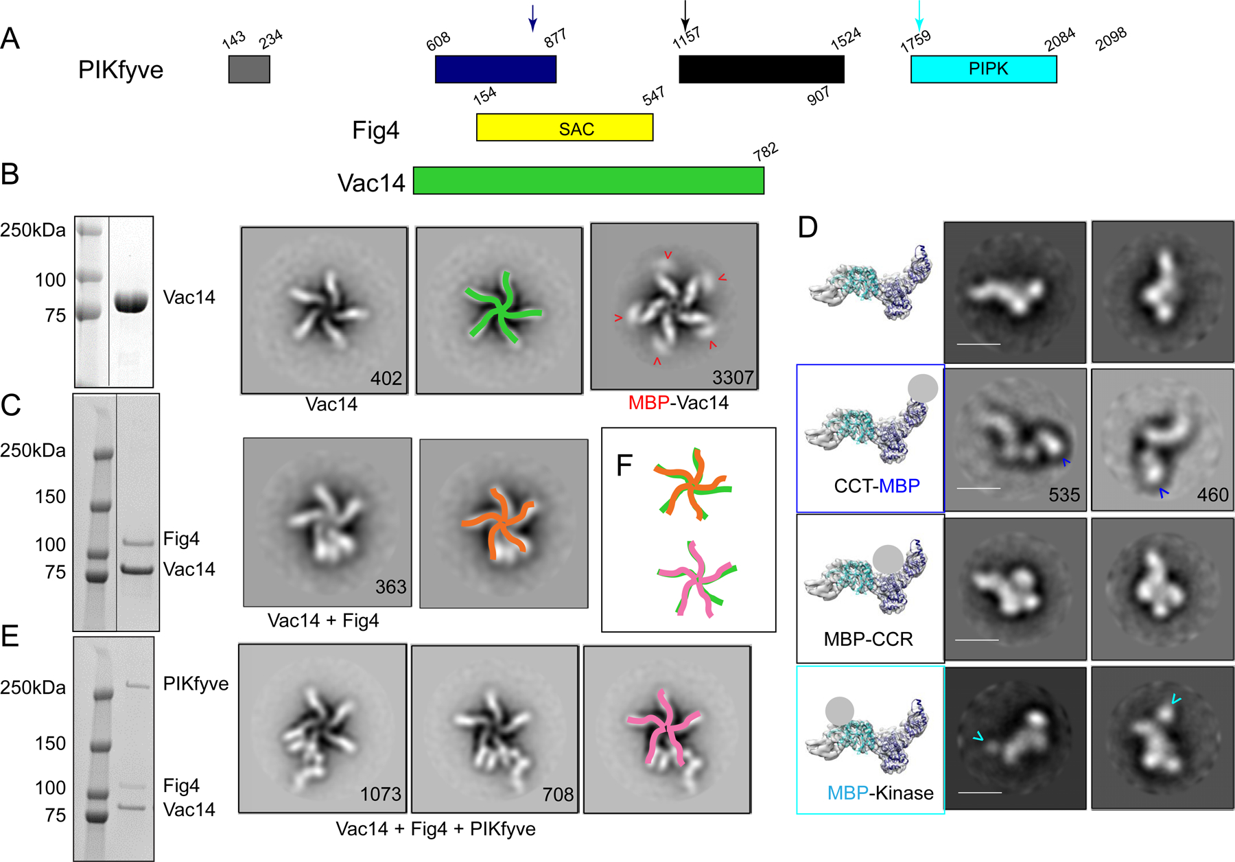
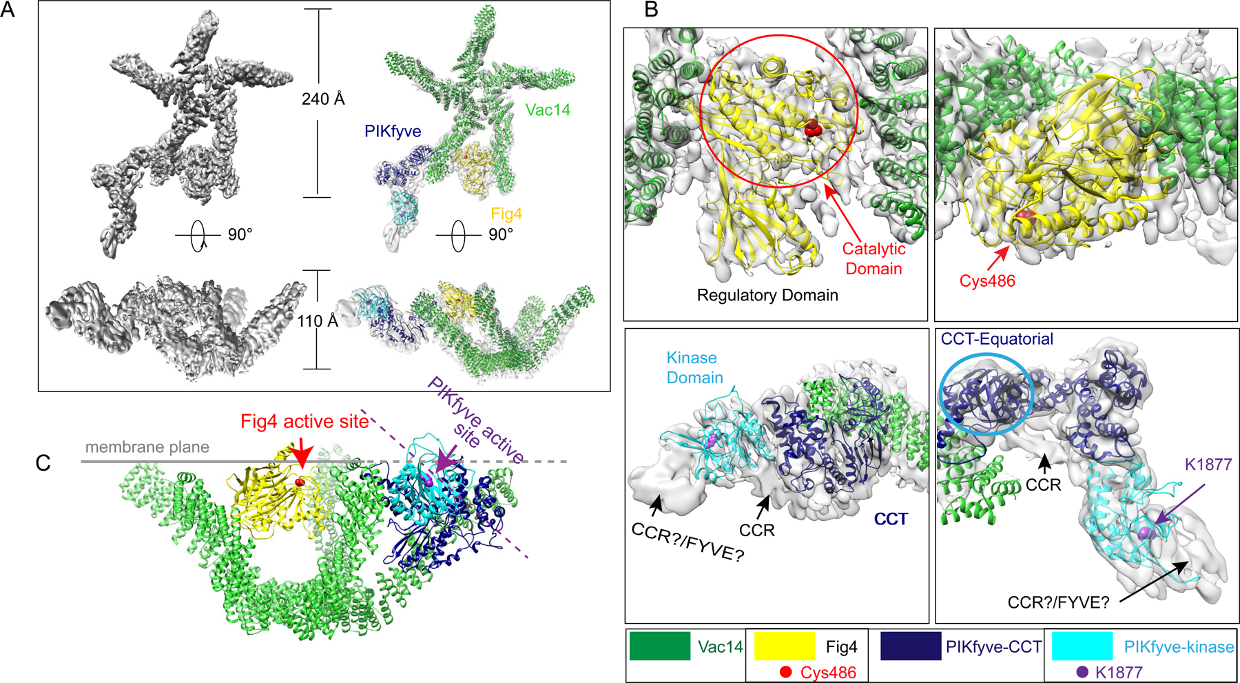
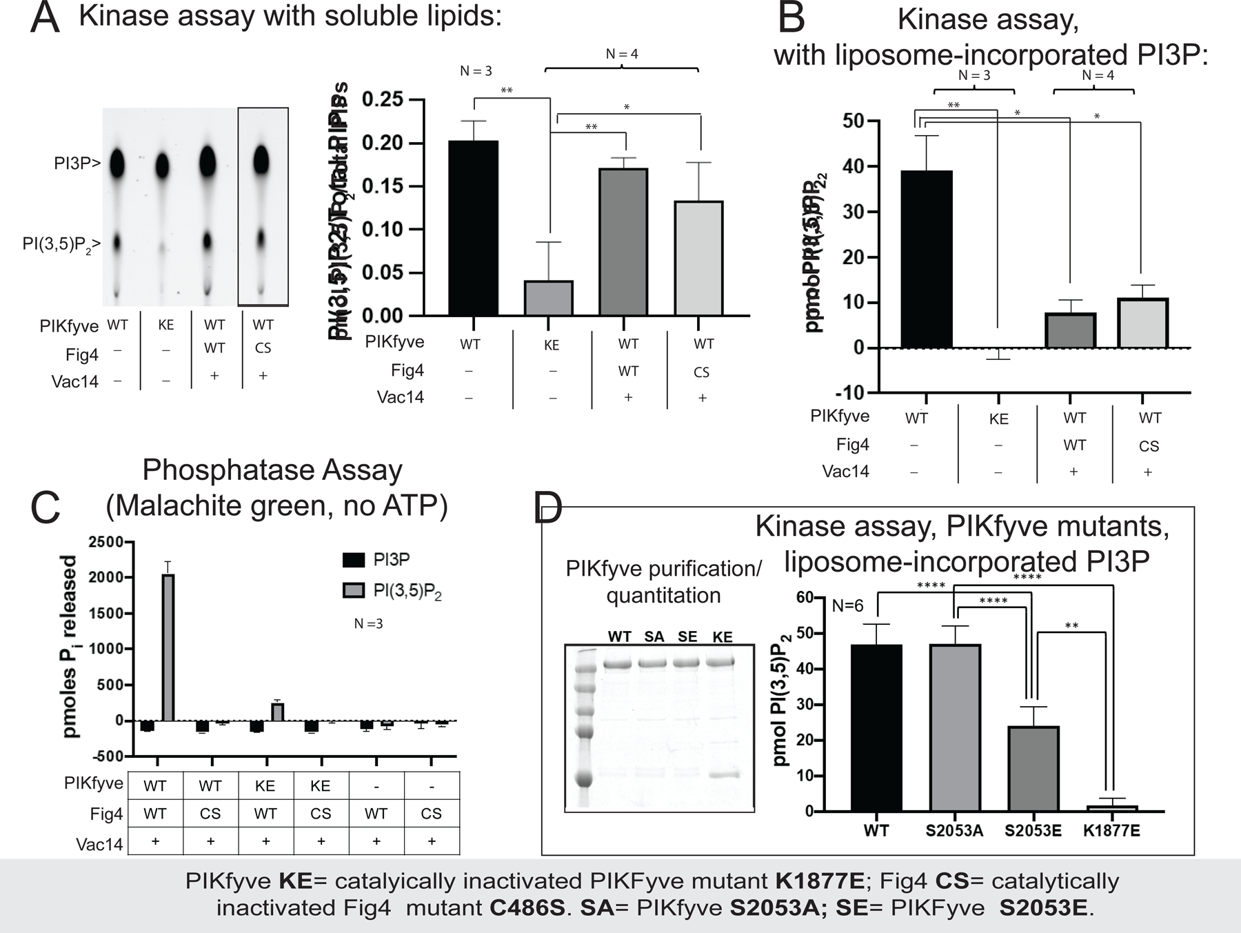
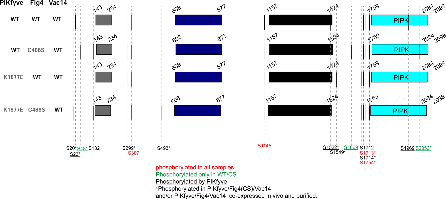
The phosphoinositide PI(3,5)P2, generated exclusively by the PIKfyve lipid kinase complex, is key for lysosomal biology. Here, we explore how PI(3,5)P2 levels within cells are regulated. We find the PIKfyve complex comprises five copies of the scaffolding protein Vac14 and one copy each of the lipid kinase PIKfyve, generating PI(3,5)P2 from PI3P and the lipid phosphatase Fig4, reversing the reaction. Fig4 is active as a lipid phosphatase in the ternary complex, whereas PIKfyve within the complex cannot access membrane-incorporated phosphoinositides due to steric constraints. We find further that the phosphoinositide-directed activities of both PIKfyve and Fig4 are regulated by protein-directed activities within the complex. PIKfyve autophosphorylation represses its lipid kinase activity and stimulates Fig4 lipid phosphatase activity. Further, Fig4 is also a protein phosphatase acting on PIKfyve to stimulate its lipid kinase activity, explaining why catalytically active Fig4 is required for maximal PI(3,5)P2 production by PIKfyve in vivo.

Keywords: lipid kinase; lipid phosphatase; phosphoinositide homeostasis.


Conflict of interest statement

Declaration of Interests The authors declare no competing interests.

Figures

Figure 1.
Figure 1.. Assembly and gross architecture of the human PIKfyve complex by negative stain EM analysis.
(A) Domain organization of PIKfyve, Fig4, and Vac14. (B) The Vac14 sample used for negative staining EM is pure per SDS-PAGE (line indicates lanes not shown). 2D class averages of Vac14 or MBP-Vac14 show that it pentamerizes. Numbers of particles for each class average are indicated. Maltose binding protein (MBP) fused to the Vac14 N-terminus is at the tip of the Vac14 “leg”. (C) The Fig4/Vac14 sample used for negative staining EM is pure per SDS-PAGE (line indicates lanes not shown). 2D class averages of Fig4/Vac14 show Fig4 between two Vac14 “legs”. (D) Negative stain EM analysis of PIKfyve or MBP+PIKfyve fusions. MBP was inserted into the PIKfyve sequence as indicated by arrows in 1A. The left-most column shows PIKfyve density from the cryo-EM maps (Fig. 2) with docked models of the CCT and kinase modules; a grey ball indicates the location of MBP in the class averages. The other columns show 2D class averages of PIKfyve by itself or with MBP-insertions. (E) The PIKfyve/Fig4/Vac14 sample used for negative staining and cryo-EM is pure per SDS-PAGE. 2D class averages of PIKfyve/Fig4/Vac14 show PIKfyve close to Fig4 at the tip of the Vac14 legs. (F) Schematic overlay of the Vac14 pentamers in Vac14, Fig4/Vac14, and PIKfyve/Fig4/Vac14, showing that the 5-fold symmetry of the Vac14 pentamer is distorted in Fig4/Vac14 and PIKfyve/Fig4/Vac14 complexes.
Figure 2.
Figure 2.. Cryo-EM reconstruction of the human PIKfyve complex at medium- low resolution.
(A) The composite reconstruction alone and with model. The left panels show the maps only; the right panels also show docked protein models colored according to panel 1A. (B) Enlarged views showing the fit of the Fig4 Sac homology module (top panels) and the PIKfyve CCT and kinase modules (bottom panels) to the map. Two different views for Fig4 and PIKfyve are related by 90° rotations. (C) Model for PIKfyve complex interacting with membrane. The Fig4 active site is oriented to face the membrane, whereas the PIKfyve active site is twisted away from the membrane by ~45° and so cannot access membrane incorporated phosphoinositides.
Figure 3.
Figure 3.. Complex formation affects the enzymatic activities of PIKfyve and Fig4 with respect to phosphoinositide lipids.
(A) PIKfyve alone and in the PIKfyve/Fig4/Vac14 complex phosphorylate soluble C6-C6-BODIPY-FL-PI3P with similar efficiency, independent of whether the lipid phosphatase Fig4 is catalytically active (WT) or not (C486S). The reaction was monitored by thin layer chromatography; representative image shown at left (lanes rearranged, as indicated by line, to match graph). (B) PIKfyve alone phosphorylates liposome incorporated PI3P in a radiometric assay more efficiently that the PIKfyve/Fig4/Vac14 complex. The PIKfyve active site may be sterically constrained in the complex and so unable to access membrane-incorporated PI3P. (C) In a malachite green assay monitoring dephosphorylation of soluble di-C8 PI(3,5)P2, robust Fig4 lipid phosphatase activity was observed only for Fig4 in a complex with WT PIKfyve. Complexes comprising a catalytically inactivated PIKfyve mutant (K1877E) were significantly less active; binary complex lacking PIKfyve was the least active. (D) PIKfyve lipid kinase activity is inhibited when Ser2053 in the kinase activation loop is mutated to glutamate to mimic its phosphorylation but not when it is changed to alanine. Experiments were carried out at least 3 times, as indicated. Error bars are standard deviations, and significance was determined by Welch’s t-test.
Figure 4.
Figure 4.. PIKfyve autophosphorylates itself, and Fig4 is also a protein phosphatase that dephosphorylates PIKfyve.
To reconstitute ternary complexes, WT PIKfyve or a catalytically inactivated mutant (K1877E) were mixed in equimolar amounts with Fig4 (WT or C486S)/Vac14 complexes. After incubation with ATP to allow phosphorylation, samples were resolved by SDS-PAGE. Bands for PIKfyve excised from the gels was analyzed by MS/MS to identify phosphosites. MS/MS analysis was also used to identify PIKfyve phosphosites in complexes produced by co-expressing all three proteins, with either WT or catalytically inactivated Fig4 (C486S). Importantly, Ser2053 in the kinase activation loop is phosphorylated in the presence of WT PIKfyve but not K1877E mutant and is dephosphorylated in complexes containing WT Fig4 but not C486S mutant. This suggests a model where Fig4 activates PIKfyve by dephosphorylating PIKfyve’s activation loop Ser2053, explaining why maximal PI(3,5)P2 production in vivo requires catalytically active Fig4.

References

    1. Botelho RJ, Efe JA, Teis D, and Emr SD (2008). Assembly of a Fab1 phosphoinositide kinase signaling complex requires the Fig4 phosphoinositide phosphatase. Mol Biol Cell 19, 4273–4286. - PMC - PubMed
    1. Chow CY, Landers JE, Bergren SK, Sapp PC, Grant AE, Jones JM, Everett L, Lenk GM, McKenna-Yasek DM, Weisman LS, et al. (2009). Deleterious variants of FIG4, a phosphoinositide phosphatase, in patients with ALS. Am J Hum Genet 84, 85–88. - PMC - PubMed
    1. Chow CY, Zhang Y, Dowling JJ, Jin N, Adamska M, Shiga K, Szigeti K, Shy ME, Li J, Zhang X, et al. (2007). Mutation of FIG4 causes neurodegeneration in the pale tremor mouse and patients with CMT4J. Nature 448, 68–72. - PMC - PubMed
    1. Currinn H, Guscott B, Balklava Z, Rothnie A, and Wassmer T (2016). APP controls the formation of PI(3,5)P(2) vesicles through its binding of the PIKfyve complex. Cell Mol Life Sci 73, 393–408. - PMC - PubMed
    1. Duex JE, Nau JJ, Kauffman EJ, and Weisman LS (2006a). Phosphoinositide 5- phosphatase Fig 4p is required for both acute rise and subsequent fall in stress-induced phosphatidylinositol 3,5-bisphosphate levels. Eukaryot Cell 5, 723–731. - PMC - PubMed

Publication types

MeSH terms

Substances