

'Vanishing carpal bones' in a patient with congenital adrenal hyperplasia - A diagnostic dilemma
- PMID: 33100741
- PMCID: PMC7577888
- DOI: 10.1016/j.jor.2020.10.001
'Vanishing carpal bones' in a patient with congenital adrenal hyperplasia - A diagnostic dilemma
Abstract
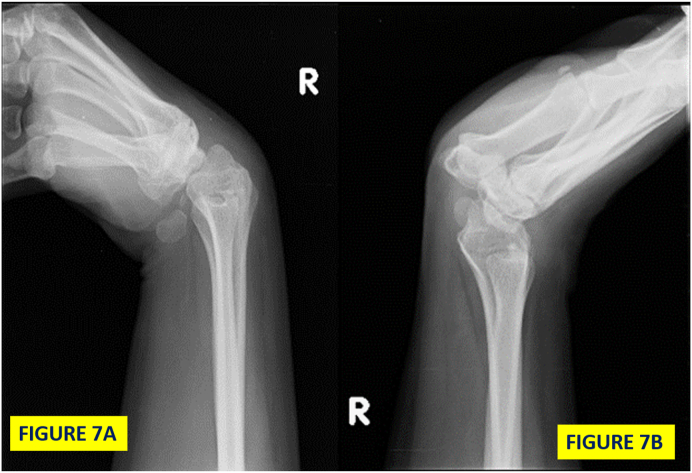
The vanishing bone disease, also known as Gorhams disease usually involves long bones. Isolated carpal bone involvement is uncommon. A 14 year old male presented with pain in the right wrist joint since 4 months. He was a known case of Congenital Adrenal Hyperplasia on oral steroids for 3 years. X-rays showed deformed arthritic scaphoid, lunate and capitate. A proximal row carpectomy was performed to provide functional wrist mobility to the patient. Histopathological examination was consistent with Gorham's disease. In a patient with systemic disease receiving steroid therapy Gorhams disease poses a diagnostic challenge.
Keywords: Gorham's disease; Vanishing carpal bones; Wrist.
© 2020 Professor P K Surendran Memorial Education Foundation. Published by Elsevier B.V. All rights reserved.
Conflict of interest statement
No conflict of interest was noted between any of the authors.
Figures


References
-
- Gorham L.W., Stout A.P. Massive osteolysis (acute spontaneous absorption of bone, phantom bone, disappearing bone): its relation to hemangiomatosis. J Bone Joint Surg [Am] 1955;37-A:985–1004. - PubMed
-
- Gowin W., Rahmanzadeh R. Radiologic diagnosis of massive idiopathic osteolysis (Gorham-Stout Syndrome) Rontgenpraxis. 1985;38:128–134. - PubMed
-
- Horst M., Zsernaviczky J., Delling G. A rare case of so-called idiopathic osteolysis associated with a lymphangioma of the fibula. Z Orthop. 1979;117:88–95. - PubMed
-
- Florchinger A., Bottger E., Claass-Bottger F., Georgi M., Harms J. Gorham Stout syndrome of the spine: case report and review of the literature. Rofo-Fortschr Geb Rontgenstr Neuen Bildgeb Verfahr. 1998;168:68–76. - PubMed
-
- Moller G., Priemel M., Amling M., Werner M., Kuhlmey A.S., Delling G. The Gorham-Stout syndrome (Gorham's massive osteolysis): a report of six cases with histopathological findings. J Bone Joint Surg [Br] 1999;81-B:501–506. - PubMed
Publication types
LinkOut - more resources
Full Text Sources
