. 2020 Oct 10:2020:8528901.
doi: 10.1155/2020/8528901. eCollection 2020.

Guttiferone K Exerts the Anti-inflammatory Effect on Mycobacterium Tuberculosis- (H37Ra-) Infected Macrophages by Targeting the TLR/IRAK-1 Mediated Akt and NF- κ B Pathway

Affiliations

Guttiferone K Exerts the Anti-inflammatory Effect on Mycobacterium Tuberculosis- (H37Ra-) Infected Macrophages by Targeting the TLR/IRAK-1 Mediated Akt and NF- κ B Pathway

Qingwen Zhang et al. Mediators Inflamm. .

Abstract

Mycobacterium tuberculosis (Mtb) remains a great threat to global health, killing more people than any other single infectious agent and causing uncontrollable inflammation in the host. Poorly controlled inflammatory processes can be deleterious and result in immune exhaustion. The current tuberculosis (TB) control is facing the challenge of drugs deficiency, especially in the context of increasingly multidrug resistant (MDR) TB. Under this circumstance, alternative host-directed therapy (HDT) emerges timely which can be exploited to improve the efficacy of TB treatment and disease prognosis by targeting the host. Here, we established the in vitro infection model of Mtb macrophages with H37Ra strain to seek effective anti-TB active agent. The present study showed that Guttiferone K, isolated from Garcinia yunnanensis, could significantly inhibit Mtb-induced inflammation in RAW264.7 and primary peritoneal macrophages. It was evidenced by the decreased production of inflammatory mediators, including interleukin-1β (IL-1β), tumor necrosis factor-α (TNF-α), interleukin-6 (IL-6), inducible nitric oxide synthase (iNOS), and cyclooxygenase-2 (COX-2). Further studies with immunoblotting and immunofluorescence revealed that Guttiferone K obviously inhibits the nuclear factor-kappa B (NF-κB) both in RAW264.7 and primary peritoneal macrophages relying on the TLR/IRAK-1 pathway. Guttiferone K could also suppress the NLRP3 inflammasome activity and induce autophagy by inhibiting the protein kinase B (p-Akt) and mammalian target of rapamycin (mTOR) phosphorylation at Ser473 and Ser2448 in both cell lines. Thus, Guttiferone K possesses significant anti-inflammatory effect, alleviating Mtb-induced inflammation with an underlying mechanism that targeting on the TLR/IRAK-1 pathway and inhibiting the downstream NF-κB and Akt/mTOR signaling pathways. Together, Guttiferone K can be an anti-inflammatory agent candidate for the design of new adjunct HDT drugs fighting against tuberculosis.


Conflict of interest statement

The authors declare no conflict of interest.

Figures

Figure 1
Figure 1
Effect of Guttiferone K on the viability of RAW264.7 cells. (a) The chemical structure of Guttiferone K. (b) A proliferation assay was conducted to assess the cytotoxic effect of Guttiferone K on RAW264.7 cells. RAW264.7 cells were treated with various concentrations of GK (0, 5, 10, 20 μM) for 24, 48, and 72 hr. After the addition of CCK-8 reagent, the optical density of each well was determined at 450/650 nm. Data are shown as the means ± SD of three independent experiments.
Figure 2
Figure 2
Guttiferone K inhibits Mtb-induced proinflammatory cytokines production. Primary peritoneal macrophage (a) cells or RAW264.7 (b–d) were infected with H37Ra and treated with different concentrations of GK (2.5, 5, 10, or 20 μM) for 12 hr. The concentrations of IL-1β (a), TNF-α (b), and IL-6 (c) in the supernatants were measured by ELISA. (d) GK suppresses the expression of iNOS and COX2 induced by Mtb infection, and the protein levels of iNOS and COX2 were detected by Western blot. Data are shown as the means ± SD (n≧3). p < 0.05, ∗∗p < 0.01, ∗∗∗p < 0.001 (one-way ANOVA).
Figure 3
Figure 3
Guttiferone K inhibits Mtb-triggered activation of the NF-κB signaling pathway by targeting the TLR/IRAK-1 pathway. (a, b) Western blot analysis of p-IRAK1 expression in RAW264.7 and primary peritoneal macrophage cells. (a) TLR2/4 antibodies were pretreated on RAW264.7 cells before Mtb infection; then, the production of p-IRAK1 at the protein level was detected by immunoblotting and normalized to β-actin. (b) Primary peritoneal macrophage cells were infected with Mtb for 0, 30, 60, and 180 min and correspondingly treated with GK (10 μM) for 0, 30, 60, and 180 min; then, the production of p-IRAK1 at the protein level was detected by immunoblotting and normalized to β-actin. Data are representative of at least three independent experiments. (c, d) Evaluation of NF-κB expression at the protein level. Western blot analysis of p-p65 expression in RAW264.7 cells (c) or in primary peritoneal macrophage cells (d) and normalized to total p65. (e, f) GK prevents the nuclear translocation of Mtb-induced NF-κB. Confocal microscopy of RAW264.7 cells (e) or primary peritoneal macrophage cells (f) receiving different treatments immunostained with anti-pp65 (green) and DAPI (blue). Data are representative of at least three independent experiments, p < 0.05, ∗∗p < 0.01 (one-way ANOVA).
Figure 4
Figure 4
Guttiferone K has no effect on the MAPK pathway. RAW264.7 cells (a) or primary peritoneal macrophage cells (b) were infected with Mtb and treated with or without GK (10 μM) for 30, 60, or 180 min. The protein levels of JNK, p-JNK, ERK, p-ERK, p38, p-p38, and β-actin were detected by Western blot and normalized to their respective nonphosphorylated total proteins. The results are representative of at least three independent experiments. NS: not significant.
Figure 5
Figure 5
Guttiferone K significantly inhibits Mtb-induced NLRP3 inflammasome activation. (a, b) Levels of NLRP3, pro-IL-1β, and pro-caspase-1 expression in cell lysates and mature IL-1β in the supernatant were determined by Western blot in RAW264.7 (a) and primary peritoneal macrophage cells (b) and normalized to β-actin. (c) ASC or NLRP3 immunoprecipitates from primary peritoneal macrophage cells were immunoblotted for NLRP3 or ASC and reblotted for ASC or NLRP3, respectively. The results are representative of at least three independent experiments, ∗∗∗p < 0.001 (one-way ANOVA), compared with the Mtb group.
Figure 6
Figure 6
Guttiferone K can promote the activation of autophagy in Mtb-infected macrophages. (a) Western blot analysis of LC3 I/II and p62 expression in Mtb-infected RAW264.7 cells after treatment with different concentrations of GK (5, 10, 20 μM) or rapamycin (1 μg/ml) for 12 hr, normalized to LC3 I or β-actin. (b) Western blot analysis of LC3 I/II and p62 expression in Mtb-infected RAW264.7 cells after treatment with GK (10 μM) or CQ (10, 20 μM) for 12 hr, normalized to LC3 I or β-actin. The results are representative of at least three independent experiments, p < 0.05, ∗∗p < 0.01, ∗∗∗p < 0.001 (one-way ANOVA), compared with the Mtb group.
Figure 7
Figure 7
Guttiferone K restrains NLRP3 inflammasome activity depending on the autophagy process. (a) Western blot analysis of NLRP3, LC3 I/II, and p62 expression in Mtb-infected RAW 264.7 cells after treatment with GK (10 μM) or CQ (10 or 20 μM) for 12 hr. (b) Evaluation of si-BECN1 transfection efficiency by Western blot in RAW264.7 cells. (c) BECN1 knockdown downregulates LC3 and restricts the inhibitory effect of GK on NLRP3 in RAW264.7 cells. (d) Western blot analysis of IL-1β, NLRP3, LC3 I/II, and p62 expression in Mtb-infected primary peritoneal macrophage cells after treatment with different concentrations of GK (5, 10, 20 μM) or rapamycin (1 μg/ml). The results are representative of at least three independent experiments.
Figure 8
Figure 8
Guttiferone K promotes the colocalization of inflammasomes with autophagosomes. (a) Confocal microscopy of primary peritoneal macrophages with different treatments immunostained with anti-LC3 antibody (pink), anti-ASC antibody (green), and DAPI (blue). (b) Confocal microscopy of primary peritoneal macrophage cells with different treatments immunostained with anti-LC3 antibody (green), anti-NLRP3 antibody (red), and DAPI (blue). Experiments performed at least three times.
Figure 9
Figure 9
Guttiferone K inhibits the PI3K/Akt/mTOR pathway to play an anti-inflammatory role. (a) RAW264.7 cells or (b) primary peritoneal macrophage cells were infected with Mtb and treated with or without GK (10 μM) for 30, 60, or 180 min. The protein levels of Akt, p-Akt (Ser 473), mTOR, and p-mTOR were detected by Western blot and normalized to nonphosphorylated total protein. The results are representative of at least three independent experiments, p < 0.05, ∗∗p < 0.01, ∗∗∗p < 0.001 (one-way ANOVA).
Figure 10
Figure 10
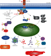
Molecular mechanism by which Guttiferone K exerts anti-inflammatory effects in Mtb-infected macrophages. Through TLR2/TLR4 recognition, Mtb intrudes into the macrophage and then accelerates IRAK-1 phosphorylation to activate its downstream signaling pathways, including NF-κB, MAPK, and Akt/mTOR, causing inflammasome activation, increased production of inflammatory mediators, and autophagy inhibition, ultimately resulting in tissue damage. Guttiferone K targets the inhibition of IRAK-1 phosphorylation and then drives the inhibition of downstream NF-κB and Akt/mTOR, reducing the expression of inflammatory factors and promoting cell autophagy to alleviate inflammation-mediated tissue damage induced by Mtb.

References

    1. Word Health Organization. Global Tuberculosis Report 2018. Geneva: WHO; 2018. https://www.who.int/tb/publications/global_report/en/
    1. Palucci I., Delogu G. Host directed therapies for tuberculosis: futures strategies for an ancient disease. Chemotherapy. 2018;63(3):172–180. doi: 10.1159/000490478. - DOI - PubMed
    1. Yang C. S. Advancing host-directed therapy for tuberculosis: new therapeutic insights from the Toxoplasma gondii. Microbial Cell. 2017;4(3):105–107. doi: 10.15698/mic2017.03.565. - DOI - PMC - PubMed
    1. Zumla A., Maeurer M. Host-directed therapies for multidrug resistant tuberculosis. International Journal of Mycobacteriology. 2016;5(Supplement 1):S21–S22. doi: 10.1016/j.ijmyco.2016.09.044. - DOI - PubMed
    1. Kaufmann S. H. E., Dorhoi A., Hotchkiss R. S., Bartenschlager R. Host-directed therapies for bacterial and viral infections. Nature Reviews Drug Discovery. 2018;17(1):35–56. doi: 10.1038/nrd.2017.162. - DOI - PMC - PubMed

MeSH terms

LinkOut - more resources