Review
. 2020 Sep 25:11:579892.
doi: 10.3389/fphys.2020.579892. eCollection 2020.

Extracellular Vesicle miRNAs in the Promotion of Cardiac Neovascularisation

Affiliations
Review

Extracellular Vesicle miRNAs in the Promotion of Cardiac Neovascularisation

Despoina Kesidou et al. Front Physiol. .

Abstract

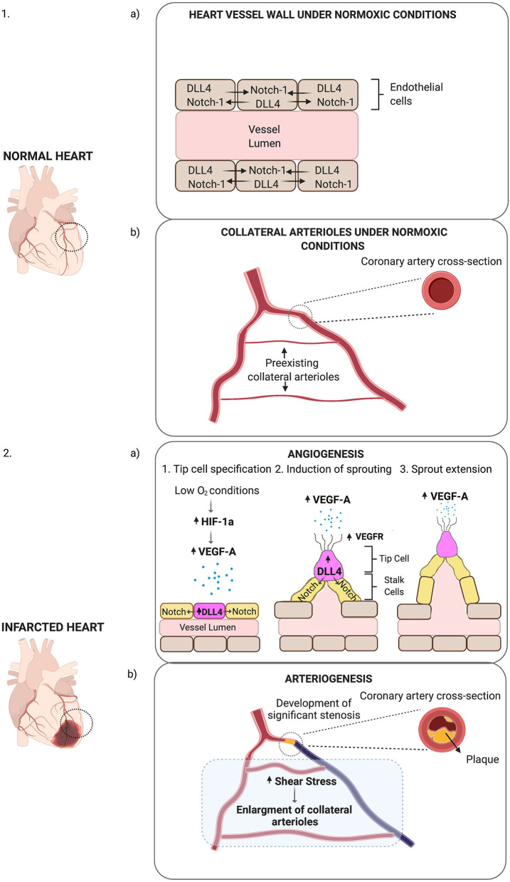
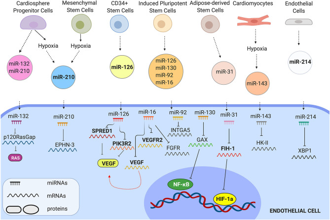
Cardiovascular disease (CVD) is the leading cause of mortality worldwide claiming almost 17. 9 million deaths annually. A primary cause is atherosclerosis within the coronary arteries, which restricts blood flow to the heart muscle resulting in myocardial infarction (MI) and cardiac cell death. Despite substantial progress in the management of coronary heart disease (CHD), there is still a significant number of patients developing chronic heart failure post-MI. Recent research has been focused on promoting neovascularisation post-MI with the ultimate goal being to reduce the extent of injury and improve function in the failing myocardium. Cardiac cell transplantation studies in pre-clinical models have shown improvement in cardiac function; nonetheless, poor retention of the cells has indicated a paracrine mechanism for the observed improvement. Cell communication in a paracrine manner is controlled by various mechanisms, including extracellular vesicles (EVs). EVs have emerged as novel regulators of intercellular communication, by transferring molecules able to influence molecular pathways in the recipient cell. Several studies have demonstrated the ability of EVs to stimulate angiogenesis by transferring microRNA (miRNA, miR) molecules to endothelial cells (ECs). In this review, we describe the process of neovascularisation and current developments in modulating neovascularisation in the heart using miRNAs and EV-bound miRNAs. Furthermore, we critically evaluate methods used in cell culture, EV isolation and administration.

Keywords: angiogenesis; cardiac; exosome (EXO); extracellular vesicles (EV); microRNA (miR); myocardial infarct; neovascularisation; regeneration.


Figures

Figure 1
Figure 1
Schematic representation of post-ischaemic neovascularisation. (1a) Under normoxic conditions, DLL4-Notch-1 signalling is balanced in ECs. (1b) Under normoxic conditions, small collateral arterioles may connect main arteries. (2a) Low O2 conditions increase HIF-1a levels, which in turn stimulates the expression of VEGF-A (represented as small blue dots). In response to VEGF-A, Dll4 expression increases on the surface of tip cells (purple cell). Dll4 receptors on tip cells bind to Notch receptors on the surface of stalk cells (yellow cells) activating Dll4/Notch signalling pathway. Dll4—Notch-1 signalling directs ECs into migrating tip or proliferating stalk cells. In response to VEGF-A, VEGFR expression increases on the filopodia of tip cells (represented as protrusions on the tip cell), which then migrate towards VEGF-A with the stalk cells proliferating behind them. This results in the expansion of the vessel lumen and the formation of sprouts on the vessel wall (angiogenesis). (2b) When a coronary artery is occluded due to the formation of plaque within the arterial wall, there is increased shear stress in the collateral arterioles. In response to shear stress, collateral arteriole diameter increases in size (arteriogenesis).
Figure 2
Figure 2
Representative examples of EV miRNAs that control EC behaviour. EVs from different cells carry angiogenic miRNAs. Once transferred in ECs, miRNAs control molecular pathways by inhibiting their mRNA-targets.

References

    1. Adamiak M., Cheng G., Bobis-Wozowicz S., Zhao L., Kedracka-Krok S., Samanta A., et al. . (2018). Induced pluripotent stem cell (iPSC)-derived extracellular vesicles are safer and more effective for cardiac repair than iPSCs. Circ. Res. 122, 296–309. 10.1161/CIRCRESAHA.117.311769 - DOI - PMC - PubMed
    1. Albanese M., Chen A. Y.-F., Hüls C., Gärtner K., Tagawa T., Keppler O. T., et al. (2020). Micro RNAs are minor constituents of extracellular vesicles and are hardly delivered to target cells. bioRxiv. 10.1101/2020.05.20.106393 - DOI - PMC - PubMed
    1. Anand S., Majeti B. K., Acevedo L. M., Murphy E. A., Mukthavaram R., Scheppke L., et al. . (2010). MicroRNA-132-mediated loss of p120RasGAP activates the endothelium to facilitate pathological angiogenesis. Nat. Med. 16, 909–914. 10.1038/nm.2186 - DOI - PMC - PubMed
    1. Arif M., Pandey R., Alam P., Jiang S., Sadayappan S., Paul A., et al. . (2017). MicroRNA-210-mediated proliferation, survival, and angiogenesis promote cardiac repair post myocardial infarction in rodents. J. Mol. Med. 95, 1369–1385. 10.1007/s00109-017-1591-8 - DOI - PMC - PubMed
    1. Arroyo J. D., Chevillet J. R., Kroh E. M., Ruf I. K., Pritchard C. C., Gibson D. F., et al. . (2011). Argonaute2 complexes carry a population of circulating microRNAs independent of vesicles in human plasma. Proc. Natl. Acad. Sci. U.S.A. 108, 5003–8. 10.1073/pnas.1019055108 - DOI - PMC - PubMed

LinkOut - more resources