Sex-Dependent Pathology in the HPA Axis at a Sub-acute Period After Experimental Traumatic Brain Injury
- PMID: 33101162
- PMCID: PMC7554641
- DOI: 10.3389/fneur.2020.00946
Sex-Dependent Pathology in the HPA Axis at a Sub-acute Period After Experimental Traumatic Brain Injury
Abstract
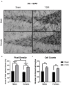
Over 2.8 million traumatic brain injuries (TBIs) are reported in the United States annually, of which, over 75% are mild TBIs with diffuse axonal injury (DAI) as the primary pathology. TBI instigates a stress response that stimulates the hypothalamic-pituitary-adrenal (HPA) axis concurrently with DAI in brain regions responsible for feedback regulation. While the incidence of affective symptoms is high in both men and women, presentation is more prevalent and severe in women. Few studies have longitudinally evaluated the etiology underlying late-onset affective symptoms after mild TBI and even fewer have included females in the experimental design. In the experimental TBI model employed in this study, evidence of chronic HPA dysregulation has been reported at 2 months post-injury in male rats, with peak neuropathology in other regions of the brain at 7 days post-injury (DPI). We predicted that mechanisms leading to dysregulation of the HPA axis in male and female rats would be most evident at 7 DPI, the sub-acute time point. Young adult age-matched male and naturally cycling female Sprague Dawley rats were subjected to midline fluid percussion injury (mFPI) or sham surgery. Corticotropin releasing hormone, gliosis, and glucocorticoid receptor (GR) levels were evaluated in the hypothalamus and hippocampus, along with baseline plasma adrenocorticotropic hormone (ACTH) and adrenal gland weights. Microglial response in the paraventricular nucleus of the hypothalamus indicated mild neuroinflammation in males compared to sex-matched shams, but not females. Evidence of microglia activation in the dentate gyrus of the hippocampus was robust in both sexes compared with uninjured shams and there was evidence of a significant interaction between sex and injury regarding microglial cell count. GFAP intensity and astrocyte numbers increased as a function of injury, indicative of astrocytosis. GR protein levels were elevated 30% in the hippocampus of females in comparison to sex-matched shams. These data indicate sex-differences in sub-acute pathophysiology following DAI that precede late-onset HPA axis dysregulation. Further understanding of the etiology leading up to late-onset HPA axis dysregulation following DAI could identify targets to stabilize feedback, attenuate symptoms, and improve efficacy of rehabilitation and overall recovery.
Keywords: astrocytosis; diffuse axonal injury; diffuse traumatic brain injury; glucocorticoid receptors; hypothalamic-pituitary-adrenal axis; microglia; neuroinflammation; sex-differences.
Copyright © 2020 Bromberg, Condon, Ridgway, Krishna, Garcia-Filion, Adelson, Rowe and Thomas.
Figures


Similar articles
-
The stress system in the human brain in depression and neurodegeneration.Ageing Res Rev. 2005 May;4(2):141-94. doi: 10.1016/j.arr.2005.03.003. Ageing Res Rev. 2005. PMID: 15996533 Review.
-
Aging with TBI vs. Aging: 6-month temporal profiles for neuropathology and astrocyte activation converge in behaviorally relevant thalamocortical circuitry of male and female rats.bioRxiv [Preprint]. 2023 Feb 7:2023.02.06.527058. doi: 10.1101/2023.02.06.527058. bioRxiv. 2023. PMID: 36798182 Free PMC article. Preprint.
-
Effects of prenatal ethanol exposure on basal limbic-hypothalamic-pituitary-adrenal regulation: role of corticosterone.Alcohol Clin Exp Res. 2007 Sep;31(9):1598-610. doi: 10.1111/j.1530-0277.2007.00460.x. Alcohol Clin Exp Res. 2007. PMID: 17760789
-
Sleep fragmentation engages stress-responsive circuitry, enhances inflammation and compromises hippocampal function following traumatic brain injury.Exp Neurol. 2022 Jul;353:114058. doi: 10.1016/j.expneurol.2022.114058. Epub 2022 Mar 28. Exp Neurol. 2022. PMID: 35358498 Free PMC article.
-
Gonadal steroid hormone receptors and sex differences in the hypothalamo-pituitary-adrenal axis.Horm Behav. 1994 Dec;28(4):464-76. doi: 10.1006/hbeh.1994.1044. Horm Behav. 1994. PMID: 7729815 Review.
Cited by
-
Age-at-Injury Determines the Extent of Long-Term Neuropathology and Microgliosis After a Diffuse Brain Injury in Male Rats.Front Neurol. 2021 Sep 8;12:722526. doi: 10.3389/fneur.2021.722526. eCollection 2021. Front Neurol. 2021. PMID: 34566867 Free PMC article.
-
Satureja khuzistanica Jamzad essential oil and pure carvacrol attenuate TBI-induced inflammation and apoptosis via NF-κB and caspase-3 regulation in the male rat brain.Sci Rep. 2023 Mar 23;13(1):4780. doi: 10.1038/s41598-023-31891-3. Sci Rep. 2023. PMID: 36959464 Free PMC article.
-
Temporal changes in the microglial proteome of male and female mice after a diffuse brain injury using label-free quantitative proteomics.Glia. 2023 Apr;71(4):880-903. doi: 10.1002/glia.24313. Epub 2022 Dec 5. Glia. 2023. PMID: 36468604 Free PMC article.
-
Sex-dependent temporal changes in astrocyte-vessel interactions following diffuse traumatic brain injury in rats.Front Physiol. 2024 Sep 25;15:1469073. doi: 10.3389/fphys.2024.1469073. eCollection 2024. Front Physiol. 2024. PMID: 39387100 Free PMC article.
-
Context is key: glucocorticoid receptor and corticosteroid therapeutics in outcomes after traumatic brain injury.Front Cell Neurosci. 2024 Mar 11;18:1351685. doi: 10.3389/fncel.2024.1351685. eCollection 2024. Front Cell Neurosci. 2024. PMID: 38529007 Free PMC article. Review.
References
-
- Centers for Disease Control and Prevention (CDC) Injury Prevention and Control: Traumatic Brain Injury National TBI Estimates. Atlanta, GA: (2013).
-
- National Center for Injury Prevention and Control Report to Congress on Mild Traumatic Brain Injury in the United States: Steps to Prevent a Serious Public Health Problem. Atlanta, GA: Centers for Disease Control and Prevention; (2003).
-
- National Women's Health Network Domestic Abuse and Brain Injury in Women. Washington, DC: Domestic Abuse and Brain Injury in Women; (2017).
Associated data
Grants and funding
LinkOut - more resources
Full Text Sources
Other Literature Sources
Miscellaneous

