

Stabilizing Cellular Barriers: Raising the Shields Against COVID-19
- PMID: 33101215
- PMCID: PMC7554589
- DOI: 10.3389/fendo.2020.583006
Stabilizing Cellular Barriers: Raising the Shields Against COVID-19
Abstract
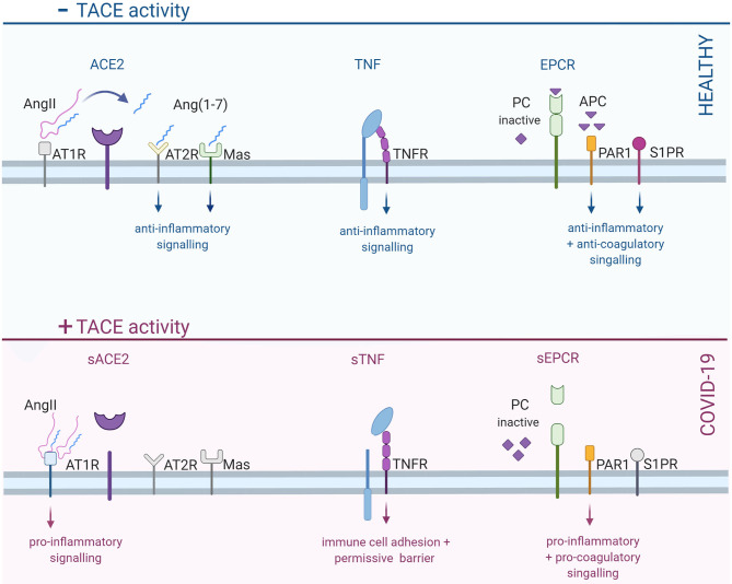
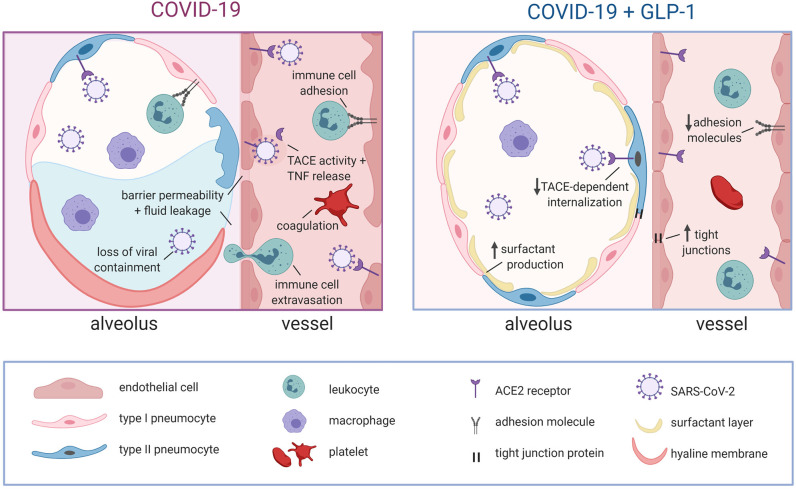
The severe acute respiratory syndrome coronavirus 2 (SARS-CoV-2) and its clinical manifestation (COVID-19; coronavirus disease 2019) have caused a worldwide health crisis. Disruption of epithelial and endothelial barriers is a key clinical turning point that differentiates patients who are likely to develop severe COVID-19 outcomes: it marks a significant escalation in respiratory symptoms, loss of viral containment and a progression toward multi-organ dysfunction. These barrier mechanisms are independently compromised by known COVID-19 risk factors, including diabetes, obesity and aging: thus, a synergism between these underlying conditions and SARS-CoV-2 mechanisms may explain why these risk factors correlate with more severe outcomes. This review examines the key cellular mechanisms that SARS-CoV-2 and its underlying risk factors utilize to disrupt barrier function. As an outlook, we propose that glucagon-like peptide 1 (GLP-1) may be a therapeutic intervention that can slow COVID-19 progression and improve clinical outcome following SARS-CoV-2 infection. GLP-1 signaling activates barrier-promoting processes that directly oppose the pro-inflammatory mechanisms commandeered by SARS-CoV-2 and its underlying risk factors.
Keywords: acute respiratory and circulatory disruption; endothelial barrier disruption; enteroendocrine; glucagon like peptide 1 (GLP-1); immune cells; lung; tumor necrosis factor (TNF); tumor necrosis factor converting enzyme (TACE).
Copyright © 2020 Hanchard, Capó-Vélez, Deusch, Lidington and Bolz.
Figures
Similar articles
-
Obesity and COVID-19.Front Endocrinol (Lausanne). 2020 Sep 30;11:581356. doi: 10.3389/fendo.2020.581356. eCollection 2020. Front Endocrinol (Lausanne). 2020. PMID: 33101213 Free PMC article. No abstract available.
-
Obesity, Diabetes and COVID-19: An Infectious Disease Spreading From the East Collides With the Consequences of an Unhealthy Western Lifestyle.Front Endocrinol (Lausanne). 2020 Sep 17;11:582870. doi: 10.3389/fendo.2020.582870. eCollection 2020. Front Endocrinol (Lausanne). 2020. PMID: 33042029 Free PMC article. Review.
-
From Influenza Virus to Novel Corona Virus (SARS-CoV-2)-The Contribution of Obesity.Front Endocrinol (Lausanne). 2020 Oct 6;11:556962. doi: 10.3389/fendo.2020.556962. eCollection 2020. Front Endocrinol (Lausanne). 2020. PMID: 33123087 Free PMC article. Review.
-
COVID-19: a conundrum to decipher.Eur Rev Med Pharmacol Sci. 2020 May;24(10):5830-5841. doi: 10.26355/eurrev_202005_21378. Eur Rev Med Pharmacol Sci. 2020. PMID: 32495923
-
Diabetes and COVID-19: Global and regional perspectives.Diabetes Res Clin Pract. 2020 Aug;166:108303. doi: 10.1016/j.diabres.2020.108303. Epub 2020 Jul 3. Diabetes Res Clin Pract. 2020. PMID: 32623038 Free PMC article.
Cited by
-
Coagulation and Inflammation in COVID-19: Reciprocal Relationship between Inflammatory and Coagulation Markers.Ann Hematol. 2024 Jun;103(6):1819-1831. doi: 10.1007/s00277-024-05630-1. Epub 2024 Feb 13. Ann Hematol. 2024. PMID: 38349409 Review.
-
The age again in the eye of the COVID-19 storm: evidence-based decision making.Immun Ageing. 2021 May 20;18(1):24. doi: 10.1186/s12979-021-00237-w. Immun Ageing. 2021. PMID: 34016150 Free PMC article.
-
Comparative Review of the State of the Art in Research on the Porcine Epidemic Diarrhea Virus and SARS-CoV-2, Scope of Knowledge between Coronaviruses.Viruses. 2024 Feb 2;16(2):238. doi: 10.3390/v16020238. Viruses. 2024. PMID: 38400014 Free PMC article. Review.
-
Cardiovascular signatures of COVID-19 predict mortality and identify barrier stabilizing therapies.EBioMedicine. 2022 Apr;78:103982. doi: 10.1016/j.ebiom.2022.103982. Epub 2022 Apr 8. EBioMedicine. 2022. PMID: 35405523 Free PMC article.
References
Publication types
MeSH terms
Substances
LinkOut - more resources
Full Text Sources
Other Literature Sources
Medical
Research Materials
Miscellaneous