

Temperature Restriction in Entomopathogenic Bacteria
- PMID: 33101227
- PMCID: PMC7554251
- DOI: 10.3389/fmicb.2020.548800
Temperature Restriction in Entomopathogenic Bacteria
Abstract
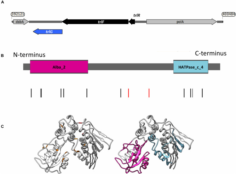
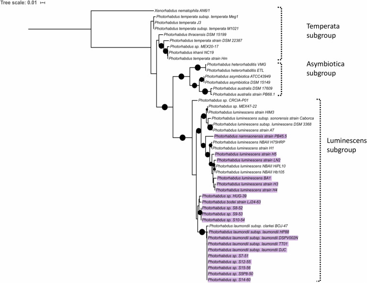
Temperature plays an important role in bacteria-host interactions and can be a determining factor for host switching. In this study we sought to investigate the reasons behind growth temperature restriction in the entomopathogenic enterobacterium Photorhabdus. Photorhabdus has a complex dual symbiotic and pathogenic life cycle. The genus consists of 19 species but only one subgroup, previously all classed together as Photorhabdus asymbiotica, have been shown to cause human disease. These clinical isolates necessarily need to be able to grow at 37°C, whilst the remaining species are largely restricted to growth temperatures below 34°C and are therefore unable to infect mammalian hosts. Here, we have isolated spontaneous mutant lines of Photorhabdus laumondii DJC that were able to grow up to 36-37°C. Following whole genome sequencing of 29 of these mutants we identified a single gene, encoding a protein with a RecG-like helicase domain that for the majority of isolates contained single nucleotide polymorphisms. Importantly, provision of the wild-type allele of this gene in trans restored the temperature restriction, confirming the mutations are recessive, and the dominant effect of the protein product of this gene. The gene appears to be part of a short three cistron operon, which we have termed the Temperature Restricting Locus (TRL). Transcription reporter strains revealed that this operon is induced upon the switch from 30 to 36°C, leading to replication arrest of the bacteria. TRL is absent from all of the human pathogenic species so far examined, although its presence is not uniform in different strains of the Photorhabdus luminescens subgroup. In a wider context, the presence of this gene is not limited to Photorhabdus, being found in phylogenetically diverse proteobacteria. We therefore suggest that this system may play a more fundamental role in temperature restriction in diverse species, relating to as yet cryptic aspects of their ecological niches and life cycle requirements.
Keywords: Photorhabdus; evolution; mutants; pathogenicity; temperature restriction.
Copyright © 2020 Hapeshi, Healey, Mulley and Waterfield.
Figures


Similar articles
-
A temperature-induced metabolic shift in the emerging human pathogen Photorhabdus asymbiotica.mSystems. 2024 Nov 19;9(11):e0097023. doi: 10.1128/msystems.00970-23. Epub 2024 Oct 24. mSystems. 2024. PMID: 39445821 Free PMC article.
-
Photorhabdus asymbiotica as an Insect and Human Pathogen.Curr Top Microbiol Immunol. 2017;402:159-177. doi: 10.1007/82_2016_29. Curr Top Microbiol Immunol. 2017. PMID: 27726002
-
Polyphasic classification of the genus Photorhabdus and proposal of new taxa: P. luminescens subsp. luminescens subsp. nov., P. luminescens subsp. akhurstii subsp. nov., P. luminescens subsp. laumondii subsp. nov., P. temperata sp. nov., P. temperata subsp. temperata subsp. nov. and P. asymbiotica sp. nov.Int J Syst Bacteriol. 1999 Oct;49 Pt 4:1645-56. doi: 10.1099/00207713-49-4-1645. Int J Syst Bacteriol. 1999. PMID: 10555346
-
Comparative analysis of the Photorhabdus luminescens and the Yersinia enterocolitica genomes: uncovering candidate genes involved in insect pathogenicity.BMC Genomics. 2008 Jan 25;9:40. doi: 10.1186/1471-2164-9-40. BMC Genomics. 2008. PMID: 18221513 Free PMC article. Review.
-
Regulation of Phenotypic Switching and Heterogeneity in Photorhabdus luminescens Cell Populations.J Mol Biol. 2019 Nov 22;431(23):4559-4568. doi: 10.1016/j.jmb.2019.04.015. Epub 2019 Apr 22. J Mol Biol. 2019. PMID: 31022406 Review.
Cited by
-
Heterorhabditis and Photorhabdus Symbiosis: A Natural Mine of Bioactive Compounds.Front Microbiol. 2022 Mar 29;13:790339. doi: 10.3389/fmicb.2022.790339. eCollection 2022. Front Microbiol. 2022. PMID: 35422783 Free PMC article. Review.
-
Insight into the emerging insect to human pathogen Photorhabdus revealing geographic differences in immune cell tropism.Front Microbiol. 2024 Sep 18;15:1425909. doi: 10.3389/fmicb.2024.1425909. eCollection 2024. Front Microbiol. 2024. PMID: 39360318 Free PMC article.
-
A temperature-induced metabolic shift in the emerging human pathogen Photorhabdus asymbiotica.mSystems. 2024 Nov 19;9(11):e0097023. doi: 10.1128/msystems.00970-23. Epub 2024 Oct 24. mSystems. 2024. PMID: 39445821 Free PMC article.
References
-
- Boemare N., Akhurst R. (1988). Biochemical and physiological characterization of colony form variants in Xenorhabdus spp. (Enterobacteriaceae). Microbiology 134 751–761. 10.1099/00221287-134-3-751 - DOI
LinkOut - more resources
Full Text Sources