. 2020 Sep 25:11:590828.
doi: 10.3389/fmicb.2020.590828. eCollection 2020.

Glucose-Mediated Protein Arginine Phosphorylation/Dephosphorylation Regulates ylxR Encoding Nucleoid-Associated Protein and Cell Growth in Bacillus subtilis

Affiliations

Glucose-Mediated Protein Arginine Phosphorylation/Dephosphorylation Regulates ylxR Encoding Nucleoid-Associated Protein and Cell Growth in Bacillus subtilis

Mitsuo Ogura. Front Microbiol. .

Abstract

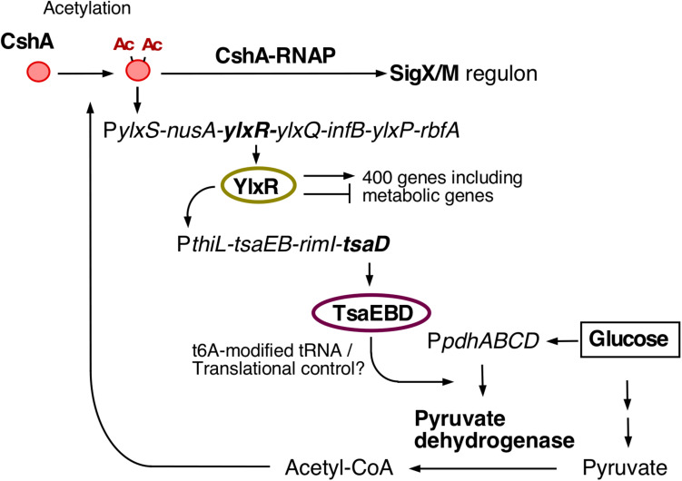
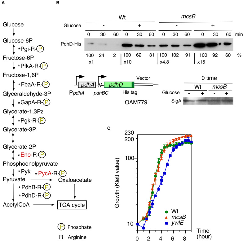
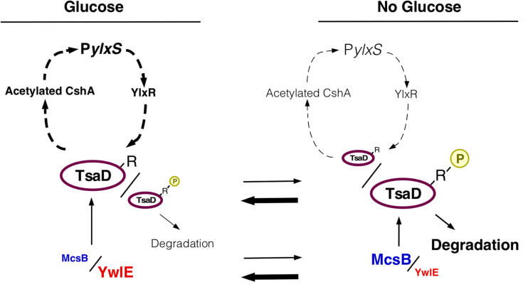
Glucose is the most favorable carbon source for many bacteria, and these bacteria have several glucose-responsive networks. We proposed new glucose responsive system, which includes protein acetylation and probable translation control through TsaEBD, which is a tRNA modification enzyme required for the synthesis of threonylcarbamoyl adenosine (t6A)-tRNA. The system also includes nucleoid-associated protein YlxR, regulating more than 400 genes including many metabolic genes and the ylxR-containing operon driven by the PylxS promoter is induced by glucose. Thus, transposon mutagenesis was performed for searching regulatory factors for PylxS expression. As a result, ywlE was identified. The McsB kinase phosphorylates arginine (Arg) residues of proteins and the YwlE phosphatase counteracts against McsB through Arg-dephosphorylation. Phosphorylated Arg has been known to function as a tag for ClpCP-dependent protein degradation. The previous analysis identified TsaD as an Arg-phosphorylated protein. Our results showed that the McsB/YwlE system regulates PylxS expression through ClpCP-mediated protein degradation of TsaD. In addition, we observed that glucose induced ywlE expression and repressed mcsB expression. It was concluded that these phenomena would cause glucose induction (GI) of PylxS, based on the Western blot analyses of TsaD-FLAG. These observations and the previous those that many glycolytic enzymes are Arg-phosphorylated suggested that the McsB/YwlE system might be involved in cell growth in glucose-containing medium. We observed that the disruption of mcsB and ywlE resulted in an increase of cell mass and delayed growth, respectively, in semi-synthetic medium. These results provide us broader insights to the physiological roles of the McsB/YwlE system and protein Arg-phosphorylation.

Keywords: ClpCP protease; YwlE phosphatase; glucose response; glycolysis; protein acetylation.


Figures

FIGURE 1
FIGURE 1
The current model for glucose-responsive system (GRS). The indicated pathways were previously identified: in vivo association of CshA with RNAP (Delumeau et al., 2011), glucose-stimulated CshA acetylation and sigX regulation (Kosono et al., 2015; Ogura and Asai, 2016), CshA-dependent PylxS regulation driving YlxR expression, which regulates metabolic genes (Ogura and Kanesaki, 2018), and transcriptional regulation of tsaEBD through the functional YlxR-binding to the promoter of tsaEBD, whose products are assembled and regulate pyruvate dehydrogenase translation (Ogura et al., 2019). Pyruvate dehydrogenase provides acetyl-CoA, which would be the acetyl moiety source for CshA acetylation. In the tsaD disruptant grown with glucose cellular acetyl-CoA pool was reduced (Ogura et al., 2019). Each pathway is supported by experimental evidence, however, considering the whole regulatory cascade there is a room for further verification. Arrows indicate transcription, translation, acetylation, enzymatic reaction, transcriptional activation or metabolic reaction. T-bars indicate transcriptional repression. Ac, acetyl moiety; RNAP, RNA polymerase.
FIGURE 2
FIGURE 2
Effects of ywlE and mcsB disruption and phosphorylated-Arg residue mutation of tsaD on PylxS-lacZ expression. (A–D) β-Gal activity in sporulation medium. Data represent means and SD from three independent experiments. The x-axis represents growth time in hours relative to the end of vegetative growth (T0). The relevant genotype and the presence of glucose or xylose are indicated. The strain lacking lacZ showed less than 1 Miller units under the condition with or without glucose. (A) PylxS-lacZ expression in mutant strains. 1, OAM741[wild] and OAM882[tsaD]; 2, OAM883[ywlE]; 3, OAM884[mcsB]; 4, OAM945[ywlE mcsB]. (B) Complementation test of the ywlE mutation using OAM885. (C) Complementation test of tsaD using OAM886[wild] and OAM946[ywlE]. (D) Complementation test of the tsaDR282K mutation using OAM887[Wt] and OAM947[ywlE]. (E) PylxS-lacZ expression on sporulation medium agar plates. Each strain (OAM741[wild], OAM884[mcsB], and OAM883[ywlE]) was inoculated onto 1.5% agar sporulation medium plates containing 100 μg/mL X-gal and spectinomycin, and incubated at 37°C. Images were taken at the indicated time.
FIGURE 3
FIGURE 3
Glucose-induction of ywlE and -repression of mcsB. (A,B) β-Gal activity in sporulation medium using CPRG (A) and ONPG (B) is shown. Data represent means and SD from three independent experiments. The x-axis is the same as in Figure 2. The presence of glucose is indicated. The strain lacking lacZ/bgaB showed less than 1 Miller units under the condition with or without glucose. (A) PctsR-bgaB expression, OAM891. The chromosomal structure of the mcsB-containing operon is shown alongside the panel. (B) PywlE-lacZ expression, OAM888. The chromosomal structures of the ywlE-containing operon and the region around the PywlE-lacZ fusion are shown alongside the panel. (C) Western blot analysis of YwlE-FLAG. The growth phase is indicated in hours relative to the end of vegetative growth (T0) in sporulation medium. Equal protein amounts of whole cell extracts were analyzed in 15% polyacrylamide gels for Western blot using anti-FLAG-tag monoclonal antibody. SigA was used as a control. (D) Model of TsaD Arg-phosphorylation control. Arrows and T-bars indicate transcriptional activation or phosphorylation/dephosphorylation, and transcriptional repression or protein degradation, respectively. R, arginine residue to be phosphorylated; circled P, phosphate residue.
FIGURE 4
FIGURE 4
Western blot analysis of TsaD-FLAG. Equal protein amounts of whole cell extracts were analyzed in 12.5% polyacrylamide gels for Western blots using anti-FLAG-tag monoclonal antibody. The chromosomal structures of OAM897 and OAM 909/910 are shown. Boxes and bent arrows show open-reading frames and promoters, respectively. The promoter region (−500/−1 relative to the translation start site) has full promoter activity (Ogura et al., 2019). (A,B) Protein stability analysis. Band intensities are shown in the graphs. After inhibiting protein synthesis, more than 100% of Arg-phosphorylated TsaD was sometimes observed. It is likely that apparent ratio of TsaD, if not degraded, to the total protein amounts increased due to other protein degradation systems. Time indicates culture sampling interval after chloramphenicol addition, which was added at T1 in sporulation medium culture. G+ and G− indicate the presence or absence of 2% glucose. Closed and open symbols indicate results from the medium containing glucose or no glucose, respectively. Means and SD (error bars) are shown from three to five biologically-independent samples. A, OAM897[wild], OAM898[ywlE], OAM899[mcsB], OAM900[clpC], and OAM901[Pspac-ywlE]. For OAM901, 1 mM IPTG (final concentration) was added. B, OAM909[wild] and OAM910[R282K mutant]. * indicates the introduced nucleotide change.
FIGURE 5
FIGURE 5
Effects of glucose on PdhD-His stability and cell growth profiles in mcsB and ywlE disruptants. (A) Glycolytic pathways and enzymes detected in Arg-phosphorylated forms. Enzymes in red letters are likely to be regulated by ClpCP-dependent degradation (Gerth et al., 2008). Asterisks show the enzymes that have been reported to be acetylated (Kosono et al., 2015; Carabetta et al., 2016). (B) PdhD-His western analysis. Cells were grown in sporulation medium with and without 2% glucose. Equal protein amounts of whole cell extracts were analyzed in 12.5% polyacrylamide gel for Western blots using anti-His-tag monoclonal antibody. Time indicates sampling interval after chloramphenicol addition, which was added at T1. Band intensities are indicated below the panels. The chromosomal structure of OAM779 is shown. Boxes and bent arrows show open-reading frames and promoters, respectively. OAM779 [Wt] and OAM908 [mcsB]. As a control, SigA is shown for time 0 samples. (C) Cell growth profiles of each mutant. Overnight culture grown in A3 medium was inoculated to 4 mL semisynthetic MC (modified competence) medium in an L-tube. Growth was monitored with a Klett calorimeter (Thermo Fisher Scientific, Waltham, MA, United States). Means and SD from three independent experiments are shown. 168[wild], OAM879[mcsB], and OAM881[ywlE].
FIGURE 6
FIGURE 6
Working hypothesis of glucose-mediated PylxS regulation through TsaD in GRS. Description of figures is the same as in Figures 1, 3D. The arrowheads with dotted line indicate indirect positive regulation (see Figure 1).

References

    1. Arrondel C., Missoury S., Snoek R., Patat J., Menara G., Collinet B., et al. (2019). Defects in t6A tRNA modification due to GON7 and YRDC mutations lead to Galloway-Mowat syndrome. Nat. Commun. 10:3967. - PMC - PubMed
    1. Blencke H. M., Homuth G., Ludwig H., Mäder U., Hecker M., Stülke J. (2003). Transcriptional profiling of gene expression in response to glucose in Bacillus subtilis: regulation of the central metabolic pathways. Metab. Eng. 5 133–149. 10.1016/s1096-7176(03)00009-0 - DOI - PubMed
    1. Bryant J. A., Sellars L. E., Busby S. J. W., Lee J. (2014). Chromosome position effects on gene expression in Escherichia coli K-12. Nucleic Acids Res. 42 11383–11392. 10.1093/nar/gku828 - DOI - PMC - PubMed
    1. Carabetta V. J., Cristea I. M. (2017). Regulation, function, and detection of protein acetylation in bacteria. J. Bacteriol. 199 107–117. - PMC - PubMed
    1. Carabetta V. J., Greco T. M., Tanner A. W., Cristea I. M., Dubnau D. (2016). Temporal regulation of the Bacillus subtilis acetylome and evidence for a role of MreB acetylation in cell wall growth. mSystems 1:e00005-16. - PMC - PubMed

LinkOut - more resources